Protective effects of Sasa quelpaertensis Leaf Residue Extract against Potassium Oxonate-induced Hyperuricemia in Mice
Mi Gyeong Jang
1,2, Hana Song
1, Ju Yeop Lee
1, Hee Chul Ko
1, Sung-Pyo Hur
3and Se Jae Kim
1,2*
1Jeju Sasa Industry Development Agency, Jeju National University, Jeju 63243, Korea
2Department of Biology, Jeju National University School, Jeju 63243, Korea
3Korea Institute of Ocean Science & Technology, Jeju 63243, Korea
Received October 15, 2018 /Revised October 30, 2018 /Accepted October 30, 2018
Leaves of Sasa quelpaertensis Nakai are used in folk medicine for their anti-inflammatory, antipyretic, and diuretic properties. To ensure efficient utilization of S. quelpaertensis leaf, we previously reported a preparation method for phytochemical-rich extract (PRE) using the leaf residue, which was pro- duced after hot water extraction. This study was undertaken to evaluate the hypouricemic potential of S. quelpaertensis leaf PRE in potassium oxonate (PO)-induced hyperuricemic mice. The admin- istration of PRE significantly reduced serum uric acid (UA), blood urea nitrogen (BUN), and serum creatinine levels and increased urine UA and creatinine levels in the PO-induced hyperuricemic mice.
It also reduced liver UA levels and xanthine oxidase (XA) activity. A histological analysis revealed that PRE administration protected against PO-induced liver damage, pointing to anti-inflammatory and cytoprotective effects in PO-induced hyperuricemic mice. We analyzed the transcriptome response to PRE administration in PO-induced hyperuricemic mice using RNA sequencing (RNA-Seq) in kidney tissues. The administration of PRE mainly enriched genes involved in mediating immune and in- flammatory responses and the metabolic pathway. A Kyoto Encyclopedia of Genes and Genomes (KEGG) analysis showed that the metabolic pathway, purine metabolism, and antibody biosynthesis were the major pathways altered in the PRE and PO groups. These results suggest a potential role for PRE in the prevention and treatment of hyperuricemia with inflammation.
Key words : Hyperuricemia, potassium oxonate, RNA sequencing, Sasa quelpaertensis, uric acid
*Corresponding author
*Tel : +82-64-754-3529, Fax : +82-64-751-4406
*E-mail : [email protected]
This is an Open-Access article distributed under the terms of the Creative Commons Attribution Non-Commercial License (http://creativecommons.org/licenses/by-nc/3.0) which permits unrestricted non-commercial use, distribution, and reproduction in any medium, provided the original work is properly cited.
Journal of Life Science 2019 Vol. 29. No. 1. 37~44 DOI : https://doi.org/10.5352/JLS.2019.29.1.37
Introduction
Uric acid (UA) is the terminal product of purine metabo- lism and is produced from hypoxanthine by xanthine oxi- dase (XO) in the liver [26]. Serum UA levels are determined by endogenous factors, such as de novo purine synthesis, tis- sue catabolism, and exogenous proteins delivered to the liv- er [12]. UA excretion is controlled by transporters in the kid- neys through renal plasma flow, glomerular filtration, and proximal tubular exchange [8, 21]. An imbalance between UA production and excretion induces hyperuricemia, which is a major causal factor for the development of gout and many other diseases such as obesity, cardiovascular and re- nal diseases, hypertension, and metabolic syndrome [3, 4].
Allopurinol (an XO inhibitor) is the synthetic drug most widely used to treat hyperuricemia [20]; however, its use can lead to side effects [5]. It is therefore necessary to search for alternative agents with few adverse effects for the treat- ment and prevention of hyperuricemia. Natural products have become a source for novel pharmaceuticals due to their potent efficacy and reduced side effects, due to the presence of complex bioactive compounds. Many studies have been conducted on natural products for the purpose of treating hyperuricemia [1, 9, 28].
Sasa species are bamboo grasses, which are widely dis-
tributed in Asian countries including China, Japan, Korea,
and Russia [19]. Their leaves have traditionally been used
in folk medicine for their anti-inflammatory, antipyretic, and
diuretic properties. Sasa quelpaertensis Nakai is a unique ge-
netic resource from Jeju Island, Korea, native to Halla
Mountain. Recently, it has been reported that S. quelpaertensis
leaves possess various health-promoting properties, includ-
ing anti-inflammatory, anti-cancer, anti-obesity properties
[10, 11]. As various applications of S. quelpaertensis leaves
become known, the leaves are increasingly used as nutrace-
uticals. We previously reported a procedure for the prepara- tion of a phytochemical-rich extract (PRE) using residue pro- duced by hot water extraction of S. quelpaertensis leaves [16].
In the current study, we investigated the hypouricemic ef- fects of PRE in potassium oxonate (PO)-induced hyper- uricemic mice. We also analyzed the effects of PRE on tran- scriptome profiles in kidney tissue by RNA sequencing (RNA-Seq).
Materials and Methods
Preparation of PRE
S. quelpaertensis leaves were collected from Mt. Halla on
Jeju Island, and washed and dried in a hot air drier at 60℃.
The dried leaves were pulverized to 100-200 mesh powder, and extracted for 4 hr with hot water (90℃). After the hot water extract was removed, the remaining residue was ex- tracted to prepare PRE with 70% ethanol for 48 hr at room temperature. PRE was filtered, concentrated, and stored at -70℃ until use.
Animals
All animals were allowed free access to water and stand- ard mouse chow, and were maintained at a regular cycle (12 hr light/dark) under room temperature (23±2℃) and rel- ative humidity (60±5%) conditions. The experimental ani- mals were acclimatized to the environment for 7 days before use in the experiments. All experiments were approved by the Institutional Animal Care and Experimental Committee of Jeju National University (No. 2016-0043).
Induction of hyperuricemia and drug administration The uricase inhibitor PO was used to induce hyper- uricemia in mice [25]. PO (250 mg/kg) was intraperitoneally administered once daily, 1 hr before the administration of the drug, for 7 consecutive days. The group was randomly divided into four groups (n=5 per group). In the normal group, mice were fed only the basic diet without PO treat- ment. The day after PO was administered, PO-treated mice were divided into three subgroups: the PO group (PO+50 mg saline/kg of body weight, BW), Allo group (PO+5 mg allopurinol/kg of BW), and PRE group (PO+50 mg PRE/kg of BW).
Blood, urine, and tissue sampling
After 6 days of drug administration, urine samples were
collected for 24 hr in a metabolic cage and centrifuged. The supernatant was collected and analyzed for UA content. On the 7th day, the mouse was anesthetized with ethyl ether.
The blood was centrifuged at 15,000 rpm for 20 minutes to separate the serum. The tissue was extracted, rapidly cooled using liquid nitrogen, and stored at -70℃.
UA, creatinine, and blood urea nitrogen analyses Levels of serum and urine UA, blood urea nitrogen (BUN), and creatinine were measured using a UA kit (Abnova, Taipei, Taiwan), BUN assay kit (Asan Pharm, Gyeonggi, Korea), and creatinine assay kit (BioAssay Systems, CA, USA), respectively, according to the manufacturers’ proto- cols.
Xanthine oxidase activity assay
Liver tissue was homogenized with 200 mM sodium phosphate buffer (pH 7.5) and centrifuged (3,000× g, 4℃) for 20 minutes. The fat layer was removed and the super- natant centrifuged (12,000× g, 4℃) for 30 min. The super- natant was used as the enzyme solution. We then added the enzyme solution and the same amount of reaction sol- ution (200 mM sodium phosphate buffer at pH 7.5 in 1 mM xanthine and 2 mM EDTA), followed by reaction at 37℃
for 10 minutes, and the absorbance at 295 nm was measured.
Histological analysis
Liver tissues were fixed with paraformaldehyde, washed, dehydrated, and embedded in paraffin. A paraffin block was then prepared and sectioned using a microtome to prepare tissue slices. Serial paraffin sections (5 µm) were stained with hematoxylin and eosin solution, and histological changes were observed with a microscope.
RNA extraction, library preparation, and RNA-Seq Kidney tissues were homogenized and total RNA was iso- lated using TRIzol reagent (Invitrogen Corp., Carlsbad, CA, USA). To reduce variation among individuals within ex- perimental groups, total RNA from kidneys of the same group was pooled together in equal amounts to generate a mixed sample. To explore gene expression profiling, RNA- Seq was performed at eBiogen Inc. (Seoul, South Korea).
Briefly, RNA quality was assessed using an Agilent 2100
BioAnalyzer with the RNA 6000 Nano Chip (Agilent Tech-
nologies, Amstelveen, The Netherlands), and RNA quantifi-
cation was performed using an ND-2000 Spectrophotometer
A B
Fig. 1. Administration of Sasa quelpaertensis leaf phytochemical-rich extract (PRE) modulated uric acid (UA) levels in potassium oxonate (PO)-induced hyperuricemic mice. (A) Serum UA; (B) urine UA. Results represent means ± standard error of the mean (SEM; n=4). Normal, normal group; Negative, PO group (PO+50 mg saline/kg of body weight, BW); Allo, allopurinol group (PO+5 mg allopurinol/kg of BW); PRE, PO + PRE group (50 mg PRE/kg of BW). *p<0.05 vs. Normal group, **p<0.01 vs. Normal group, ***p<0.001 vs. Normal group, #p<0.05 vs. Negative group, ##p<0.01 vs. Negative group, ###p<0.001 vs. Negative group.
(Thermo Inc., Wilmington, DE, USA). For control and test RNAs, a library was constructed using the QuantSeq 3’
mRNA-Seq Library Prep Kit (Lexogen, Inc., Vienna, Austria) according to the manufacturer’s instructions. High-through- put sequencing was performed as single-end 75 sequencing using NextSeq 500 (Illumina, Inc., San Diego, CA, USA).
QuantSeq 3’ mRNA-Seq reads were aligned using Bowtie2 [15]; Bowtie2 indices were either generated from the genome assembly sequence or representative transcript sequences for alignment to the genome and transcriptome. The alignment file was used to assemble transcripts, estimate their abun- dances, and detect differential gene expression. Differentially expressed genes (DEGs) were determined based on counts from unique and multiple alignments using coverage in BEDTools [22]. The RT (read count) data were processed based on the quantile–quantile normalization method using EdgeR within the R software environment (R Development Core Team, 2016) using Bioconductor [7]. Gene classification was based on searches performed using the DAVID (http://
david.abcc.ncifcrf.gov/) and Medline databases (http://www.
ncbi. nlm.nih.gov/).
Statistical analysis
Statistical analysis was performed using (SPSS for Win- dows software (ver. 12.0; SPSS Inc., Chicago, IL, USA). All data are expressed as means ± standard error (SE). Statistical differences between groups were examined using one-way analysis of variance (ANOVA) test. Differences were consid- ered statistically significant at a level of p<0.05.
Results
PRE modulated UA levels in PO-induced hyper- uricemic mice
The hypouricemic potential of PRE was assessed by meas- uring serum and urine UA levels in each experimental group. Serum UA levels were significantly higher in the PO-administrated group than in the normal group, indicat- ing that hyperuricemia was induced appropriately by PO treatment (Fig. 1A). Serum UA levels in the PRE group were lower than those in the PO group, which served as a neg- ative control, but were similar to those of the Allo group, which served as a positive control. This result suggests that PRE effectively suppressed UA production in the body. In contrast, urine UA levels were higher in the PRE and Allo groups than in the PO group (Fig. 1B), suggesting that PRE may promote the secretion of UA. These results indicate that PRE may improve PO-induced hyperuricemia by regulating UA levels throughout the body.
PRE modulated BUN and creatinine levels in PO- induced hyperuricemic mice
To evaluate whether PRE administration affected kidney
function in PO-induced hyperuricemic mice, we measured
BUN and serum and urine creatinine levels in each ex-
perimental group. As shown in Fig. 2, BUN levels in the
PRE group were significantly lower than those in the PO
group, and were similar to those in the normal and Allo
groups. Additionally, we measured creatinine levels, which
A B
C
Fig. 3. PRE administration restored PO-induced liver damage. (A) UA; (B) xanthine oxidase levels in livers from each experimental group. (C) Hematoxylin and eosin staining of hepatic paraffin sections at ×200 magnification. Results represent means ± SEM (n=4). **p<0.01 vs. Normal group, ##p<0.01 vs. Allopurinol group, ###p<0.001 vs. Negative group.
A B C
Fig. 2. PRE administration modulated blood urea nitrogen (BUN) and creatinine levels in PO-induced hyperuricemic mice. (A) Serum BUN; (B) serum creatinine; (C) urine creatinine. Results represent means ± SEM (n=4). *p<0.05 vs. Normal group,
**p<0.01 vs. Normal group, ***p<0.001 vs. Normal group, #p<0.05 vs. Negative group, ##p<0.01 vs. Negative group.
are related to the glomerular filtration rate, in the serum and urine of the experimental animals. Serum creatinine lev- els were significantly higher in the PO group, and sig- nificantly lower in the PRE group (Fig. 2B). Conversely, urine creatinine levels were significantly higher in the PRE group than in the PO group (Fig. 2C). These results indicate that PRE may improve kidney function in PO-induced hy- peruricemic mice.
PRE restored liver damage in PO-induced hyper- uricemic mice
We next investigated whether PRE administration could
restore PO-induced liver damage by measuring the amount of UA and XO activity in the liver tissues of each ex- perimental group. The amounts of UA were significantly higher in the PO group than in the normal group. However, the amount of UA in PRE group livers was lower than that in PO group livers (Fig. 3A). Consistent with the amounts of UA detected in liver tissues, XO activity, which is a major factor in UA production, was highest in the PO group (Fig.
3B). XO activities of PRE groups were reduced by 41% com-
pared to those of PO group. These data suggest that PRE
administration may improve liver function by restoring
PO-induced liver damage. These results were confirmed by
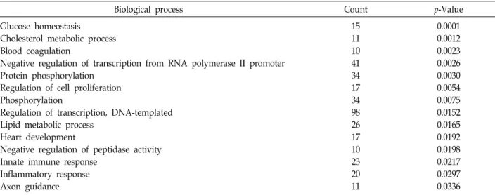
Table 1. Gene ontology (GO) analysis of differentially expressed genes (DEGs) in PRE versus PO groups
Biological process Count p-Value
Glucose homeostasis
Cholesterol metabolic process Blood coagulation
Negative regulation of transcription from RNA polymerase II promoter Protein phosphorylation
Regulation of cell proliferation Phosphorylation
Regulation of transcription, DNA-templated Lipid metabolic process
Heart development
Negative regulation of peptidase activity Innate immune response
Inflammatory response Axon guidance
15 11 10 41 34 17 34 98 26 17 10 23 20 11
0.0001 0.0012 0.0023 0.0026 0.0030 0.0054 0.0075 0.0152 0.0165 0.0192 0.0198 0.0217 0.0297 0.0336
Significant GO terms (biological processes) associated with the identified DEGs. Count: number of genes in set with annotation.
p-Value: Modified Fisher Extract p-Value.
histology of the liver tissue. As shown in Fig. 3C, PRE ad- ministration improved PO-induced liver damage. These re- sults indicate that PRE may improve hyperuricemia by in- hibiting XO activity, thus reducing UA production and pro- tecting liver tissue from destruction by excessive UA.
PRE modulated transcriptome profiles in PO-in- duced hyperuricemic mice
To obtain a global view of the transcriptome response to PRE administration in PO-induced mice, we performed com- parative RNA-Seq analyses of kidney transcriptomes. To identify DEGs in kidneys from the PO and PRE groups, we compared gene expression data from each group using the ExDEGA software (eBiogen). A total of 856 DEGs (456 upre- gulated, 384 downregulated) were detected in the kidneys of PRE versus PO groups. Gene ontology (GO) analysis was performed to determine the DEG signatures. This analysis revealed that enriched genes were mainly involved in the regulation of transcription, DNA templates, the innate im- mune response, the inflammatory response, and immune system processes (in order of the number of counted DEGs;
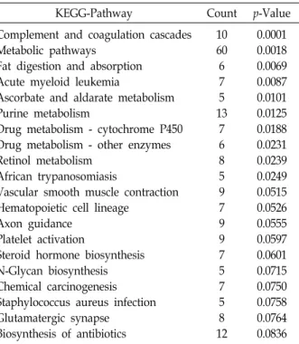
p<0.05; Table 1). Pathway analysis using the Kyoto Encyclo-
pedia of Genes and Genomes (KEGG) database showed that 10 pathways were significantly enriched in the identified DEGs (p<0.05; Table 2). These genes were mainly involved in complement and coagulation cascades, metabolic path- ways, fat digestion and absorption, acute myeloid leukemia, ascorbate and aldarate metabolism, purine metabolism, and drug metabolism.
Discussion
Recently, morbidity due to hyperuricemia has rapidly in- creased worldwide in all age groups. Therefore, there is a growing demand for natural hypouricemic agents that have fewer side effects than therapeutic drugs in current use. In the current study, we investigated the hypouricemic activ- ities of S. quelpaertensis leaves using PRE, an ethanol extract of residue produced by hot water extraction, in PO-induced hyperuricemic mice. PO has been shown to induce a sig- nificant elevation of XO activity and the amount of UA amount in mouse livers, and to impair renal function [2].
We found that PRE decreased serum UA levels in PO-in- duced hyperuricemic mice and was effective in reducing liv- er UA levels in hyperuricemic mice. PRE also significantly inhibited liver XO. Thus, it seems likely that PRE reduced serum UA levels by inhibiting liver XO activity. XO is an enzyme that generates superoxide radicals with xanthine as a substrate to generate UA; i.e., it acts as an oxidizing agent [2, 29]. Since PRE has exhibited antioxidant and XO in- hibition activities in vitro, PRE may exert a similar function to that of allopurinol.
The glomerular filtration rate is an indicator of kidney function. When kidney function deteriorates and the ex- cretion capacity of waste products decreases, the blood crea- tinine level increases and urinary excretion decreases [6, 30].
PRE appears to have the potential to restore kidney function;
this activity was affected by PO-induced hyperuricemia in
the current study, because PRE controlled BUN and crea-
Table 2. Pathway analysis of differentially expressed genes (DEGs) in PRE versus PO groups
KEGG-Pathway Count p-Value
Complement and coagulation cascades Metabolic pathways
Fat digestion and absorption Acute myeloid leukemia
Ascorbate and aldarate metabolism Purine metabolism
Drug metabolism - cytochrome P450 Drug metabolism - other enzymes Retinol metabolism
African trypanosomiasis
Vascular smooth muscle contraction Hematopoietic cell lineage
Axon guidance Platelet activation
Steroid hormone biosynthesis N-Glycan biosynthesis Chemical carcinogenesis Staphylococcus aureus infection Glutamatergic synapse
Biosynthesis of antibiotics
10 60 6 7 5 13
7 6 8 5 9 7 9 9 7 5 7 5 8 12
0.0001 0.0018 0.0069 0.0087 0.0101 0.0125 0.0188 0.0231 0.0239 0.0249 0.0515 0.0526 0.0555 0.0597 0.0601 0.0715 0.0750 0.0758 0.0764 0.0836 Significant pathway associated with the identified DEGs. Count:
number of genes in set with annotation. p-Value: Modified Fisher Extract p-Value.
tinine levels, which are critical renal function indicators.
There is evidence that excess UA is associated with kidney disease, cardiovascular disease, and metabolic disease [30, 18]. It has been reported that liver injury increases XO activ- ity in the liver and serum and may be involved in organ- ismal defenses [24]. Taken together, the results of the current study indicate that PRE restored PO-induced liver damage, and subsequently reduced XO activity and liver UA levels.
The data analysis in the present study indicated that PRE exhibited hypouricemic activities in PO-induced hyper- uricemic mice through UA production and increasing UA excretion.
In recent years, the goal of clinical gout treatment has been to reduce serum UA levels and the inflammatory re- sponse, because UA crystallization within joints and tissues can drive an inflammatory response. The leaves of Sasa spe- cies have various health-promoting properties, including an- tioxidant, anti-inflammatory, anti-cancer, and anti-obesity ef- fects [10, 11, 13, 14, 23]. PRE is a mixture of phytonutrients including polysaccharides, amino acids, and polyphenols, including tricin and p-coumaric acid, which have higher an- tioxidant and anti-inflammatory activities [16]. p-Coumaric acid, a major compound in S. quelpaertensis extracts, has the
potential to prevent or improve insulin resistance and type 2 diabetes by modulating glucose and lipid metabolism [27].
Thus, PRE is expected to exhibit beneficial activities against metabolic diseases, such as hyperuricemia, obesity, car- diovascular and renal diseases, and hypertension [3, 4].
To elucidate the molecular mechanism underlying PRE hypouricemia action, we further investigated the tran- scriptome response to PRE administration using RNA-Seq.
GO and KEGG pathway analyses revealed that PRE regu- lated the expression of genes involved in transcription regu- lation, DNA templates, glucose homeostasis, the innate im- mune response, the inflammatory response, and immune system processes. These results are consistent with those of our previous studies, which demonstrated that PRE played an important role in lipid metabolism and glucose regulation by influencing the metabolic processes associated with the AMPK signaling pathway [11, 27]. It has also been demon- strated the PRE is involved in the regulation of inflammatory responses and immune system processes by inhibiting NF-κ B activity [10]. Therefore, PRE may improve PO-induced hy- peruricemia in part by regulating immune response and in- flammatory signaling in hyperuricemic mice. The tran- scriptome data and identified genes obtained in this study will serve as a molecular basis for understanding the mecha- nisms through which PRE improves PO-induced hyper- uricemia.
In summary, we examined the hypouricemia effects of PRE in hyperuricemia mice. These actions may be attributed to the synergistic effects of UA production inhibition and uricosuric activities of PRE. PRE administration in PO-in- duced hyperuricemic mice mainly enriched genes for im- mune and inflammatory response mediation and the meta- bolic pathway. These results suggest that PRE has potential applications in the prevention and treatment of hyper- uricemia with inflammation.
Acknowledgement
This research was supported by Basic Science Research Program through the National Research Foundation of Korea (NRF) by the Ministry of Education, Science and Technology (2017R1D1A3B03029845), Republic of Korea.
References
1. Chen, G., Tan, M. L., Li, K. K., Leung, P. C. and Ko, C.
H. 2015. Green tea polyphenols decreases uric acid level
through xanthine oxidase and renal urate transporters in hy- peruricemic mice. J. Ethnopharmacol. 175, 14-20.
2. Duke, E. J., Joyce, P. and Ryan, J. P. 1973. Characterization of alternative molecular forms of xanthine oxidase in the mouse. Biochem. J. 131, 187-190.
3. Edwards, N. L. 2009. The role of hyperuricemia in vascular disorders. Curr. Opin. Rheumatol. 21, 132-137.
4. Feig, D. I., Kang, D. H. and Johnson, R. J. 2008. Uric acid and cardiovascular risk. N. Engl. J. Med. 359, 1811-1821.
5. Freig, D. I., Soletsky, B. and Johnson, R. J. 2008. Effect of allopurinol on blood pressure of adolescents with newly di- agnosed essential hypertension: A randomized trial. JAMA.
300, 924-932.
6. Fan, C. Y., Wang, M. X., Ge, C. X., Wang, X., Li, J. M. and Kong, L. D. 2014. Betaine supplementation protects against high-fructose-induced renal injury in rats. J. Nutr. Biochem.
25, 353-362.
7. Gentleman, R. C., Carey, V. J., Bates, D. M., Bolstad, B., Dettling, M., Dudoit, S., Ellis, B., Gautier, L., Ge, Y., Gentry, J., Homik, K., Hothorn, T., Buber, W., Lacus, S., Lrizarry, R., Leisch, F., Li, C., Maechler, M., Rossini, A. J., Sawitzki, G., Smith, C., Smyth, G., Tierney, L., Yang, J. Y. and Zhang, J. 2004. Bioconductor: open software development for com- putational biology and bioinformatics. Genome Biol. 5, R80.
8. George, J. and Struthers, A. D. 2009. Roles of urate, xanthine oxidase and effects of allopurinol in vascular oxidative stress. Vasc. Health. Risk Manag. 5, 265-272.
9. Hidetomo, K., Satomi, K., Rie, A., kouki, S., Atsuko, O., Tadashi, T. and Katsuyoshi, S. 2017. Rosehip inhibits xan- thine oxidase activity and reduces serum urate levels in a mouse model of hyperuricemia. Biomed. Rep. 6, 539-544 10. Hwang, J. H., Choi, S. Y., Ko, H. C., Jang, M. G., Jin, Y.
J., Kang, S. I., Park, J. G., Chung, W. S. and Kim, S. J. 2007.
Anti-inflammatory effect of the hot water extract from Sasa quelpaedensis leaves. Food Sci. Biotech. 16, 728-733.
11. Kang, S. I., Shin, H. S., Kim, H. M., Hong, Y. S., Yoon, S.
A., Kang, S. W., Kim, J. H., Ko, H. C. and Kim, S. J. 2012.
Anti-obesity properties of a Sasa quelpaertensis extract in high-fat diet-induced obese mice. Biosci. Biotechnol. Biochem.
76, 755-761.
12. Kikuchi, H., Kogure, S., Apai, R., Saino, K., Ohkubo, A., Tsuda, T. and Sunaga, K. 2017. Rosehip inhibits xanthine oxidase activity and reduces serum urate levels in a mouse model of hyperuricemia. Biomed. Rep. 6, 539-544.
13. Kim, J. H., Kang, S. I., Shin, H. S., Yoon, S. A., Kang, S.
W., Ko, H. C. and Kim, S. J. 2013. Sasa quelpaertensis and p-coumaric acid attenuate oleic acid-induced lipid acculula-
tion in HepG2 cells. Biosci. Biotechnol. Biochem. 77, 1595-1598.
14. Kim, K. M., Kim, Y. S., Lim, J. Y., Min, S. J., Shin, J. H., Ko, H. C., Kim, S. J., Lim, Y. and Kim, Y. 2014. Sasa quel- paertensis leaf extract suppresses dextran sulfate sodium–
induced colitis in mice by inhibiting the proinflammatory mediators and mitogen-activated protein kinase phosphor- ylation. Nutr. Res. 34, 894-905.
15. Langmead, B. and Salzberg, S. L. 2012. Fast gapped-read alignment with Bowtie 2. Nat. Mathods 9, 357-359.
16. Lee, J. Y., Ko, H. C., Jang, M. G. and Kim, S. J. 2016.
Preparation and characterization of phytochemical-rich ex- tract from Sasa quelpaertensis leaf. J. Life Sci. 26, 1330-1335.
17. Li, J. M., Zhang, X., Wang, X., Xie, Y. C. and Kong, L. D.
2011. Protective effects of cortex fraxini coumarines against oxonate-induced hyperuricemia and renal dysfunction in mice. Eur. J. Pharmacol. 666, 196-204.
18. Lin, K. C., Lin, H. Y. and Chou, P. 2000. The interaction between uric acid level and other risk factors on the devel- opment of gout among asymptomatic hyperuricemic men in a prospective study. J. Rheumatol. 27, 1501-1505.
19. Okabe, S., Takeuchi, K., Takagi, K. and Shibata, M. 1975.
Stimulatory effect of the water extract of bamboo grass (Folin solution) on gastric acid secretion in pylorus-ligated rats. Jap. J. Pharm. 25, 608-609.
20. Pacher, P., Nivorozhkin, A. and Szabo, C. 2006. Therapeutic effects of xanthine oxidase inhibitors: Renaissance half a century after the discovery of allopurinol. Pharmacol. Rev.
58, 87-114.
21. So, A. and Thorens, B. 2010. Uric acid transport and disease.
J. Clini. Investig. 120, 1791-1799.
22. Quinlan, A. R. and Hall, I. M. 2010. BEDTools: a flexible suite of utilities for comparing genomic features. Bioinfor- matics 26, 841-842.
23. Ren, M., Reilly, R. T. and Sacchi, N. 2001. Sasa health exerts a protective effect on Her2/NeuN mammary tumorigenesis.
Anticancer Res. 24, 2879-2884.
24. Tubaro, E., Banci, F., Lotti, B. and Croce, C. 1976. Xanthine oxidase activation in animal liver during infectious proc- esses. Arzneimittel-Forschung 26, 2185-2186.
25. Wang, M. X., Liu, Y. L., Yang, Y., Zhang, D. M. and Kong, L. D. 2015. Nuciferine restores potassium oxonate-induced hyperuricemia and kidney inflammation in mice. Eur. J.
Pharm. 747, 59-70.
초록:생쥐에서 제주조릿대 잎 잔사 추출물의 고요산 혈증 저감 효과
장미경
1,2․송하나
1․이주엽
1․고희철
1․허성표
3․김세재
1,2*
(1제주대학교 제주조릿대 RIS사업단, 2제주대학교 생물학과, 3한국해양과학기술원)