217
Copyrights © 2014 The Korean Society of Radiology
INTRODUCTION
Carpal tunnel syndrome (CTS) is a common disorder caused by compression of the median nerve within the carpal tunnel.
The disorder is most frequently idiopathic. However, the space occupying lesion or systemic disease could contribute to CTS (1). Extraskeletal chondroma is a rare, benign, cartilaginous, soft tissue tumor, usually occurring in the hands and the feet (2-4).
There are few reports in the literature on carpal tunnel syn- drome caused by extraskeletal chondroma (5). We present a case of carpal tunnel syndrome, as a result of an extraskeletal chondroma arising within the carpal tunnel, with descriptions on the radiological and pathological findings of the mass. Au- thors also discuss the differential diagnosis of the calcified space occupying lesions that may occur in carpal tunnel.
CASE REPORT
A 64-year-old female patient presented with a five-year histo- ry of a progressively painful and tingling sensation in her hand.
A clinical examination showed wasting in her thenar eminence and altered sensation in the median nerve distribution. Tinel’s and Phalen’s tests of the median nerve at the carpal tunnel level were both positive. There was no specific past medical or family history. Radiographs of the wrist, including carpal tunnel view, demonstrated a well-defined, oval, homogeneously calcified le- sion within the carpal tunnel, just ventral to the capitate (Fig.
1A-C). A ultrasound (US) study demonstrated a well-defined, echogenic lesion with posterior acoustic shadowing, measuring 15 × 8 × 5 mm in size. The calcified mass was surrounded by the flexor tendons, and was located in the floor of carpal tunnel (Fig. 1D, E). The mass was not movable with finger motion in US. The vascularity of the lesion was not detected on Doppler US scan. The median nerve flattened at the carpal tunnel, with a hy- poechoic swollen appearance at the proximal side of the tunnel (Fig. 1D, F). In the views of imaging, our differential diagnosis of the lesion included hydroxyapatite deposition disease (HADD) and extraarticular synovial chondromatosis. The patient under- went carpal tunnel release and total excision of the lesion, with complete decompression of the median nerve. There was no
Case Report
pISSN 1738-2637 / eISSN 2288-2928 J Korean Soc Radiol 2014;70(3):217-220 http://dx.doi.org/10.3348/jksr.2014.70.3.217
Received September 23, 2013; Accepted February 10, 2014 Corresponding author: Ok Hwa Kim, MD
Department of Radiology, Haeundae Paik Hospital, Inje University College of Medicine, 875 Haeun-daero, Haeundae-gu, Busan 612-896, Korea.
Tel. 82-51-797-0380 Fax. 82-51-797-0379 E-mail: [email protected]
This is an Open Access article distributed under the terms of the Creative Commons Attribution Non-Commercial License (http://creativecommons.org/licenses/by-nc/3.0) which permits unrestricted non-commercial use, distri- bution, and reproduction in any medium, provided the original work is properly cited.
Carpal tunnel syndrome caused by extraskeletal chondroma has been scarcely re- ported in the literature. Authors report a case of carpal tunnel syndrome as a result of an extraskeletal chondroma arising within the carpal tunnel, and describe the ra- diological and pathological findings of the mass. We also discuss the differential di- agnosis of the calcified space, occupying lesions that may occur in carpal tunnel.
Index terms Chondroma Extraskeletal Carpal Tunnel Radiography Ultrasonography
Extraskeletal Chondroma Causing Carpal Tunnel Syndrome: A Case Report
1수근관증후군을 유발한 골외연골종: 증례 보고1
Ok Hwa Kim, MD
1, Yeon Mee Kim, MD
2Departments of 1Radiology, 2Pathology, Haeundae Paik Hospital, Inje University College of Medicine, Busan, Korea
Extraskeletal Chondroma Causing Carpal Tunnel Syndrome
218
J Korean Soc Radiol 2014;70(3):217-220 jksronline.orgcupying lesions or systemic diseases might cause CTS. Systemic diseases may include diabetes, amyloid deposits, and hypothy- roidism. The space occupying the lesion may include inflamma- tion, trauma, tumors, or anatomical anomalies. Among these, tenosynovitis of the flexor tendons and ganglion are the most often encountered (1). Both increase in content or decrease in size of the carpal canal may elevate the pressure within the car- pal tunnel and cause symptoms. In patients with idiopathic CTS, symptoms are typically bilateral and particularly prevalent in middle-aged woman. When patients present with an atypical feature, the possibility of another cause of CTS, such as space occupying lesion, should be considered (1). Extraskeletal chon- droma is an unusual and benign soft tissue mass. It is small and usually is a well-defined nodule of cartilage that is unattached to the bones. The mass constitutes approximately 1.5% of all be- nign soft tissue tumors. It mainly affects the patients of 30 to 60 years of age. But the mass may be encountered over a wide age connection between the lesion and the carpal bones. Histologi-
cally, the excised tumor was encapsulated by a thick fibrous cap- sule and showed nodular configuration (Fig. 1G). Microscopi- cally, the tumor consisted of mature chondrocytes, which were irregularly divided by interlobular fibrous and myxoid septa.
Calcium deposits surrounded benign looking chondrocytes and/or intercellular stroma. A mixed hypocellular zone, with myxoid background in the center and hypercellular zone in the periphery, was seen (Fig. 1G, H). These histopathologic features were consistent with the benign catilagenous tumor. The tumor was diagnosed as an extraskeletal calcified chondroma. Six weeks following the surgery, the patient had no residual painful tingling sensation in her fingers.
DISCUSSION
CTS are most frequently idiopathic. However, some space oc-
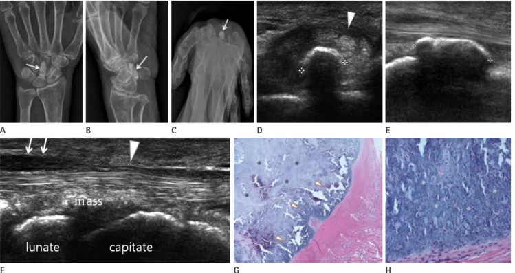
Fig. 1. A 64-year-old woman with carpal tunnel syndrome caused by an extraskeletal chondroma.
A-C. AP (A) and oblique (B) radiographs of the wrist demonstrate a small, well-defined, oval, densely calcified mass (arrow) in the volar wrist.
On carpal tunnel radiograph (C), the mass is located within the carpal tunnel (arrow).
D-F. Transverse (D) and longitudinal (E) ultrasound (US) of the ventral wrist demonstrate a well-defined, echogenic lesion with posterior acoustic shad- owing, consisting with a calcified lesion (cursors), in the deep portion of the carpal tunnel. Transverse (D) and paramedian longitudinal (F) US demon- strate that the median nerve flattens at the carpal tunnel (arrowhead in D, F) and appears to be hypoechoic swollen at the proximal portion (arrows in F).
G, H. At low power photomicrograph (G), the tumor is encapsulated by a thick fibrous capsule and shows the nodular configuration (arrows).
The hypocellular zone with myxoid background in the center (asterisks) and the hypercellular zone in the periphery (arrowheads) are seen (H&E stain, × 40). At high power photomicrograph (H), the peripheral zone of the tumor consists of mature benign looking chondrocytes with diffuse calcium depositions in the intercellular stroma (asterisks) (H&E stain, × 400).
E
A B C D
F G H
Ok Hwa Kim, et al
219
jksronline.org J Korean Soc Radiol 2014;70(3):217-220
chondromatosis, crystal deposition diseases such as HADD, cal- cium pyrophosphate deposition disease, gout, and tumoral cal- cinosis (8). Typically, periosteal chondroma is juxtacortical, and is attached to the bone from within and beneath periosteum.
Radiologically, it presents as a sharply margined bone surface tumor, often with calcification and mineralization of the chon- droid matrix, which are classically associated with saucerization of the underlying bone. Extraarticular synovial chondromatosis is usually characterized by the multiple, small, metaplastic carti- laginous or osseo-cartilaginous nodules, attached to the synovi- um of the tendon sheath or the extraarticular bursa, whereas ex- traskeletal chondroma tends to form a well-defined solitary mass (9). Unlike extraskeletal chondroma, the calcification tends to be amorphous, nodular, or multilobulated, and have relatively poor margins in crystal deposition diseases (10). Malignant soft tissue tumors showing calcification also should be distin- guished. They may include synovial sarcorma, extraskeletal myxoid chondrosarcoma, and extraskeletal osteosarcoma. Syno- vial sarcoma usually shows indistinct margin, less frequently has calcifications often at the periphery, and is spotty (4). Extraskel- etal chondrosarcoma and extraskeletal osteosarcoma are ex- tremely rare in the hands and the feet. They tend to be larger in deep soft tissues, and they have more irregular calcifications or ossification than that of extraskeletal chondroma (8).
In summary, extraskeletal chondroma may rarely occur in carpal tunnel and result in CTS. The radiographic and the US features of extraskeletal chondroma seem to be non-specific and are various, particularly depending on the amount of calcifica- tion of the tumor. But, when a small, slowly growing, well-de- marcated, calcified soft tissue mass is detected, particularly in the distal extremities including the carpal tunnel, extraskeletal chondroma should be included in the differential diagnosis of the lesion.
REFERENCES
1. Peetrons PA, Derbali W. Carpal tunnel syndrome. Semin Musculoskelet Radiol 2013;17:28-33
2. Kransdorf MJ, Meis JM. From the archives of the AFIP. Ex- traskeletal osseous and cartilaginous tumors of the ex- tremities. Radiographics 1993;13:853-884
3. Chung EB, Enzinger FM. Chondroma of soft parts. Cancer range (2-4). This benign tumor is usually discovered as a solitary
lesion in the soft tissue of the hand and the foot. Clinically, the mass usually grows slowly, occasionally causing pain or tender- ness. Lesions are typically well demarcated and lobulated, rarely exceeding 2 cm in greater dimension, firm and rubbery on pal- pation, and often mobile. The extraskeletal chondroma is not re- ported to metastasize or to undergo malignant transformation, and the rate of local recurrence is reported to be 15–20% (3).
Local excision appears to be the treatment of choice (3). The his- topathological appearance of extraskeletal chondroma is vari- able, ranging from an immature pattern dominated by chondro- blasts to a mature form with chondrocytes. It contains matrix of hyaline cartilage, fibrosis, calcification or ossification, or myxoid content. A granuloma-like reaction may be seen within the stro- ma. Radiologically, calcification is observed in 33–70% of the cases, and may have curvilinear, punctuate, mixed pattern, and dystrophic or focal dense pattern. Extrinsic bone erosion or sclerosis can be associated (2-4). There are scant literatures on US findings of extraskeletal chondroma. Bianchi et al. (6) re- ported that extraskeletal chondroma is revealed as a well-de- fined hypoechoic mass with good posterior acoustic enhance- ment. Park et al. (7) reported a case of soft tissue chondroma represented by a well-demarcated, hypervascular, hypoechoic mass with posterior acoustic enhancement on US. There were no radiological or histological calcifications reported in masses in the previous studies. In this case, the mass radiographically showed the dense calcification. It appeared as a well-defined, hy- povascular, echogenic lesion with posterior acoustic shadowing on US (Fig. 1D-G). These discrepant US findings seemed to be the results of the various degrees of calcification in the mass from case to case. So, conventional and Doppler US features of extraskeletal chondroma can be varied, and can depend on the amount of calcification of the mass and the response of the sur- rounding tissue. Based on the literatures, the tumors have non- specific findings on MR imaging, and have shown high signal intensity on T2-weighted images (T2WI), intermediate signal intensity on T1-weighted images (T1WI), and hypointensity of corresponding areas both on T1WI and T2WI, when significant calcification is seen (2). When extraskeletal chondroma occurs in carpal tunnel, especially in the presence of calcifications as in our case, it should be differentiated from other benign soft tissue le- sions, including periosteal chondroma, extraarticular synovial
Extraskeletal Chondroma Causing Carpal Tunnel Syndrome
220
J Korean Soc Radiol 2014;70(3):217-220 jksronline.org8. Bansal M, Goldman AB, DiCarlo EF, McCormack R. Soft tis- sue chondromas: diagnosis and differential diagnosis.
Skeletal Radiol 1993;22:309-315
9. Fetsch JF, Vinh TN, Remotti F, Walker EA, Murphey MD, Sweet DE. Tenosynovial (extraarticular) chondromatosis: an analysis of 37 cases of an underrecognized clinicopatholog- ic entity with a strong predilection for the hands and feet and a high local recurrence rate. Am J Surg Pathol 2003;27:
1260-1268
10. Chen CK, Chung CB, Yeh L, Pan HB, Yang CF, Lai PH, et al.
Carpal tunnel syndrome caused by tophaceous gout: CT and MR imaging features in 20 patients. AJR Am J Roent- genol 2000;175:655-659
1978;41:1414-1424
4. Zlatkin MB, Lander PH, Begin LR, Hadjipavlou A. Soft-tis- sue chondromas. AJR Am J Roentgenol 1985;144:1263- 1267
5. Cumming D, Massraf A, Jones JW. Extraskeletal chondro- ma as a cause of carpal tunnel syndrome: a case report.
Hand Surg 2005;10:327-330
6. Bianchi S, Zwass A, Abdelwahab IF, Olivieri M, Marinaro E.
Sonographic evaluation of soft tissue chondroma. J Clin Ultrasound 1996;24:148-150
7. Park JC, Lee YH, Jung KJ. Subungual hypervascular soft tissue chondroma mimicking a glomus tumor: a case re- port. J Korean Soc Ultrasound Med 2009;28:185-188
수근관증후군을 유발한 골외연골종: 증례 보고1
김옥화
1· 김연미
2골외연골종에 의한 수근관증후군의 발생에 대한 보고는 매우 드물다. 저자들은 수근관내의 연부조직에서 기원해 수근관 증후군을 일으킨 골외연골종 한 예를 영상의학적 소견 및 병리학적 소견과 함께 보고하고자 한다. 또한, 저자들은 감별 진 단을 위해 수근관내에서 석회화를 보일 수 있는 질환에 대해 영상의학적 고찰을 하고자 한다.
인제대학교 의과대학 해운대백병원 1영상의학과, 2병리과