Sonographic Growth Charts for Kidney Length in Normal Korean Children: A Prospective Observational Study
Kidney length is the most useful parameter for clinical measurement of kidney size, and is useful to distinguish acute kidney injury from chronic kidney disease. In this prospective observational study of 437 normal children aged between 0 and < 13 years, kidney length was measured using sonography. There were good correlations between kidney length and somatic values, including age, weight, height, and body surface area. The rapid growth of height during the first 2 years of life was intimately associated with a similar increase in kidney length, suggesting that height should be considered an important factor correlating with kidney length. Based on our findings, the following regression equation for the reference values of bilateral kidney length for Korean children was obtained: kidney length of the right kidney (cm) = 0.051 × height (cm) + 2.102; kidney length of the left kidney (cm) = 0.051 × height (cm) + 2.280. This equation may aid in the diagnosis of various kidney disorders.
Keywords: Kidney; Length; Height; Child; Ultrasonography Min-su Oh,1 Geol Hwang,1
Sanghoon Han,2 Hyun Sik Kang,1 Seung Hyo Kim,3 Young Don Kim,3 Ki-Soo Kang,3 Kyung-Sue Shin,3 Mu Sook Lee,4 Guk Myung Choi,4 and Kyoung Hee Han3
1Department of Pediatrics, Jeju National University Hospital, Jeju, Korea; 2Department of Internal Medicine, Jeju National University School of Medicine, Jeju, Korea; 3Department of Pediatrics, Jeju National University School of Medicine, Jeju, Korea; 4Department of Diagnostic Radiology, Jeju National University School of Medicine, Jeju, Korea Received: 5 January 2016
Accepted: 9 April 2016 Address for Correspondence:
Kyoung Hee Han, MD
Department of Pediatrics, Jeju National University School of Medicine, 15 Aran 13-gil, Jeju 63241, Korea
E-mail: [email protected]
http://dx.doi.org/10.3346/jkms.2016.31.7.1089 • J Korean Med Sci 2016; 31: 1089-1093
INTRODUCTION
Congenital anomalies of the kidney and urinary tract are rela- tively common, accounting for approximately 20%-30% of all anomalies identified in the prenatal period (1). Among the con- genital anomalies of the kidney and urinary tract, reflux nephro- pathy, obstructive uropathy, and renal agenesis/hypoplasia/
dysplasia are responsible for 30%-50% of end-stage renal dis- ease in children (2). Renal hypoplasia is defined as congenitally small kidneys with a reduced number of nephrons but normal architecture, and can be clinically diagnosed based on renal ul- trasonography findings as a small-sized kidney of 2 standard deviations below the expected mean of an age-matched nor- mal individual (3). In addition, mild renal hypoplasia at the lower expected normal range is associated with hypertension and chronic kidney disease (CKD) in adults (3). Therefore, it is important to clinically diagnose renal hypoplasia in children.
To diagnose renal hypoplasia in children, the size of the kid- neys should be measured. While renal volume is the most ac- curate parameter of kidney size, kidney length is the most use- ful parameter for clinical measurement of kidney size because it is simple to obtain and is minimally affected by interobserver variability (4,5). Kidney length is also useful to distinguish pa- tients with acute kidney injury from those with CKD (6). It is
known that the renal length and calculated volume are influ- enced by the overall body size of the individual, including the age, height, weight, and body surface area (BSA) (7-9). Since 1980, several researchers have reported the renal length in nor- mal children; however, Korean reference data from national studies are lacking. Therefore, we conducted the present study to determine the renal size in children by ultrasound. Here, we present our results along with a review of the available literature on kidney size measurements.
MATERIALS AND METHODS
This prospective observational study involved 437 healthy chil- dren aged less than 13 years who were born at full-term and were examined in the National Health Screening Program for infants and children in Korea, who visited for vaccination or were discharged after admission for unrelated health problems between July 2012 and May 2014. Subjects whose weight-for- age and height-for-age were in the 5th to 95th percentiles based on the 2007 Korean National Growth Charts (10), without any clinical history of precocious puberty; urinary tract infection;
chronic illness, including neurologic, oncologic, immunologic, gastroenterologic, or nephrologic conditions; and abnormal kidney ultrasonography findings (e.g., single kidney, dysplastic Pediatrics
kidney, renal cyst, hydronephrosis, ureter duplication) were in- cluded.
The examinations were prospectively carried out using kid- ney ultrasonography by two pediatric radiologists. All 437 pa- tients were scanned using an iU22 ultrasound machine (Philips, Bothell, WA, USA) with 4-9 MHz convex transducers. The larg- est longitudinal dimension of both kidneys was measured in the longitudinal view, passing through the renal hilum, with the subjects in the prone or supine oblique position (Fig. 1).
The relationships of renal length with age, sex, body weight, height, BSA, and body mass index (BMI) were analyzed, and renal growth curves according to age, height, weight and BSA were obtained.
Statistical analysis was performed using SPSS version 18.0 (SPSS Inc., Chicago, IL, USA). Statistical significance was con- sidered at P < 0.05. Linear regression analysis was used to as- sess the correlations of renal length with age, sex, body weight, height, BSA, and BMI. Right-left asymmetry in renal length was assessed using the paired t-test.
Ethics statement
The study protocol was approved by the institutional review board in Jeju National University Hospital (IRB file no. 2012-05- 007-004). Written informed consent was obtained from all sub- jects.
RESULTS
There were 247 (56.5%) and 190 (43.5%) males and females, re- spectively. The age distribution ranged from 3 days to 12.7 years.
There were 228 (52.2%) and 209 (47.8%) subjects aged less and more than 2 years, respectively. The left kidney was significantly longer than the right (P < 0.001); the mean values of the right and left kidneys were 6.8 ± 1.5 cm and 7.0 ± 1.5 cm, respectively.
There was no consistent difference in kidney length accord- ing to sex. However, there were good correlations between kid-
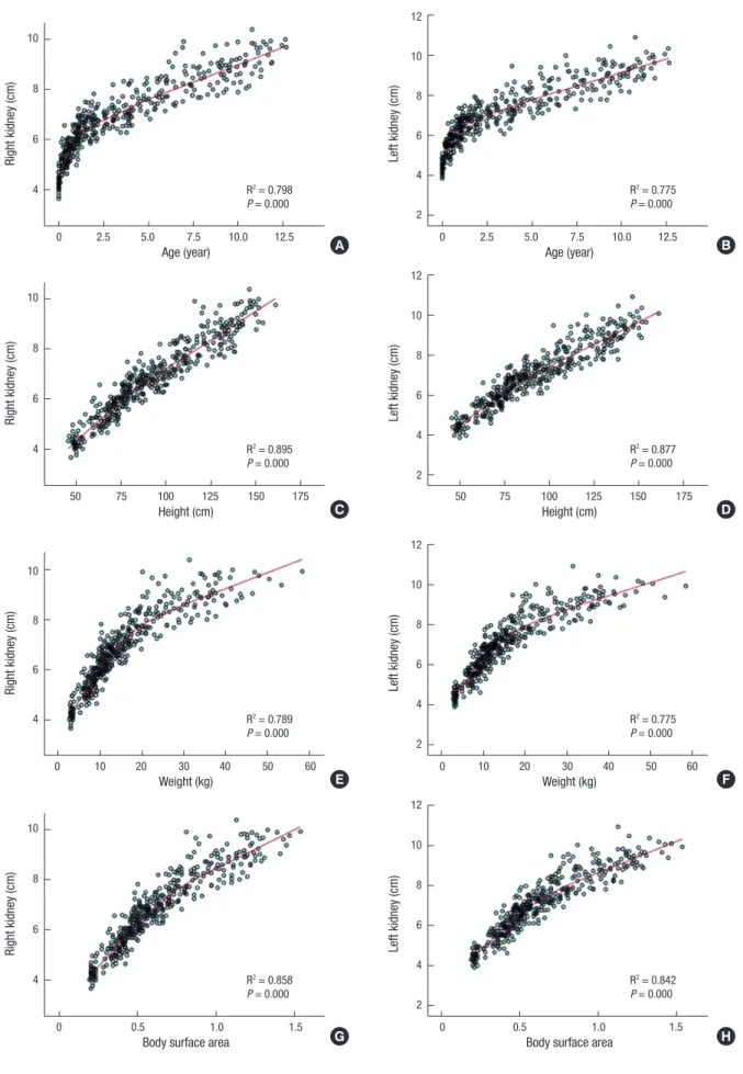
ney length and other somatic values, including age, weight, hei- ght, and BSA (Fig. 2; linear regression analysis, P < 0.01 for all).
On the other hand, the correlation between BMI and kidney length was not significant, with low R2 correlation coefficients of 0.093 and 0.097 for the right and left kidney, respectively.
The renal length curve for age showed different patterns be- fore and after 20 months of age (Fig. 2). In other words, the kid- ney was growing rapidly during the first 24 months, while the growth rate slowed down thereafter. The strongest influencing factor among these somatic values was height. Using this pa- rameter, the following linear regression equation was obtained:
right kidney length (cm) = 0.051 × height (cm) + 2.102; left kid- ney length (cm) = 0.051 × height (cm) + 2.280.
DISCUSSION
Sonography can be easily and safely performed, without radia- tion exposure in children. Renal sonography can provide im- portant information regarding for example renal anomalies and indirect renal function. For this reason, accurate reference values of renal length measured by sonography are important in children. A major strength of the present study is the analysis of a pediatric healthy cohort; therefore, the measurement val- ues can be considered representative. Moreover, the most valu- able point of the present study is that almost 50% of the enrolled children were aged less than 24 months; hence, more accurate reference values could be obtained for these subjects.
Many studies have shown that height is the strongest factor related with kidney length (7,11-13). Furthermore, according to the current study, the renal growth curve might be biphasic, with 20 months as its starting point, indicating that height should be considered an important factor correlating with kidney length (Fig. 2). In fact, a previous study concluded that the reason for this important relationship is that the rapid growth in height during the first 2 years of life is intimately associated with a simi- lar increase in kidney length (7). Our renal growth curve showed a biphasic pattern for the relationship with age, and a linear pat- tern for the relationship with height. Therefore, our linear regres- sion equation for bilateral renal length according to height is simpler than similar equations from prior studies (11,13,14).
Of note, our study was a descriptive study. Therefore, there was no null hypothesis about pre-specified estimated 95% con- fidence intervals and total width (precision) for determining the appropriate sample size. The tolerance probability is 0.900 that a sample size of 437 from approximately 85,000 children aged less than 13 years on Jeju Island, according to the 2010 Census, will produce a two-sided 95% confidence interval with a distance from the mean to the limits that is ≤ 0.060 if the pop- ulation standard deviation is 0.600.
In terms of renal growth according to age, it is known that the renal volume changes around 1 year of age (9), with a rapid but Fig. 1. Maximum longitudinal length of the left kidney passing through the renal hi-
lum (arrow).
Fig. 2. Renal growth curve according to the child’s age, height, weight, and body surface area.
Right kidney (cm)
Age (year)
0 2.5 5.0 7.5 10.0 12.5 10
8
6
4 R2 = 0.798
P = 0.000
Left kidney (cm)
Age (year)
0 2.5 5.0 7.5 10.0 12.5 12
10 8 6 4 2
R2 = 0.775 P = 0.000
Right kidney (cm)
Weight (kg)
0 10 20 30 40 50 60 10
8
6
4 R2 = 0.789
P = 0.000
Left kidney (cm)
Weight (kg)
0 10 20 30 40 50 60 12
10 8 6 4 2
R2 = 0.775 P = 0.000
Right kidney (cm)
Body surface area
0 0.5 1.0 1.5
10
8
6
4 R2 = 0.858
P = 0.000
Left kidney (cm)
Body surface area
0 0.5 1.0 1.5
12 10 8 6 4 2
R2 = 0.842 P = 0.000
Right kidney (cm)
Height (cm)
50 75 100 125 150 175 10
8
6
4 R2 = 0.895
P = 0.000
Left kidney (cm)
Height (cm)
50 75 100 125 150 175 12
10 8 6 4 2
R2 = 0.877 P = 0.000 A
C
E
G
B
D
F
H
gradually decreasing growth rate observed in the initial 7 months after birth, followed by a constant and slower rate (15). In infants under 12 months, kidney length has been reported to be poorly estimated with an age-based simple formula (16). In other stud- ies, the renal length has been shown to increase rapidly until 24 months, while the growth rate is reduced thereafter (13,17), wher- eas our data showed a statistically significant difference before and after 20 months of age.
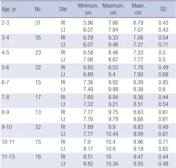
The reference values for the renal lengths according to age are presented in Tables 1 and 2. Compared with in a previous study (13), our data provide more accurate values in subjects aged less than 24 months, owing to the fact that these subjects were subdivided according to the age distribution. According to our study, the average renal lengths tended to be slightly larger than the previously reported values (13), by as much as 1-2 mm in subjects aged less than 24 months. Especially, these differenc- es tended to be greater in subjects aged more than 4-5 years. It can be speculated that renal growth is affected by the propor- tion of their current height, which was taller in the present study (18). Of note, although our findings differed from those obtained in an Indian study in 2012, they showed no differences com- pared to the American reference values in 2011 (16,19). We be- lieve that this lack of difference is due to the fact that the height growth of Korean children has recently been reported to be sim- ilar to that of western children (16,18).
The other values of age and weight also showed good corre- lations with kidney length in children from 2 to 12 years of age in the present study. The left kidney was significantly longer than the right, as has been previously reported (11,13,14). On the oth- er hand, there were no significant sex-specific differences.
In conclusion, we here reported the reference values of kid- ney length for Korean children. We believe that these data may Table 1. Longitudinal lengths of both kidneys in individuals aged less than 24 months
Age, mon No. Site Minimum,
cm Maximum,
cm Mean,
cm SD
< 1 36 Rt
Lt
3.66 3.85
5.07 5.08
4.31 4.47
0.31 0.30
1-3 22 Rt
Lt 4.29
4.35 6.04
5.81 5.13
5.25 0.42 0.39
3-5 14 Rt
Lt
4.89 4.89
6.57 6.59
5.43 5.56
0.43 0.47
5-7 29 Rt
Lt
4.65 4.57
6.09 6.68
5.43 5.63
0.43 0.49
7-9 15 Rt
Lt 5.15
5.14 6.05
6.40 5.64
5.81 0.27 0.39
9-11 16 Rt
Lt
5.32 5.50
6.60 6.96
5.89 6.13
0.39 0.47
11-15 42 Rt
Lt
4.99 5.24
7.24 7.36
6.23 6.40
0.46 0.52
15-18 21 Rt
Lt 5.70
5.76 7.51
7.47 6.54
6.67 0.44 0.40
18-24 33 Rt
Lt
5.86 5.77
7.83 7.93
6.69 6.84
0.48 0.53 SD, standard deviation; Rt, right; Lt, left.
Table 2. Longitudinal lengths of both kidneys in individuals aged between 2 and 13 years
Age, yr No. Site Minimum,
cm
Maximum, cm
Mean,
cm SD
2-3 31 Rt
Lt 5.96
6.07 7.66
7.84 6.79
7.07 0.43
0.43
3-4 35 Rt
Lt
6.29 6.07
8.33 9.46
7.06 7.27
0.54 0.71
4-5 23 Rt
Lt 6.58
7.06 8.46
8.67 7.53
7.77 0.5
0.5
5-6 22 Rt
Lt 6.83
6.89 8.53
9.4 7.78
7.93 0.49
0.68
6-7 15 Rt
Lt
7.36 7.49
9.92 9.99
8.39 8.38
0.85 0.8
7-8 17 Rt
Lt 7.69
7.32 8.94
9.21 8.36
8.51 0.44
0.54
8-9 13 Rt
Lt 7.77
7.76 9.75
9.79 8.63
8.68 0.61
0.61
9-10 22 Rt
Lt
7.89 7.77
9.9 10.44
8.83 9.09
0.49 0.61
10-11 15 Rt
Lt 7.9
8.17 10.4
10.9 8.96
9.19 0.71
0.65
11-13 16 Rt
Lt
8.51 8.82
10 10.34
9.47 9.55
0.44 0.48 SD, standard deviation; Rt, right; Lt, left.
help facilitate the diagnosis of renal hypoplasia or pediatric CKD and help estimate the appropriate donor kidney length in chil- dren by our simple linear regression equation in which only the height of the donor needs to be applied.
ACKNOWLEDGMENT
This study was supported by a grant from the Jeju National Uni- versity Hospital Research Fund (2012).
DISCLOSURE
The authors have no potential conflicts of interest to disclose.
AUTHOR CONTRIBUTION
Literature review: Han S, Lee MS, Choi GM, Han KH. Study con- cept and design: Choi GM, Han KH. Data collection and/or pro- cessing: Oh MS, Hwang G, Han S, Kang HS, Kim SH, Kim YD, Kang KS, Shin KS, Han KH. Statistical analysis and/or interpre- tation: Oh MS, Hwang G, Han KH. Writing: Han S, Han KH. Crit- ical revision and final approval: all authors.
ORCID
Min-su Oh http://orcid.org/0000-0001-5327-0934 Geol Hwang http://orcid.org/0000-0002-2299-6780 Sanghoon Han http://orcid.org/0000-0003-0976-9134 Hyun Sik Kang http://orcid.org/0000-0002-7495-7871
Seung Hyo Kim http://orcid.org/0000-0001-7277-3748 Young Don Kim http://orcid.org/0000-0003-2141-9882 Ki-Soo Kang http://orcid.org/0000-0001-6374-8356 Kyung-Sue Shin http://orcid.org/0000-0002-7179-9599 Mu Sook Lee http://orcid.org/0000-0002-0382-5564 Guk Myung Choi http://orcid.org/0000-0003-0370-484X Kyoung Hee Han http://orcid.org/0000-0002-6830-7311 REFERENCES
1. Queisser-Luft A, Stolz G, Wiesel A, Schlaefer K, Spranger J. Malformations in newborn: results based on 30,940 infants and fetuses from the Mainz congenital birth defect monitoring system (1990-1998). Arch Gynecol Obstet 2002; 266: 163-7.
2. Seikaly MG, Ho PL, Emmett L, Fine RN, Tejani A. Chronic renal insuffi- ciency in children: the 2001 Annual Report of the NAPRTCS. Pediatr Nep- hrol 2003; 18: 796-804.
3. Cain JE, Di Giovanni V, Smeeton J, Rosenblum ND. Genetics of renal hy- poplasia: insights into the mechanisms controlling nephron endowment.
Pediatr Res 2010; 68: 91-8.
4. Emamian SA, Nielsen MB, Pedersen JF. Intraobserver and interobserver variations in sonographic measurements of kidney size in adult volun- teers. A comparison of linear measurements and volumetric estimates.
Acta Radiol 1995; 36: 399-401.
5. Schlesinger AE, Hernandez RJ, Zerin JM, Marks TI, Kelsch RC. Interob- server and intraobserver variations in sonographic renal length measure- ments in children. AJR Am J Roentgenol 1991; 156: 1029-32.
6. Faubel S, Patel NU, Lockhart ME, Cadnapaphornchai MA. Renal relevant radiology: use of ultrasonography in patients with AKI. Clin J Am Soc Ne- phrol 2014; 9: 382-94.
7. Dinkel E, Ertel M, Dittrich M, Peters H, Berres M, Schulte-Wissermann H.
Kidney size in childhood. Sonographical growth charts for kidney length
and volume. Pediatr Radiol 1985; 15: 38-43.
8. Konuş OL, Ozdemir A, Akkaya A, Erbaş G, Celik H, Işik S. Normal liver, spleen, and kidney dimensions in neonates, infants, and children: evalu- ation with sonography. AJR Am J Roentgenol 1998; 171: 1693-8.
9. Han BK, Babcock DS. Sonographic measurements and appearance of normal kidneys in children. AJR Am J Roentgenol 1985; 145: 611-6.
10. Moon JS, Lee SY, Nam CM, Choi JM, Choe BK, Seo JW, Oh K, Jang MJ, Hwang SS, Yoo MH, et al. 2007 Korean National Growth Charts: review of developmental process and an outlook. Korean J Pediatr 2008; 51: 1-25.
11. Kim JH, Kim MJ, Lim SH, Kim J, Lee MJ. Length and volume of morpho- logically normal kidneys in Korean children: ultrasound measurement and estimation using body size. Korean J Radiol 2013; 14: 677-82.
12. Zerin JM, Blane CE. Sonographic assessment of renal length in children:
a reappraisal. Pediatr Radiol 1994; 24: 101-6.
13. Kim IO, Cheon JE, Lee YS, Lee SW, Kim OH, Kim JH, Kim HD, Sim JS. Kid- ney length in normal Korean children. J Korean Soc Ultrasound Med 2010;
29: 181-8.
14. Lee MJ, Son MK, Kwak BO, Park HW, Chung S, Kim KS. Kidney size esti- mation in Korean children with Technesium-99m dimercaptosuccinic acid scintigraphy. Korean J Pediatr 2014; 57: 41-5.
15. Mesrobian HG, Laud PW, Todd E, Gregg DC. The normal kidney growth rate during year 1 of life is variable and age dependent. J Urol 1998; 160:
989-93.
16. Akhavan A, Brajtbord JS, McLeod DJ, Kabarriti AE, Rosenberg HK, Stock JA. Simple, age-based formula for predicting renal length in children. Urol- ogy 2011; 78: 405-10.
17. Haugstvedt S, Lundberg J. Kidney size in normal children measured by sonography. Scand J Urol Nephrol 1980; 14: 251-5.
18. Ryoo NY, Shin HY, Kim JH, Moon JS, Lee CG. Change in the height of Ko- rean children and adolescents: analysis from the Korea National Health and Nutrition Survey II and V. Korean J Pediatr 2015; 58: 336-40.
19. Otiv A, Mehta K, Ali U, Nadkarni M. Sonographic measurement of renal size in normal Indian children. Indian Pediatr 2012; 49: 533-6.