Impact of Framingham Risk Score, Flow-Mediated Dilation, Pulse Wave Velocity, and Biomarkers for Cardiovascular Events in Stable Angina
Although the age-adjusted Framingham risk score (AFRS), flow-mediated dilation (FMD), brachial-ankle pulse wave velocity (baPWV), high-sensitivity C-reactive protein (hsCRP), fibrinogen, homocysteine, and free fatty acid (FFA) can predict future cardiovascular events (CVEs), a comparison of these risk assessments for patients with stable angina has not been reported. We enrolled 203 patients with stable angina who had been scheduled for coronary angiography (CAG). After CAG, 134 patients showed significant coronary artery disease. During 4.2 yr follow-up, 36 patients (18%) showed CVEs, including myocardial infarction, de-novo coronary artery revascularization, in-stent restenosis, stroke, and cardiovascular death. ROC analysis showed that AFRS, FMD, baPWV, and hsCRP could predict CVEs (with AUC values of 0.752, 0.707, 0.659, and 0.702, respectively, all P < 0.001 except baPWV P = 0.003). A Cox proportional hazard analysis showed that AFRS and FMD were independent predictors of CVEs (HR, 2.945; 95% CI, 1.572-5.522;
P = 0.001 and HR, 0.914; 95% CI, 0.826-0.989; P = 0.008, respectively). However, there was no difference in predictive power between combining AFRS plus FMD and AFRS alone (AUC 0.752 vs. 0.763; z = 1.358, P = 0.175). In patients with stable angina, AFRS and FMD are independent predictors of CVEs. However, there is no additive value of FMD on the AFRS in predicting CVEs.
Keywords: Framingham Risk Score; Flow-Mediated Dilation; Cardiovascular Event Kyoung-Ha Park, Sang Jin Han,
Hyun-Sook Kim, Min-Kyu Kim, Sang Ho Jo, Sung-Ai Kim, and Woo Jung Park
Cardiovascular Division, Department of Internal Medicine, Hallym University Medical Center, Anyang, Korea
Received: 24 March 2014 Accepted: 26 June 2014 Address for Correspondence:
Kyoung-Ha Park, MD
Cardiovascular Division, Department of Internal Medicine, Hallym University Medical Center, 22 Gwanpyeong-ro 170beon-gil, Dongan-gu, Anyang 431-796, Korea Tel: +82.31-380-3749, Fax: +82.31-386-2269 E-mail: [email protected].
http://dx.doi.org/10.3346/jkms.2014.29.10.1391 • J Korean Med Sci 2014; 29: 1391-1397
INTRODUCTION
Coronary artery disease (CAD) is one of the major predictors of future cardiovascular events (CVEs). Flow-mediated dilation (FMD), which represents endothelial function, reflects not only the presence of CAD but also predicts the risk of CVEs in pa- tients with CAD (1, 2). Brachial-ankle pulse wave velocity (baP- WV) is also a predictor of future CVEs for the general popula- tion and for patients with CAD (3, 4). In addition, biomarkers such as high-sensitivity C-reactive protein (hsCRP), fibrinogen, homocysteine, and free fatty acid (FFA) correlate well with a fu- ture CVE (5-9). The Framingham risk scoring system has been used to help identify future cardiovascular risk based on a large cohort study (10). However, there are few data on whether the Framingham risk score is still valid for secondary prevention after a diagnosis of stable angina. von Birgelen et al. (11) report- ed that a positive relationship exists between initial Framing- ham risk score and future plaque progression in patients with stable coronary artery disease. In addition, the Framingham risk score was a strong predictor of CVEs, including stroke (12).
Although the Framingham risk score, FMD, baPWV, and bio- markers (such as hsCRP, fibrinogen, homocysteine, and FFA)
could predict future CVEs, a comparison of these risk assess- ments for future CVEs in patients with stable coronary artery disease has not been reported.
The aim of this study was to assess the inter-relationship be- tween the Framingham risk score, FMD, baPWV, and biomark- ers for future CVEs in patients with stable angina.
MATERIALS AND METHODS
Study population, study endpoint, and follow-up
Patients were eligible for enrollment in the study if they were aged between 30 and 75 yr, had stable angina and were sched- uled to undergo coronary angiography (CAG). Patients were excluded if they had acute coronary syndrome, significant val- vular heart disease (more than a moderate degree), left ventric- ular dysfunction (left ventricular ejection fraction < 55%), an ankle-brachial index < 0.9, or an inability to follow the protocol.
The end point of this study was a composite of CVE, includ- ing myocardial infarction, de-novo coronary revascularization, in-stent restenosis, stroke, and cardiovascular death. All events were based on clinical diagnoses made by the patient’s physi- cian. Clinical follow-up was performed in the office or via tele- Cardiovascular Disorders
phone at one month, six months, and one year and then annu- ally thereafter.
Before evaluation of the baPWV and FMD, which were eval- uated on the morning of the planned CAG, the patients were instructed not to exercise and to discontinue substances that might affect the baPWV and FMD, such as caffeine, food, to- bacco, or vasoactive medication, for at least 12 hr before the study. The studies were done in a quiet temperature-controlled room (22°C-24°C). All of the patients had been given clopido- grel (loading dose, if needed) before CAG, because there was a chance that they would need to be undergo percutaneous cor- onary intervention (PCI).
In this study, significant coronary artery disease (sCAD) was defined as lumen diameter stenosis > 50% in one or more ma- jor coronary arteries as determined by CAG. The CAG was in- terpreted by one cardiologist who was blinded to the patients’
clinical data. If a patient had sCAD, a decision regarding the treat- ment option (medical treatment, PCI, or a coronary artery by- pass graft operation) was made by the attending physician.
Measurement of FMD
An experienced vascular sonographer who was blinded to the patients’ information obtained ultrasound images using a Vivid 7 system (GE Vingmed Ultrasound, Horten, Norway) with a 12- MHz linear array transducer. FMD was measured according to the recommendations of Corretti and colleagues (13). In brief, a landmark 10 cm above the proximal wrist crease of the left radi- al artery (RA) was used for the vascular ultrasound measure- ment. After the baseline diameter of the RA was measured, a blood pressure cuff was inflated on the forearm up to 220 mmHg for five minutes and then deflated; after one, two, and three min- utes, the RA diameter was measured to obtain the post-occlu- sion value. All images were recorded digitally by capturing the RA in the longitudinal plane with an electrocardiogram. One cardiologist, who was blinded to the participants’ clinical data interpreted the ultrasound results using an off-line method. The maximal RA diameter image for analysis was evaluated during the end diastole in the cardiac cycle (the onset of the R wave on electrocardiography). Measurements were taken at seven points, and the maximal and minimal values were discarded. The mean value from the remaining five measurements was used for fur- ther analysis. Thirty randomly selected images were reanalyzed to assess intra-observer variation plotting by two independent measurements; the standard error of the estimate of the intra- observer variability was 5.6%.
Measurement of brachial-ankle pulse wave velocity The baPWV was measured using a volume-plethysmographic apparatus (VP-2000, Colin Co., Komaki, Japan). Cuffs were con- nected to both plethysmographic and oscillometric sensors and were placed around both the arms and ankles while the subject
remained in the supine position. Electrocardiogram electrodes were placed on both wrists and a microphone to detect heart sounds was placed on the left edge of the sternum to detect the second heart sound. The time interval between the wave front of the brachial waveform and that of the ankle waveform was defined as the time interval between the brachium and ankle (DTba). The distance between sampling points of the baPWV was calculated automatically according to the height of the pa- tient. The path length from the suprasternal notch to the bra- chium (Lb) was obtained from superficial measurements and was expressed using the following equation: Lb = (0.2195 × hei- ght of the subject [cm]-2.0734). The path length from the supra- sternal notch to the ankle (La) was obtained from superficial measurements and was expressed using the following equa- tion: La = (0.8129 × height of the subject [cm]+12.328). Finally, the following equation was used to obtain baPWV: baPWV = ([La-Lb]/DTba). In this study, the left side baPWV was used for the analyses.
Age-adjusted Framingham risk score
Although the Framingham risk score has been computed in many different ways since it was first introduced, we used the version described by Wilson et al. (14) that reports the age-ad- justed Framingham risk score (AFRS) using categorical vari- ables such as age, sex, blood pressure, total cholesterol, high- density lipoprotein cholesterol, cigarette smoking, and diabe- tes. This version divides the subject’s Framingham risk score by the estimat ed average risk of the same age and sex group, thus providing the relative risk in the next 10 yr.
Statistical analysis
The continuous variables were summarized as mean ± SD. Cat- egorical variables were presented as numbers or percentages.
To compare the predictive power of the CVE, a receiver operat- ing characteristic (ROC) analysis was performed, and the area under curve (AUC) was calculated for each variable; the AUCs of the two equations were compared according to the proce- dure described by DeLong et al. (15). In addition, the Cox pro- portional hazard analysis was performed to assess independent risk predictors for a CVE. Because the beneficial effects of aspi- rin and statin have been well proven for patients with coronary artery disease (16, 17), we added these variables on the analy- sis. As a subgroup analysis, we also performed ROC analysis and Cox proportional hazard analysis for the coronary events of the patients with sCAD. Data were analyzed using standard sta- tistical software (SPSS version 17.0; SPSS Inc., Chicago, IL, USA and MedCalc version 13.0; MedCalc Software, Mariakerke, Bel- gium). A probability value of < 0.05 was considered statistically significant.
Ethics statement
This study was approved by the institutional review board of the Hallym University Medical Center, and all patients gave their written informed consent.
RESULTS
Between March 2008 and June 2009, 224 patients with stable angina were assessed for the study. Of these, 21 patients were excluded because of significant valvular heart disease (n = 3), LV dysfunction (n = 13), and an ankle-brachial index < 0.9 (n = 5), leaving 203 subjects enrolled in the study. The baseline clini- cal characteristics and the values of the AFRS, FMD, baPWV, hsCRP, fibrinogen, homocysteine, FFA, and prescribed medi- cations are shown in Table 1. Of the 203 patients, 134 (66%) had sCAD while 69 patients showed mild or intermediate stenosis (non-sCAD; lumen diameter stenosis ≤ 50%) on CAG. Among the 134 patients with sCAD, 82 patients underwent percutane- ous coronary intervention. Three patients were treated with
coronary artery bypass graft surgery and 49 patients decided to get medical treatment because of relatively less-tight lesions or small vessel disease (< 2.5 mm lumen diameter), based on the decision of the attending physician. The duration of mean fol- low-up was 50 ± 13 months, and 18 patients were lost at follow- up. During the study period, two patients presented with myo- cardial infarction and were treated with PCI, 19 patients under- went coronary revascularization due to the progression of CAD, eight patients showed in-stent restenosis, and seven patients suffered from stroke; in total, 36 CVEs occurred.
The results of the ROC analysis for CVEs with the AFRS, FMD, baPWV, hsCRP, fibrinogen, homocysteine, and FFA are shown in Table 2. The predictive power of the AFRS was shown to be best and was significantly better than the fibrinogen (z = 2.463, P = 0.014), homocysteine (z = 2.206, P = 0.027), and FFA (z = 2.288, P = 0.022). However, the predictive power of the AFRS was not statistically different from the FMD (z = 0.759, P = 0.448), baPWV (z = 1.794, P = 0.073), and hsCRP (z = 0.865, P = 0.387).
Including aspirin and statin, the Cox proportional hazard anal-
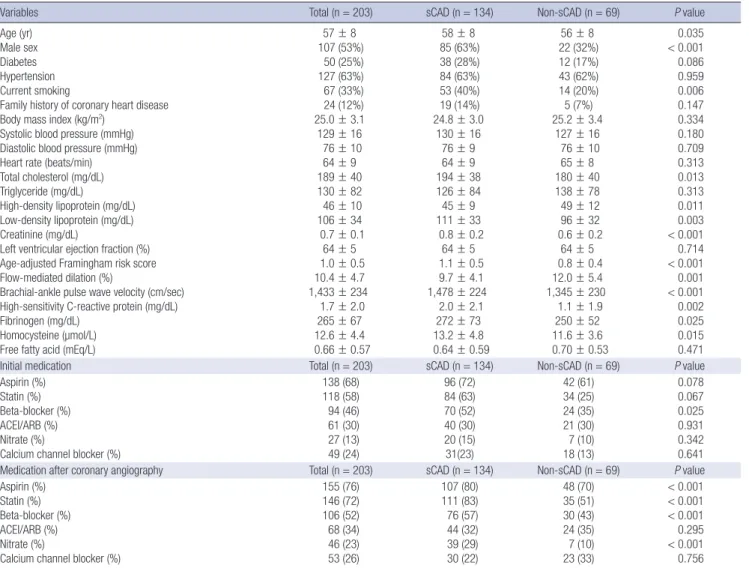
Table 1. Baseline clinical characteristics and medications
Variables Total (n = 203) sCAD (n = 134) Non-sCAD (n = 69) P value
Age (yr) Male sex Diabetes Hypertension Current smoking
Family history of coronary heart disease Body mass index (kg/m2)
Systolic blood pressure (mmHg) Diastolic blood pressure (mmHg) Heart rate (beats/min) Total cholesterol (mg/dL) Triglyceride (mg/dL) High-density lipoprotein (mg/dL) Low-density lipoprotein (mg/dL) Creatinine (mg/dL)
Left ventricular ejection fraction (%) Age-adjusted Framingham risk score Flow-mediated dilation (%)
Brachial-ankle pulse wave velocity (cm/sec) High-sensitivity C-reactive protein (mg/dL) Fibrinogen (mg/dL)
Homocysteine (µmol/L) Free fatty acid (mEq/L)
57 ± 8 107 (53%) 50 (25%) 127 (63%) 67 (33%) 24 (12%) 25.0 ± 3.1 129 ± 16
76 ± 10 64 ± 9 189 ± 40 130 ± 82 46 ± 10 106 ± 34 0.7 ± 0.1
64 ± 5 1.0 ± 0.5 10.4 ± 4.7 1,433 ± 234 1.7 ± 2.0 265 ± 67 12.6 ± 4.4 0.66 ± 0.57
58 ± 8 85 (63%) 38 (28%) 84 (63%) 53 (40%) 19 (14%) 24.8 ± 3.0
130 ± 16 76 ± 9 64 ± 9 194 ± 38 126 ± 84 45 ± 9 111 ± 33
0.8 ± 0.2 64 ± 5 1.1 ± 0.5 9.7 ± 4.1 1,478 ± 224
2.0 ± 2.1 272 ± 73 13.2 ± 4.8 0.64 ± 0.59
56 ± 8 22 (32%) 12 (17%) 43 (62%) 14 (20%) 5 (7%) 25.2 ± 3.4
127 ± 16 76 ± 10 65 ± 8 180 ± 40 138 ± 78 49 ± 12 96 ± 32 0.6 ± 0.2
64 ± 5 0.8 ± 0.4 12.0 ± 5.4 1,345 ± 230 1.1 ± 1.9 250 ± 52 11.6 ± 3.6 0.70 ± 0.53
0.035
< 0.001 0.086 0.959 0.006 0.147 0.334 0.180 0.709 0.313 0.013 0.313 0.011 0.003
< 0.001 0.714
< 0.001 0.001
< 0.001 0.002 0.025 0.015 0.471
Initial medication Total (n = 203) sCAD (n = 134) Non-sCAD (n = 69) P value
Aspirin (%) Statin (%) Beta-blocker (%) ACEI/ARB (%) Nitrate (%)
Calcium channel blocker (%)
138 (68) 118 (58) 94 (46) 61 (30) 27 (13) 49 (24)
96 (72) 84 (63) 70 (52) 40 (30) 20 (15) 31(23)
42 (61) 34 (25) 24 (35) 21 (30) 7 (10) 18 (13)
0.078 0.067 0.025 0.931 0.342 0.641
Medication after coronary angiography Total (n = 203) sCAD (n = 134) Non-sCAD (n = 69) P value
Aspirin (%) Statin (%) Beta-blocker (%) ACEI/ARB (%) Nitrate (%)
Calcium channel blocker (%)
155 (76) 146 (72) 106 (52) 68 (34) 46 (23) 53 (26)
107 (80) 111 (83) 76 (57) 44 (32) 39 (29) 30 (22)
48 (70) 35 (51) 30 (43) 24 (35) 7 (10) 23 (33)
< 0.001
< 0.001
< 0.001 0.295 < 0.001 0.756 ACEI/ARB, angiotensin converting enzyme inhibitor/angiotensin receptor blocker; sCAD, significant coronary artery disease.
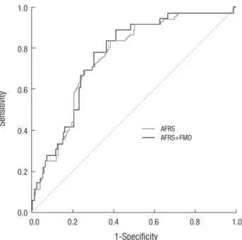
ysis was performed with the AFRS, FMD, baPWV, and hsCRP which significantly predicted CVEs on the ROC analysis. The AFRS and FMD were independent predictors for CVEs (HR, 2.945; 95% CI, 1.572-5.522, P = 0.001 and HR, 0.914; 95% CI, 0.826-0.989, P = 0.008, respectively; Table 3). The area under the ROC curves of combined parameters of AFRS plus FMD indi- cating a future CVE was 0.763 (95% CI, 0.698-0.820; P < 0.001).
However, there was no difference in the power between com- bining the AFRS plus FMD and AFRS alone for predicting a CVE (z = 1.358, P = 0.175; Fig. 1). In the 134 patients with sCAD, we also evaluated for a coronary event, which removed stroke from CVEs. In patients with sCAD, the clinical results may be affect- ed by the lesion characteristics and number of diseased vessels on CAG or by treatment strategy (medical treatment or PCI) (18). According to the ACC/AHA coronary artery lesion classifi- cation, 12 patients had type A, 37 patients had type B1, 37 pa- tients had type B2, and 48 patients had type C lesions (19). In terms of the number of diseased vessels, 79 patients had one vessel disease, 33 patients had two and 22 patients had three.
During the study period, 19 patients underwent PCI due to pro- gression of CAD, and eight patients showed in-stent restenosis;
in total, 27 coronary events occurred. In the 134 patients with sCAD, the ROC analysis for coronary events with the AFRS, FMD, baPWV, hsCRP, fibrinogen, homocysteine, and FFA are shown in Table 4. The predictive power of the AFRS was significantly better than the fibrinogen (z = 2.006, P = 0.045) and homocys- teine (z = 2.353, P = 0.019). However, the predictive power of the AFRS was not statistically different from the FMD (z = 0.505, P = 0.613), baPWV (z = 1.541, P = 0.123), hsCRP (z = 1.026, P = 0.305), and FFA (z = 1.577, P = 0.115). In addition to the class of
the coronary artery lesion, number of coronary artery disease, and PCI, the Cox proportional hazard analysis was performed with the AFRS, FMD, baPWV, and hsCRP, which significantly predicted coronary events on the ROC analysis. The AFRS was an independent predictor for coronary events and the FMD show- ed marginal significance (Table 5). The area under the ROC curves Table 2. Receiver operating characteristic analysis for cardiovascular event
Findings AUC value 95% CI P value
Age-adjusted Framingham risk score 0.752 0.670-0.834 < 0.001 Flow-mediated dilation 0.707 0.615-0.799 < 0.001 Brachial-ankle pulse wave velocity 0.659 0.563-0.755 0.003 High-sensitivity C-reactive protein 0.702 0.611-0.794 < 0.001
Fibrinogen* 0.603 0.504-0.702 0.053
Homocysteine* 0.602 0.501-0.704 0.054
Free fatty acid* 0.596 0.500-0.692 0.070
*Significantly decreased predictive power of cardiovascular event compared with age- adjusted Framingham risk score (P < 0.05). AUC, area under curve; CI, confidence interval.
Table 3. Cox proportional hazard analysis for cardiovascular events
Findings Hazard ratio 95% CI P value
Age-adjusted Framingham risk score 2.945 1.572-5.522 0.001
Flow-mediated dilation 0.914 0.826-0.989 0.008
Brachial-ankle pulse wave velocity 1.000 0.999-1.010 0.624 High-sensitivity C-reactive protein 1.047 0.938-1.154 0.429
Aspirin 0.967 0.291-3.003 0.918
Statin 0.578 0.183-1.809 0.347
CI, confidence interval.
Table 4. Receiver operating characteristic analysis for coronary event in the 134 pa- tients with significant coronary artery disease
Coronary events AUC value 95% CI P value
Age-adjusted Framingham risk score 0.731 0.631-0.831 < 0.001
Flow-mediated dilation 0.696 0.584-0.807 0.002
Brachial-ankle pulse wave velocity 0.627 0.504-0.750 0.042 High-sensitivity C-reactive protein 0.654 0.543-0.765 0.013
Fibrinogen* 0.590 0.478-0.701 0.150
Homocysteine* 0.531 0.414-0.649 0.616
Free fatty acid 0.606 0.500-0.713 0.089
*Significantly decreased predictive power of cardiovascular event compared with age- adjusted Framingham risk score (P < 0.05). AUC, area under curve; CI, confidence interval.
Table 5. Cox proportional hazard analysis for coronary event in the 134 patients with significant coronary artery disease
Coronary events Hazard ratio 95% CI P value
Age-adjusted Framingham risk score 2.646 1.261-5.551 0.010
Flow-mediated dilation 0.908 0.822-1.004 0.060
Brachial-ankle pulse wave velocity 1.000 0.998-1.002 0.758 High-sensitivity C-reactive protein 1.163 0.990-1.367 0.067 Coronary artery lesion class 1.318 0.823-2.113 0.251 Number of diseased coronary artery 1.241 0.774-1.991 0.370 Percutaneous coronary intervention 0.876 0.362-2.120 0.769 CI, confidence interval.
Fig. 1. Receiver operating characteristic curves of the AFRS and the AFRS plus FMD for the prediction of cardiovascular events. AFRS, age-adjusted Framingham risk score; FMD, flow-mediated dilation.
Sensitivity
1-Specificity
0.0 0.2 0.4 0.6 0.8 1.0 1.0
0.8
0.6
0.4
0.2
0.0
AFRS AFRS+FMD
of combined parameters of AFRS plus FMD indicating coronary events was 0.736 (95% CI, 0.653-0.808; P < 0.001). However, there was no difference in the power between combining the AFRS plus FMD and the AFRS alone for predicting coronary events (0.736 vs. 0.731; z = 0.474, P = 0.635).
DISCUSSION
The principal finding of this study was that in patients with sta- ble angina, the AFRS and FMD were independent predictors of a CVE. However, FMD could not improve the prediction of a fu- ture CVE beyond that offered by the AFRS.
The Framingham risk score is a conventional means of pre- dicting the risk of a CVE in the general population (10). Althou- gh the major risk factors are important in primary prevention, there has been limited data about whether the Framingham risk score is still valid for secondary prevention after a diagnosis of stable angina. In this study, the AFRS was the most effective measurement in predicting a future CVE in patients with stable angina. Our results might be supported by a report that a posi- tive linear relationship exists between initial Framingham risk score and future clinical events, with plaque progression mea- sured by serial intravascular ultrasound in patients with stable coronary artery disease (11). In addition, reports indicate that the major risk factors in primary prevention still have crucial value in secondary prevention (20-22).
It is well known that FMD could offer a noninvasive assess- ment of preclinical CVE in subjects with and without cardiovas- cular risk factors (23, 24). In addition, FMD is strongly associat- ed with the presence and extent of CAD, and impaired FMD could independently predict the occurrence of in-stent reste- nosis after percutaneous coronary intervention (1, 25). In this study, the FMD had good predictive power for a CVE in the ROC analysis, but there was no additional benefit of predicting fu- ture CVEs when combining FMD with the AFRS compared to the AFRS alone. Likewise, when the Cox proportional hazard analysis was performed for the 134 patients with sCAD for fu- ture coronary events, the AFRS was still an independent predic- tor and FMD showed marginal significance. However there was no additional benefit of predicting coronary events when com- paring FMD plus AFRS to the AFRS alone. A possible explana- tion was that FMD was closely related to the principal cardio- vascular risk factors and the estimated 10-yr risk of CAD in pa- tients with low cardiovascular risk (26). It means that the merit of FMD could be attenuated for future CVE risk assessment as a measurement of secondary prevention, not only in patients with stable angina but also in patients with sCAD.
The baPWV, which is known to be an indicator of arterial stiff- ness and vascular damage, is a well-known predictor of future CVEs in the general population (3). In addition, the value of baPWV increased in patients with CAD and could be an inde-
pendent predictor of the prognosis of patients with acute coro- nary syndrome (4, 27). In this study, ROC analysis showed that baPWV could predict a CVE, but there was no difference in pow- er between the AFRS alone and the AFRS plus baPWV (with AUC values of 0.752 and 0.748, respectively; z = 0.267, P = 0.789). This could be supported by our pervious report that in patients with stable angina the baPWV could predict the presence of sCAD, but there was no additional benefit in predictive power when the baPWV was combined with the Framingham risk score (28).
In many large prospective studies, biomarkers such as CRP, fibrinogen, homocysteine, and FFA have been proposed to en- hance current risk score algorithms and be associated with an increased risk of CVE (7-9, 29, 30). CRP correlates with the risk of cardiovascular disease not only in patients with stable angina but also in those who have never been diagnosed with CAD (5, 6). However, among the biomarkers of the study, the ROC anal- ysis showed that only hsCRP had a significant predictive power for a CVE, but there was no additional value in the power be- tween the AFRS plus hsCRP and the AFRS alone for predicting a CVE (with AUC 0.744 vs. 0.752, z = 0.204, P = 0.839). In addi- tion, the Cox hazard analysis showed that hsCRP was not an in- dependent predictor of a CVE. In terms of fibrinogen, homo- cysteine, and FFA, these biomarkers showed marginal signifi- cance or non-significance in predicting a CVE in the ROC anal- ysis of the study. The post hoc analysis showed that there was no additional value in adding the above biomarkers to the AFRS compared with the AFRS alone for a CVE. Our results suggest that although these novel surrogate markers are associated with a future CVE, they contribute little to the prediction of a CVE when the conventional risk factors (age, sex, total and high-den- sity lipoprotein cholesterol levels, blood pressure, anti-hyper- tensive medication use, smoking status, and diabetes) are well considered in patients with stable angina. In addition, this could be explained by several studies that have not shown reclassifi- cation in predicting a CVE with the addition of conventional biomarkers compared to conventional risk factors (14, 31, 32).
Therefore, it cannot be emphasized enough that precise evalu- ation and control of the conventional risk factors are most im- portant in the management of patients with stable angina.
There are several potential limitations in our study. First, this study only included patients with stable angina and excluded patients with acute coronary syndrome. Therefore, our results may not be generalized to all patients with coronary heart dis- ease. Second, many of the patients were taking statin or anti- hypertensive medications that may affect the results of FMD.
Although the patients were instructed to discontinue vasoactive medications 12 hr before the study, we could not completely exclude the interaction of these drugs. Third, the measurement method for the FMD was rather old and the fixed time interval of measurement was somewhat long (one, two, and three min- utes). In addition, we did not use the automated edge detection
system that is frequently found in the literature at present (33).
However, according to Corretti et al. (13), nearly 70% of the NO is released in the first 60 sec after cuff release, and most maxi- mal dilation occurs around one minute after cuff release. In ad- dition, previous and recent studies have given reliable results using the FMD data measured at 60 sec (34, 35). Fourth, many clinical reports have shown that anti-hypertensive medication and lipid-lowering medication have the beneficial effects of improving endothelial function and change of endothelial func- tion could predict future CVEs (36-39). However, we did not perform follow-up FMD and cannot describe the relationships between used medications and changes of the endothelial func- tion. Fifth, we selected hsCRP, fibrinogen, homocysteine, and FFA to predict a CVE on the basis of previous clinical studies.
However, we acknowledge that other biomarkers that were not included in this study might have resulted in additional infor- mation. Finally, the number of patients was relatively small and the duration of follow-up was somewhat short. In addition, a large proportion of events related to coronary revascularization.
Therefore, further studies with larger numbers of patients and longer-term follow-up will be needed.
In patients with stable angina, AFRS and FMD are indepen- dent predictors of CVEs but the baPWV and biomarkers, such as hsCRP, fibrinogen, homocysteine, and FFA show no signifi- cance. However, there is no additive value of FMD on the AFRS in predicting future CVEs.
DISCLOSURE
The authors report no conflicts of interest to disclose.
ORCID
Kyoung-Ha Park http://orcid.org/0000-0002-5935-8715 Sang Jin Han http://orcid.org/0000-0003-4451-468X Hyun-Sook Kim http://orcid.org/0000-0002-7469-7993 Min-Kyu Kim http://orcid.org/0000-0002-1873-6513 Sang Ho Jo http://orcid.org/0000-0002-2063-1542 Sung-Ai Kim http://orcid.org/0000-0002-3411-0529 Woo Jung Park http://orcid.org/0000-0003-3896-5169
REFERENCES
1. Neunteufl T, Katzenschlager R, Hassan A, Klaar U, Schwarzacher S, Glo- gar D, Bauer P, Weidinger F. Systemic endothelial dysfunction is related to the extent and severity of coronary artery disease. Atherosclerosis 1997;
129: 111-8.
2. Kitta Y, Obata JE, Nakamura T, Hirano M, Kodama Y, Fujioka D, Saito Y, Kawabata K, Sano K, Kobayashi T, et al. Persistent impairment of endo- thelial vasomotor function has a negative impact on outcome in patients with coronary artery disease. J Am Coll Cardiol 2009; 53: 323-30.
3. Yamashina A, Tomiyama H, Arai T, Hirose K, Koji Y, Hirayama Y, Yama-
moto Y, Hori S. Brachial-ankle pulse wave velocity as a marker of ath- erosclerotic vascular damage and cardiovascular risk. Hypertens Res 2003; 26: 615-22.
4. Orlova IA, Nuraliev EY, Yarovaya EB, Ageev FT. Prognostic value of chan- ges in arterial stiffness in men with coronary artery disease. Vasc Health Risk Manag 2010; 6: 1015-21.
5. de Ferranti S, Rifai N. C-reactive protein and cardiovascular disease: a review of risk prediction and interventions. Clin Chim Acta 2002; 317:
1-15.
6. Gach O, Legrand V, Biessaux Y, Chapelle JP, Vanbelle S, Pierard LA. Long- term prognostic significance of high-sensitivity C-reactive protein before and after coronary angioplasty in patients with stable angina pectoris.
Am J Cardiol 2007; 99: 31-5.
7. Fibrinogen Studies Collaboration, Danesh J, Lewington S, Thompson SG, Lowe GD, Collins R, Kostis JB, Wilson AC, Folsom AR, Wu K, et al.
Plasma fibrinogen level and the risk of major cardiovascular diseases and nonvascular mortality: an individual participant meta-analysis.
JAMA 2005; 294: 1799-809.
8. Homocysteine Studies Collaboration. Homocysteine and risk of ischemic heart disease and stroke: a meta-analysis. JAMA 2002; 288: 2015-22.
9. Breitling LP, Rothenbacher D, Grandi NC, März W, Brenner H. Prog- nostic usefulness of free fatty acids in patients with stable coronary heart disease. Am J Cardiol 2011; 108: 508-13.
10. Grundy SM, Balady GJ, Criqui MH, Fletcher G, Greenland P, Hiratzka LF, Houston-Miller N, Kris-Etherton P, Krumholz HM, LaRosa J, et al.
Primary prevention of coronary heart disease: guidance from Framing- ham. A statement for healthcare professionals from the AHA Task Force on Risk Reduction. Circulation 1998; 97: 1876-87.
11. von Birgelen C, Hartmann M, Mintz GS, van Houwelingen KG, Depper- mann N, Schmermund A, Böse D, Eggebrecht H, Neumann T, Gössl M, et al. Relationship between cardiovascular risk as predicted by established risk scores versus plaque progression as measured by serial intravascular ultrasound in left main coronary arteries. Circulation 2004; 110: 1579- 85.
12. Pencina MJ, D’Agostino RB Sr, Larson MG, Massaro JM, Vasan RS. Pre- dicting the 30-year risk of cardiovascular disease: the Framingham heart study. Circulation 2009; 119: 3078-84.
13. Corretti MC, Anderson TJ, Benjamin EJ, Celermajer D, Charbonneau F, Creager MA, Deanfield J, Drexler H, Gerhard-Herman M, Herrington D, et al. Guidelines for the ultrasound assessment of endothelial-depen- dent flow-mediated vasodilation of the brachial artery: a report of the International Brachial Artery Reactivity Task Force. J Am Coll Cardiol 2002; 39: 257-65.
14. Wilson PW, Nam BH, Pencina M, D’Agostino RB Sr, Benjamin EJ, O’Don- nell CJ. C-reactive protein and risk of cardiovascular disease in men and women from the Framingham Heart Study. Arch Intern Med 2005; 165:
2473-8.
15. DeLong ER, DeLong DM, Clarke-Pearson DL. Comparing the areas under two or more correlated receiver operating characteristic curves: a nonparametric approach. Biometrics 1988; 44: 837-45.
16. Troche CJ, Tacke J, Hinzpeter B, Danner M, Lauterbach KW. Cost-effec- tiveness of primary and secondary prevention in cardiovascular diseas- es. Eur Heart J 1998; 19: C59-65.
17. De Backer G, Ambrosioni E, Borch-Johnsen K, Brotons C, Cifkova R, Dallongeville J, Ebrahim S, Faergeman O, Graham I, Mancia G, et al.
European guidelines on cardiovascular disease prevention in clinical practice. Third Joint Task Force of European and Other Societies on Car- diovascular Disease Prevention in Clinical Practice. Eur Heart J 2003;
24: 1601-10.
18. Lopes NH, Paulitsch Fda S, Gois AF, Pereira AC, Stolf NA, Dallan LO, Ramires JA, Hueb WA. Impact of number of vessels disease on outcome of patients with stable coronary artery disease: 5-year follow-up of the Medical, Angioplasty, and bypass Surgery study (MASS). Eur J Cardio- thorac Surg 2008; 33: 349-54.
19. Ryan TJ, Faxon DP, Gunnar RM, Kennedy JW, King SB 3rd, Loop FD, Peterson KL, Reeves TJ, Williams DO, Winters WL Jr, et al. Guidelines for percutaneous transluminal coronary angioplasty. A report of the Amer- ican College of Cardiology/American Heart Association Task Force on Assessment of Diagnostic and Therapeutic Cardiovascular Procedures (Subcommittee on Percutaneous Transluminal Coronary Angioplasty).
Circulation 1988; 78: 486-502.
20. Smith SC Jr, Blair SN, Criqui MH, Fletcher GF, Fuster V, Gersh BJ, Gotto AM, Gould KL, Greenland P, Grundy SM, et al. Preventing heart attack and death in patients with coronary disease. Circulation 1995; 92: 2-4.
21. Grover SA, Paquet S, Levinton C, Coupal L, Zowall H. Estimating the benefits of modifying risk factors of cardiovascular disease: a compari- son of primary vs secondary prevention. Arch Intern Med 1998; 158: 655- 62.
22. Genest J, Pedersen TR. Prevention of cardiovascular ischemic events:
high-risk and secondary prevention. Circulation 2003; 107: 2059-65.
23. Celermajer DS, Sorensen KE, Bull C, Robinson J, Deanfield JE. Endo- thelium-dependent dilation in the systemic arteries of asymptomatic subjects relates to coronary risk factors and their interaction. J Am Coll Cardiol 1994; 24: 1468-74.
24. Anderson TJ. Prognostic significance of brachial flow-mediated vasodi- lation. Circulation 2007; 115: 2373-5.
25. Patti G, Pasceri V, Melfi R, Goffredo C, Chello M, D’Ambrosio A, Monte- santi R, Di Sciascio G. Impaired flow-mediated dilation and risk of re- stenosis in patients undergoing coronary stent implantation. Circulation 2005; 111: 70-5.
26. Witte DR, Westerink J, de Koning EJ, van der Graaf Y, Grobbee DE, Bots ML. Is the association between flow-mediated dilation and cardiovascu- lar risk limited to low-risk populations? J Am Coll Cardiol 2005; 45: 1987- 93.
27. Tomiyama H, Koji Y, Yambe M, Shiina K, Motobe K, Yamada J, Shido N, Tanaka N, Chikamori T, Yamashina A. Brachial -- ankle pulse wave ve- locity is a simple and independent predictor of prognosis in patients with acute coronary syndrome. Circ J 2005; 69: 815-22.
28. Park KH, Kim MK, Kim HS, Park WJ, Cho GY, Choi YJ. Clinical signifi- cance of Framingham risk score, flow-mediated dilation and pulse wave
velocity in patients with stable angina. Circ J 2011; 75: 1177-83.
29. Ridker PM. C-reactive protein and the prediction of cardiovascular events among those at intermediate risk: moving an inflammatory hypothesis toward consensus. J Am Coll Cardiol 2007; 49: 2129-38.
30. Rana JS, Cote M, Després JP, Sandhu MS, Talmud PJ, Ninio E, Wareham NJ, Kastelein JJ, Zwinderman AH, Khaw KT, et al. Inflammatory bio- markers and the prediction of coronary events among people at interme- diate risk: the EPIC-Norfolk prospective population study. Heart 2009;
95: 1682-7.
31. Folsom AR, Chambless LE, Ballantyne CM, Coresh J, Heiss G, Wu KK, Boerwinkle E, Mosley TH Jr, Sorlie P, Diao G, et al. An assessment of in- cremental coronary risk prediction using C-reactive protein and other novel risk markers: the atherosclerosis risk in communities study. Arch Intern Med 2006; 166: 1368-73.
32. Wang TJ, Gona P, Larson MG, Tofler GH, Levy D, Newton-Cheh C, Jac- ques PF, Rifai N, Selhub J, Robins SJ, et al. Multiple biomarkers for the prediction of first major cardiovascular events and death. N Engl J Med 2006; 355: 2631-9.
33. Soga J, Nishioka K, Nakamura S, Umemura T, Jitsuiki D, Hidaka T, Ter- agawa H, Takemoto H, Goto C, Yoshizumi M, et al. Measurement of flow- mediated vasodilation of the brachial artery: a comparison of measure- ments in the seated and supine positions. Circ J 2007; 71: 736-40.
34. Warnholtz A, Wild P, Ostad MA, Elsner V, Stieber F, Schinzel R, Walter U, Peetz D, Lackner K, Blankenberg S, et al. Effects of oral niacin on endo- thelial dysfunction in patients with coronary artery disease: results of the randomized, double-blind, placebo-controlled INEF study. Atheroscle- rosis 2009; 204: 216-21.
35. Kuvin JT, Patel AR, Sliney KA, Pandian NG, Rand WM, Udelson JE, Karas RH. Peripheral vascular endothelial function testing as a noninvasive indicator of coronary artery disease. J Am Coll Cardiol 2001; 38: 1843-9.
36. Treasure CB, Klein JL, Weintraub WS, Talley JD, Stillabower ME, Kosin- ski AS, Zhang J, Boccuzzi SJ, Cedarholm JC, Alexander RW. Beneficial effects of cholesterol-lowering therapy on the coronary endothelium in patients with coronary artery disease. N Engl J Med 1995; 332: 481-7.
37. Rajagopalan S, Harrison DG. Reversing endothelial dysfunction with ACE inhibitors: a new trend. Circulation 1996; 94: 240-3.
38. Sola S, Mir MQ, Cheema FA, Khan-Merchant N, Menon RG, Parthasara- thy S, Khan BV. Irbesartan and lipoic acid improve endothelial function and reduce markers of inflammation in the metabolic syndrome: results of the Irbesartan and Lipoic Acid in Endothelial Dysfunction (ISLAND) study. Circulation 2005; 111: 343-8.
39. Modena MG, Bonetti L, Coppi F, Bursi F, Rossi R. Prognostic role of re- versible endothelial dysfunction in hypertensive postmenopausal wom- en. J Am Coll Cardiol 2002; 40: 505-10.