ⓒ 2018 Korean Association of Physical Anthropologists
This is an Open Access article distributed under the terms of the Creative Commons Attribution Non-Commercial License(http://creativecommons.org/ licenses/by-nc/3.0) which permits unrestricted non-commercial use, distribution, and reproduction in any medium, provided the original work is properly cited.
ISSN 2287-626X (Online)·ISSN 1225-150X (Print) Korean J Phys Anthropol Vol. 31, No. 3(2018) pp.99~103
https://doi.org/10.11637/kjpa.2018.31.3.99
서 론
얕은손가락굽힘근은 아래팔 앞칸에 있는 근육 중 중간 층에 위치한 가장 큰 근육이다. 이 근육은 보통 두 갈래로 일어난다. 위팔자갈래는 위팔뼈안쪽위관절융기와 자뼈 갈 고리돌기의 안쪽모서리에서 일어나고, 노갈래는 노뼈 몸통 앞면의 빗선에서 일어난다. 정중신경과 자동맥이 얕은손가 락굽힘근 두 갈래 사이를 지난다. 얕은손가락굽힘근 두 갈 래는 하나의 근육힘살을 형성한 후 아래팔 먼쪽끝에서 네 개의 힘줄로 분리되며 이 힘줄은 2개의 층(얕은층-가운데 와 넷째손가락으로 향하는 힘줄, 깊은층-집게와 새끼손가 락으로 향하는 힘줄)으로 배열되어 있고 손목굴을 통과한 후 안쪽의 네 손가락으로 달린다. 각 손가락의 첫마디뼈바 닥 근처에서 얕은손가락굽힘근 힘줄은 다시 둘로 나뉘어 져 깊은손가락굽힘근 힘줄 주위를 돌아 손등쪽으로 진행 하여 중간마디뼈 양쪽 모서리에 닿는다. 얕은손가락굽힘근 은 안쪽 네 손가락의 손허리손가락관절과 몸쪽손가락뼈사
이관절 그리고 손목관절을 굽힌다[1].
아래팔뒤칸 근육의 변이는 오랜 기간 동안 보고되었고 흔하게 관찰되는 변이기도 하다. 그러나 아래팔앞칸 근육 의 변이는 덧근육(additional muscle)의 출현도 매우 드물 뿐만 아니라 근육변이가 뚜렷한 증상을 나타내지 않는 경 우가 대부분이므로 보고 또한 흔하지 않다[2]. 그 중 얕은 손가락굽힘근의 다양한 변이에 대해서는 상세히 기술되어 왔다. 힘줄의 개수와 닿는곳 또는 근육힘줄간의 비정상적 인 연결 등뿐만 아니라[3,4], 굽힘근을 연결하는 Gantzer 근육, 특히 긴엄지굽힘근 또는 깊은손가락굽힘근에 연결된 Gantzer근육에 대한 다수의 보고들이 있다[5-8].
본 증례에서는 얕은손가락굽힘근 얕은층과 깊은층이 하 나의 근육힘살을 이루지 않고 분리된 형태 변이, 근육가지 의 구조적 변이와 이와 연계된 힘줄의 변이 및 긴엄지굽힘 근으로 향하는 Gantzer근육 관찰 등 여러가지 복합적인 변 이를 보고하고, 비교해부학적, 발생학적 관점에서 고찰 하 고자 한다.
증례보고
일개 의과대학 의학과 해부학실습 중, 일반적인 방부법
얕은손가락굽힘근의 복합변이 : 증례 보고
최우뢰
1, 조영석
2, 남광일
21전남대학교 의과대학 의학과, 2의과대학 해부학교실
(2018년 8월 28일 접수, 2018년 9월 13일 수정접수, 2018년 9월 13일 게재승인)
간추림 : 얕은손가락굽힘근은 아래팔 앞칸에 있는 근육중 중간층에 위치한 근육으로 갈래의 수와 이는곳, 근육가지
(muscle slip)의 분포와 연결, 그리고 손가락에 닿는곳까지 다양한 변이가 보고되었다. 본 증례에서는 일개 의과대학
해부학실습 중 시신에서 관찰된 얕은손가락굽힘근의 복합적 변이를 보고하고자 한다. 얕은손가락굽힘근 힘줄이 얕
은층과 깊은층으로 분리된 변이, 근육가지의 구조적 변이와 이와 연계된 힘줄의 변이 및 긴엄지굽힘근으로 향하는
Gantzer근육의 발견 등 여러가지 복합적인 변이를 확인하였다. 얕은손가락굽힘근에서의 이러한 복합적인 변이는 매
우 드문 경우로서, 본 증례 보고에서는 변이와 관련된 비교해부학적, 발생학적 의의를 고찰하였다.
찾아보기 낱말 : 얕은손가락굽힘근, 변이, 덧근육, Gantzer근육, 두힘살근 힘줄
저자 (들)는 ‘의학논문 출판윤리 가이드라인’을 준수합니다.
저자 (들)는 이 연구와 관련하여 이해관계가 없음을 밝힙니다.
교신저자 : 남광일 (전남대학교 의과대학 해부학교실) 전자우편 : [email protected]
Case Report
으로 처리된 한국인 남성 시신(56세, 췌장암)의 왼쪽 아래 팔 앞칸을 해부하던 과정에서 얕은손가락굽힘근을 중심으
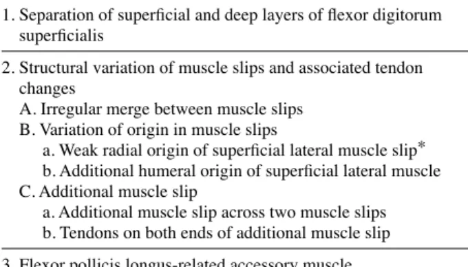
로 복합적인 변이를 관찰하였다. 해부진행은 아래팔 중심 선을 절개한 뒤 피부, 피부밑조직, 깊은근막의 순서로 노 출하였고, 얕은손가락굽힘근을 확보하여 관찰을 진행하였 다. 관찰을 통해 발견한 변이들을 다음과 같이 정리하였다 (Table 1). 얕은손가락굽힘근의 두 층(얕은층, 깊은층)은 하 나의 근육힘살을 형성하여 손가락으로 향하는 것으로 알 려져 있으나, 본 시신에서는 얕은층과 깊은층이 분리된 상 태로 진행하는 변이를 확인하였다. 일반적으로 얕은손가락 굽힘근은 두 갈래에서 일어나 하나의 근육힘살을 이루고 아래팔 먼쪽에서 각 손가락으로 향하는 4개의 힘줄을 형 성한다. 각 힘줄에 연결된 근육가지는 하나로 합쳐진 근육 덩어리의 형태를 띠고 있어, 각 근육가지의 주행이나, 일 어나는 곳, 닿는곳 등 근육가지에 대한 자세한 구조는 파 악하기 어렵다. 그러나 본 시신에서는 근육가지가 분리되 어 주행하고 있어 자세한 형태를 관찰 할 수 있었다. 얕은 층-안쪽, 깊은층-안쪽, 깊은층-가쪽 근육가지는 위팔자갈 래에서 일어나 각각 넷째, 새끼, 집게손가락에 닿고, 얕은 Table 1. Concomitant variations in flexor digitorum superficialis
in this report.
1. Separation of superficial and deep layers of flexor digitorum superficialis
2. Structural variation of muscle slips and associated tendon changes
A. Irregular merge between muscle slips B. Variation of origin in muscle slips
a. Weak radial origin of superficial lateral muscle slip*
b. Additional humeral origin of superficial lateral muscle C. Additional muscle slip
a. Additional muscle slip across two muscle slips b. Tendons on both ends of additional muscle slip 3. Flexor pollicis longus-related accessory muscle (Gantzer’s muscle)
* Superficial lateral muscle slip points to the slip connected to the middle finger.
Fig. 1. Cadaveric dissection showing left flexor digitorum superficialis. Superficial layer(A) and deep layer(B) of left flexor digitorum su- perficialis. SM, superficial-medial muscle slip; SL, superficial-lateral muscle slip; HH, humeral head of superficial-lateral muscle slip; RH, radial head of superficial-lateral muscle slip; MN, median nerve; RN, radial nerve; BR, brachioradialis(cut); DM, deep-medial muscle slip;
DL, deep-lateral muscle slip; AM, additional muscle slip; AT, additional tendon; GM, Gantzer’s muscle; FPL, flexor pollicis longus.
층-가쪽 근육가지는 위팔자갈래와 노갈래에서 동시에 일 어나 가운데손가락에 닿았다(Fig. 1). 얕은손가락굽힘근의 노갈래는 노뼈 빗선에서 넓게 일어난다. 그러나 본 시신 에서 얕은층-가쪽 근육가지가 노뼈 빗선의 일부에만 좁게 연결된 형태로 위축되어 나타났고, 대신 추가적으로 얕은 층-가쪽 근육가지가 정중신경의 가쪽으로 위팔뼈에서 일 어나는 것을 관찰하였다(Fig. 1A). 얕은손가락굽힘근의 깊 은층-안쪽 근육가지 가운데 지점과 깊은층-가쪽 근육가지 의 먼쪽 1/4 지점을 가로지르는 덧근육가지를 발견하였다.
이 덧근육가지는 약 7cm 길이의 방추형 모양이고 정중신 경의 가지가 분포하였다. 또한, 이 근육가지로 인해 깊은 층-안쪽 근육가지의 힘줄구조 역시 변이가 생겼는데, 깊은 층-안쪽 근육가지 가운데 부위에 힘줄이 형성되어 두힘살 근(digastric) 형태를 보였고, 깊은층-가쪽 근육가지는 아래 팔 먼쪽 부위에 덧근육가지의 힘줄이 존재함으로써 다른 근육가지보다 먼저 몸쪽 부위에서부터 집게손가락으로 향 하는 힘줄이 시작되는 양상을 보였다. 마지막으로, 얕은손 가락굽힘근 깊은층-안쪽 근육가지의 앞쪽에서 일어나 긴 엄지굽힘근의 중간지점으로 닿는 Gantzer근육이 관찰되 었다. 이 근육은 길이가 약 9cm 의 방추형 모형이며, 정중 신경의 가지가 분포하고 있는 것을 확인할 수 있었다(Fig.
1B).
고 찰
지금까지 얕은손가락굽힘근의 변이에 관련된 연구는 증 례보고가 주를 이루고[3,9,10], 일부 비교해부학적 분석 [11-13], 기능적 연관성에 대한 연구[14-17]와 소수의 변이 발생 원인에 관한 고찰[6,18,19]이 보고되었다. 본 증례에 서는 복합변이의 발생원인을 비교해부학적 관점과 발생학 적 관점으로 설명하고자 한다.
비교해부학적 관점에서는, 얕은손가락굽힘근을 양서류 의 아래팔에서 관찰할 수 있는 두 층의 근육이 하나의 구 조물로 합쳐진 진화적 구조물로 설명하고 있다. 또한 이 과정과 함께 손 부위에 존재하였던 짧은 근육 역시 힘줄로 대체된 것으로 본다. 이로 인해 손은 운동기능에서 물건을 쥐는 도구로 기능적 변화를 이루었고, 아래팔 역시 추진력 을 전달하는 손목의 굽힘 역할과 더불어, 손 부위의 잡는 힘 제공과 손허리손가락관절과 몸쪽손가락뼈사이관절의 굽힘작용을 추가하게 되었다[11-13]. 본 증례에서 얕은손 가락굽힘근의 얕은층과 깊은층의 융합이 완전하게 이루어 지지 않고 분리되어있다는 점이 퇴화의 흔적이라고 볼 수 있다(Fig. 1). 또한, 근육가지 사이 다리 역할을 하는 근육
구조(Fig. 1B)가 남아있는 것 역시, 근육구조 변이 과정의 중간 단계라고 추측할 수 있다.
한편, 근육의 변이를 발생학적 관점으로 설명하고 있는 연구에서는, 태아에서도 성인과 같이 근육의 변이가 발견 되어야 하는 것을 전제로 한다고 설명한다[6]. 또한 태아 에서의 Gantzer근육 발생 비율(32%)이 성인의 비율(39%) 과 크게 차이 나지 않는다는 보고가 있다. 이 연구에서는 근육 구조의 발생 단계를 사지싹, 중배엽 응축, 근육덩어 리, 분할의 단계로 나누었고, 이 중 분할단계에서 각 근육 의 세부 구조가 갖춰지는데 불완전한 분할이 여러 개의 근 육가지로 나타날 수 있다고 기술하고 있다[18]. 또 다른 연 구에서는 근육구조의 윤곽을 구성하는 힘줄원기는 기계적 인 자극에 의해서 유도된다고 보고하였다. 힘줄세포라고 불리는 힘줄섬유아세포는 기계적 수용성이 있어서 기계적 인 자극에 따라 힘줄의 생성 및 항상성을 유지한다. 다시 말해, 기계적 부하는 세포바깥 환경의 원섬유 구조를 변화 시키고, 이는 기질단백질의 조성을 변화시켜 힘줄원기 형 성을 촉진한다. 결국, 발생과정에서 기계적인 자극은 힘줄 구조의 재편성을 유발할 수 있고, 이는 근육구조에까지 영 향을 미칠 수 있다고 말하고 있다[19]. 본 증례에서는 얕 은손가락굽힘근 깊은층의 덧근육가지 양 끝에서 힘줄 구 조의 변화를 관찰할 수 있었다(Fig. 1B). 발생학적 관점으 로 볼 때, 외부의 물리적 자극으로 인해 굽힘근 덩어리의 중간에 힘줄원기가 형성되었다면, 이로 인해 얕은손가락 굽힘근에 덧근육가지가 형성되는 것은 물론 근육원시세포 의 딴곳 주행을 통해 이차적으로 각 근육가지의 분리를 유 도했을 것이다. 더욱이 왼쪽 아래팔에서 다양한 변이는 복 합적으로 나타났음에도 불구하고, 오른쪽 아래팔은 정상적 구조를 갖는 것 역시, 유전적인 요인과 같은 내재적 원인 보다는 외부 원인으로 인해 신체의 일부에만 변이가 형성 된 것을 뒷받침한다.
얕은손가락굽힘근의 변이에 관한 연구는 많은 선행 연 구자들에 의해 진행되어 왔지만, 아래팔 부위에 대한 깊이 있는 연구자료는 충분하지 않다. 가장 큰 이유는 아래팔 의 근육 변이가 손 부위에 비해 임상적으로 큰 의미를 갖 지 않기 때문일 것이다. 또한, 변이와 신체 기능 간의 정확 한 인과관계를 파악하기 위해서는 초음파스캔이나 MRI 등 고가의 장비를 사용해야 한다는 비용적 어려움도 있다 [15]. 이러한 제한적 상황 때문에, 여러 변이가 복합적으로 나타난 본 증례는 근육 변이의 형성 원인을 밝힐 수 있는 매우 가치 있는 추가적인 연구라고 할 수 있다. 또한 본 증 례 이외에 복합적인 변이가 일어나는 증례에 대한 깊이 있 는 연구를 통해 비교 분석을 시도한다면 변이의 원인을 이 해할 수 있는 좋은 자료가 될 수 있을 것으로 사료된다.
REFERENCES
1. Richard LD, A. Wayne V, Adam WM. Gray’s anatomy for students. 3th ed. Philadelphia: Elsevier/ Churchill Living- stone; 2013. p. 779.
2. Rodrigues V, Nayak SB, Rao MK, Vollala V, Somayaji N, Rao AS. Abnormal muscle in the anterior compartment of the forearm: a case report. Cases J. 2009; 2:9125-8.
3. Yesilada AK, Tatlıdede HS, Cakmak E. Anomalous large uniquemuscle belly of flexor digitorum superficialis and the absence of palmaris longus in the forearm. J Plast Reconstr Aesthet Surg. 2013; 66:137-9.
4. Han DK, Won HS, Liu HF, Chung IH, Kim IB. Separate muscle bundles of the flexor digitorum superficialis overly- ing the ulnar nerve. Folia Morphol. 2015; 74: 434-8.
5. Pai MM, Nayak SR, Krishnamurthy A, Vadgaonkar R, Prabhu LV, Ranade AV, et al. The accessory heads of flexor pollicis longus and flexor digitorum profundus: incidence and morphology. Clin Anat. 2008; 21:252-8.
6. Takkallapalli A, Sanjay K, Krishnamurthy A, Dattatray D, Neelee J. A unique variation of flexor digitorum superfi- cialis muscle and its clinical significance. J Life Sci. 2012;
4:39-43.
7. Lee SW, Lee JH, Lee HS. Double Gantzer’s muscles by four muscle bellies and its clinical significance: A case re- port. Korea J Phys Anthropol. 2017; 30:67-70.
8. Oh CS, Chung IH, Koh KS. Anatomical study of the acces- sory head of the flexor pollicis longus and the anterior inter- osseous nerve in asians. Clin Anat. 2000; 13:434-8.
9. Nayak SR, Ramanathan L, Prabhu LV, Raju S. Additional flexor muscles of the forearm: case report and clinical sig- nificance. Singapore Med J. 2007; 48:e231-3.
10. Saghir N, Saghir R, Shahid S, Hachach-Haram N, Johal K, Sojitra N. A unilateral variation in the flexor digitorum
superficialis with two distinct muscle bellies and associated tendons to the ring finger. J Plast Reconstr Aesthet Surg.
2016; 69:869-870.
11. Dixit SG, Kakar S. An uncommon variation of flexor dig- itorum superficialis indicis, a case report: Anatomical and clinical relevance. Clin Anat. 2010; 23:889-90.
12. Amrita G, Virendra K. Bilateral absence of flexor digito- rum superficialis(FDS) tendon of the little finger: Clinical significance. J Clin Diagn Res. 2014; 8:135-6.
13. Elliot D, Khandwala AR, Kulkarni M. Anomalies of the flexor digitorum superficialis muscle. J Hand Surg Br.
1999; 24:570-4.
14. Tan JS, Oh L, Louis DS. Variations of the flexor digitorum superficialis as determined by an expanded clinical exam- ination. J Hand Surg Am. 2009; 34:900-6.
15. Godwin Y, Wheble GA, Feig C. Assessment of the pres- ence of independent flexor digitorum superficialis function in the small fingers of professional string players: is this an example of natural selection? J Hand Surg Eur Vol. 2014;
39:93-100.
16. Puhaindran ME, Sebastin SJ, Lim AY, Xu WX, Chen YM.
Absence of flexor digitorum superficialis tendon in the lit- tle finger is not associated with decreased grip strength. J Hand Surg Eur Vol. 2008; 33:205-7.
17. Methot J, Chinchalkar SJ, Richards RS. Contribution of the ulnar digits to grip strength. Can J Plast Surg. 2010;
18:e10-4.
18. Kara A, Elvan O, Yildiz S, Ozturk H. Accessory head of flexor pollicis longus muscle in fetuses and adult cadavers and its relation to anterior interosseous nerve. Clin Anat.
2012; 25:601-8.
19. Galloway MT, Lalley AL, Shearn JT. The role of mechani- cal loading in tendon development, maintenance, injury, and repair. Bone Joint Surg Am. 2013; 95:1620-8.
Concomitant Variations in Flexor Digitorum Superficialis: A Case Report
Woo-Roe Choe
1, Young-Suk Cho
2, Kwang Il Nam
21Department of Medicine, 2Departments of Anatomy, Chonnam National University Medical School
Abstract :
The flexor digitorum superficialis(FDS) muscle is located in the intermediate layer of the muscles in the anterior compartment of the forearm. Variable but individual variations have been reported in the FDS regarding the number of head and the origin, distribution and interconnections of muscle slip and insertion to finger. In this case, we report a concomitant complex variation in FDS which was observed in a cadaver during a routine dissection classes for the undergraduate medical students. It includes the variation which is the separation of the tendon of FDS into the super- ficial and deep layers, the structural variations in muscle slips and associated tendon variations, the finding of Gantzer’muscle leading to flexor pollicis longus muscle. These complex variations in FDS are very rare case and this report summarizes the related phylogenetic and embryological significance.
Keywords : Flexor digitorum superficialis, Variation, Additional muscle, Gantzer’ muscle, Additional tendon
Correspondence to : Kwang IL Nam (Departments of Anatomy, Chonnam National University Medical School) E-mail : [email protected]