Identification of the Mutations in the Prostaglandin Transporter Gene, SLCO2A1 and Clinical Characterization in Korean Patients with Pachydermoperiostosis
Pachydermoperiostosis (PDP), or primary hypertrophic osteoarthropathy, is a rare genetic disease affecting both skin and bones. Both autosomal dominant with incomplete penetrance and recessive inheritance of PDP have been previously confirmed. Recently, hydroxyprostaglandin dehydrogenase (HPGD) and solute carrier organic anion transporter family member 2A1 (SLCO2A1) were reported as pathogenic genes responsible for PDP.
Both genes are involved in prostaglandin E2 (PGE2) degradation. We aimed to identify responsible genes for PDP and the clinical features in Korean patients with PDP. Six affected individuals and their available healthy family members from three unrelated Korean families with PDP were studied. All of the patients displayed complete phenotypes of PDP with finger clubbing, pachydermia, and periostosis. Mutation analysis revealed a novel heterozygous mutation in the SLCO2A1 gene at nucleotide 302 causing a substitution of the amino acid isoleucine to serine at codon 101 (p.IIe101Ser) in affected individuals. We also identified known SLCO2A1 mutations, one homozygous for c.940+1G>A, and another compound heterozygous for c.940+1G>A and c.1807C>T (p.Arg603*) from two PDP families. Genetic analyses of the PDP patients showed no abnormality in the HPGD gene. Our study further supports the role of mutations in the SLCO2A1 gene in the pathogenesis of PDP and could provide additional clues to the genotype-phenotype relations of PDP.
Keywords: Pachydermoperiostosis; Primary Hypertrophic Osteoarthropathy; SLCO2A1 Gene; Mutation
Sihoon Lee,1* So Young Park,2* Hyun Jin Kwon,1 Chul-Ho Lee,3 Ok-Hwa Kim,4 and Yumie Rhee5
1Department of Internal Medicine and Laboratory of Genomics and Translational Medicine, Gachon University School of Medicine, Incheon, Korea;
2Department of Internal Medicine, Cheil General Hospital and Women’s Healthcare Center, Dankook University College of Medicine, Seoul, Korea;
3Division of Clinical Genetics, Department of Pediatrics, Severance Children’s Hospital, Severance Hospital, Yonsei University College of Medicine, Seoul, Korea; 4Department of Radiology, Woorisoa Children’s Hospital, Seoul, Korea; 5Department of Internal Medicine, Endocrine Research Institute, Yonsei University College of Medicine, Seoul, Korea
* Sihoon Lee and So Young Park contributed equally to this work.
Received: 19 November 2015 Accepted: 15 January 2016 Address for Correspondence:
Yumie Rhee, MD
Department of Internal Medicine, Endocrine Research Institute, Yonsei University College of Medicine, 50-1 Yonsei-ro, Seodaemun-gu, Seoul 03722, Korea
E-mail: [email protected]
Funding: This research was supported by the Basic Science Research Program through the National Research Foundation of Korea (NRF) funded by the Ministry of Science, ICT and Future Planning, Korea (NRF-2013R1A1A1A05005629 to S.L.) and in part by a Research Grant from the Korean Diabetes Association to S.L.
http://dx.doi.org/10.3346/jkms.2016.31.5.735 • J Korean Med Sci 2016; 31: 735-742
INTRODUCTION
Pachydermoperiostosis (PDP), or primary hypertrophic osteo- arthropathy, is a cluster of genetic syndromes characterized by three major symptoms; digital clubbing, periostosis, and pachy- dermia. Additional manifestations including excessive sweat- ing, acne, arthropathy, and acro-osteolysis of long bones have been reported (1). The phenotypic spectrum of PDP is broad, and three clinical presentations of PDP are generally recognized:
the complete form involving all three major symptoms, the in- complete form with periostosis without pachydermia, and the
‘forme fruste’ with pachydermia and minimal or no skeletal anomalies (2).
Both autosomal dominant with incomplete penetrance and
recessive inheritance have been suggested in PDP (3). The pre- cise incidence and prevalence are still unknown. Uppal et al. (4) recently discovered homozygous mutations in the hydroxypros- taglandin dehydrogenase (HPGD) gene in a subset of patients with PDP. In addition, exome analysis of PDP in Japanese, Chi- nese, Caucasian, and other races has revealed homozygous and compound heterozygous mutations in the solute carrier organic anion transporter family member 2A1 (SLCO2A1) gene (5-8). Both HPGD and SLCO2A1 genes are involved in prosta- glandin E2 (PGE2) degradation. Increased levels of PGE2 in pa- tients with causal genetic mutations could contribute to the pathogenesis of PDP (9).
To date, one family of PDP related to SLCO2A1 gene muta- tion has been reported in Korea (10). In this study, we describe Endocrinology, Nutrition & Metabolism
complete types of PDP carrying SLCO2A1 mutations including a novel mutation in Koreans with review of literature.
MATERIALS AND METHODS Subjects
All participants were of Korean ethnicity. Six patients with PDP and their family members from three unrelated families were recruited.
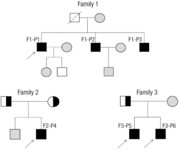
Family 1
The case of this family (Fig. 1 and 2A-C) has been previously re- ported (11). The 56-year-old proband, who was born to healthy non-consanguineous parents, visited the clinic for genetic coun- seling of his son. At 19 years of age, the proband presented with enlarged hands and feet, knee joints swelling and pain and was then referred to a neurosurgeon for suspicion of acromegaly.
He underwent hypophysectomy, which showed no tumor le- sion in the specimen. Attention was aroused only after perios-
Fig. 1. Pedigrees of affected individuals with SLCO2A1 mutations. In the pedigree, arrows indicate the proband. Filled black, patients with PDP; Half-black, healthy members with a heterozygous mutation; Gray, healthy members not doing genotyping test.
Family 1
F1-P1 F1-P2 F1-P3
Family 2 Family 3
F2-P4 F3-P5 F3-P6
Fig. 2. Clinical features of affected individuals with SLCO2A1 mutations. (A-C) Clinical pictures of family 1 proband. (A) Thickening and furrowing of the facial skin. (B-C) Digital clubbing and swelling of the ankle joint. (D-H) Clinical pictures of family 2 proband. (D) Thickening and greasiness of facial skin. (E) Digital clubbing. (F-G) Cortical hyperostosis of long bones. (H) Diffusely increased uptake in the whole axial and appendicular long bones shown by whole body bone scan. (I-J) 18F-fluoride PET scan images of femur and tibia show increased cortical/periosteal uptake in the proband of family 3.
A
F G H I J
B C D E
teal thickening was observed in the skeletal survey of the hands and forearm bones, indicating primary hypertrophic osteoar- thropathy as well as facial furrowing. His two brothers, who were also diagnosed as PDP, showed similar clinical features to the proband.
Family 2
The 19-year-old proband (Fig. 1 and 2D-H) was born to a healthy non-consanguineous couple. Beginning at 17 years of age, he noticed inappropriately thickened lower legs and swelling of ankle joints. He also complained of hyperhidrosis, acne, and difficulty of squatting. He had atrial septal defect (ASD), patent ductus arteriosus (PDA), and valvular heart disease and under- went an operation for PDA at the age of 1 month. A physical ex- amination revealed enlargement of the terminal phalanges and facial furrowing. Skeletal survey showed periosteal thickening of the long bones. The radionuclide whole body bone scan (WBBS) showed diffusely increased uptake in the whole axial and ap- pendicular long bones, suspicious of metabolic bone disease.
Results of laboratory analyses, including growth hormone, in- sulin-like growth factor 1, and thyroid-stimulating hormone were normal. His parents were not clinically affected.
Family 3
The case of this family was recently reported during preparation of this manuscript (10). Two siblings (Fig. 1) of ages 23 and 19 years presented with progressive pachydermia and enlargement of their hands and feet since puberty. They also complained of
hyperhidrosis, acne, and frontal furrowing. The brothers suffer- ed from watery diarrhea; however, colonoscopic finding was not remarkable. The younger patient had a history of the opera- tion for PDA in infancy; whereas his older brother had no histo- ry of PDA or other congenital heart diseases. Skeletal survey showed periosteal thickening of the long bones. The 18F-fluo- ride positron emission tomography (PET) scan showed incre- ased cortical/periosteal uptake of long bones (Fig. 2I and J). La- boratory tests including growth hormone and insulin-like growth factor 1 were within normal limits. Their parents were healthy without any similar features.
Mutation analysis of the associated genes
A mutation analysis of the affected individuals and their family members was performed by direct sequencing of PCR products amplified from genomic DNA. Genomic DNA was extracted from peripheral blood leukocytes using conventional methods.
The DNA sequences of SLCO2A1 and HPGD were obtained from the available online database (NM_005630.2 and NM_000860.4, respectively). Primers were designed using Primer 3 software (http://frodo.wi.mit.edu/cgi-bin/primer3/). All exons and their exon-intron boundaries in SLCO2A1 and HPGD were ampli- fied via PCR. Direct sequencing was performed using the Big- DyeTM Terminator v3.1 Cycle Sequencing Kit (Applied Biosys- tems, Waltham, MA, USA).
Ethics statement
The study was approved by the institutional review board (IRB) Table 1. Clinical phenotypes of Korean patients with pachydermoperisostasis
Phenotypes
Cases
F1-P1 F1-P2 F1-P3 F2-P4 F3-P5
Kim et al. (10)
F3-P6 Kim et al. (10)
Current age, yr 56 54 52 19 23 19
Onset age, yr 19 17 20 17 puberty 13
Gender M M M M M M
SLCO2A1 mutations c.302T>G c.302T>G c.302T>G c.940+1G>A
c.1807C>T
c.940+1G>A c.940+1G>A
c.940+1G>A c.940+1G>A Triad
Digital clubbing Periostosis Pachydermia
+ + +
+ + +
+ + +
+ + +
+ + +
+ + + Skin
Palmar and plantar hyperhidrosis Acne
Seborrhoea and eczema
+ + +
- + +
- + +
- + +
- + +
- + + Skeletal
History of bone fractures Swelling of large joints Painful joints on exercise Hydrarthrosis
- + + +
- + + +
- + + +
- + + +
- + + +
- + + + Others
Anemia Hypoalbuminemia Patent ductus arteriosus
- - -
- - -
- - -
- - +
- - -
- - + M, male; +, positive; -, negative or unknown.
Fig. 3. Localization and sequence chromatogram of identified SLCO2A1 mutations. Upper: The positions of the mutations in the exons of SLCO2A1 in this study. Lower: SL- CO2A1 mutations in PDP families. family 1, (A) c.302T>G; family 2, (B) c.940+1G>A and (C) c.1807C>T; family 3, (C) c.940+1G>A.
SLCO2A1, Chromosome 3q21 exon
c.302T>G c.940+1G>A
1 2 3 4 5 6 7 8 9 10 11 12 13 14
c.1807C>T c.302T>G (p.Ile101Ser) c.940+1G>A (p.R288Gfs*7) c.1807C>T (p.R603*)
A B C
of Gachon University Gil Medical Center, Korea (IRB No. GIR- BA2151). Informed consent was obtained from all subjects be- fore participation.
RESULTS
Clinical features of PDP patients
Based on major diagnostic criteria (digital clubbing, periostosis, and pachydermia) (1), all patients showed major clinical find- ings and conformed to the diagnosis of PDP. They were catego- rized as having the complete form and developed major symp- toms before the age of 20 years and subsequently suffered from progressive skin thickening with seborrhea, swelling of periar- ticular tissue, and joint discomfort or arthralgia. Their parents did not show clinical signs of PDP. The clinical phenotypes of PDP patients are summarized in Table 1.
Identification of SLCO2A1 as a gene responsible for PDP PCR was performed for screening of SLCO2A1 and HPGD mu- tations followed by a direct sequence analysis, and no HPGD mutation was found in affected individuals. The results of these genetic analyses are shown in Fig. 3.
In family 1, DNA sequencing of the proband revealed a novel heterozygous mutation in the SLCO2A1 gene at nucleotide 302 (c.302T>G) causing a substitution of the amino acid isoleucine (Ile) to serine (Ser) at codon 101 (p.Ile101Ser) in exon 3 as well as a known polymorphism (A396T) in exon 14. The two broth- ers of the proband were also heterozygous for c.302T>G. The proband’s son, who seemed normal in appearance, did not car- ry the mutations.
In family 2, the proband showed compound heterozygous for 2 different SLCO2A1 mutations; c.940+1G>A and c.1807C>T.
The c.940+1G>A mutation was located in the splice donor site of intron 7, which resulted in the loss of exon 7 and a truncation
of prostaglandin transporter (5,12), and c.1807C>T in exon 13 introduced a stop codon at position 603 (p.Arg603*). The pati- ents’ father and mother were heterozygous for c.940+1G>A and c.1807C>T, respectively. In family 3, the brothers were homozy- gous for c.940+1G>A. The patients’ father was heterozygous for c.940+1G>A, and evaluation of the mother was not available.
DISCUSSION
PDP or primary hypertrophic osteoarthropathy is characterized by pachydermia, periostosis and finger clubbing; however, the phenotypic spectrum of PDP is known to be broad (1). The di- agnosis of PDP is based on the typical clinical features and ra- diological data. PDP needs to be differentiated from acromega- ly and secondary hypertrophic osteoarthropathy that is associ- ated with pulmonary or cardiac disease. For the differential di- agnosis, hormone tests including growth hormone and thyroid stimulating hormone, absence or presence of thickening and furrowing of facial skin, and sella turcica imaging might be help- ful (13).
Since PDP was first described in 1868 (1), mutations in HPGD and SLCO2A1 have been identified in families affected with PDP (4-6). So far, 43 different SLCO2A1 mutations have been report- ed and are summarized in Table 2. Our study identified a novel mutation, c.302T>G, as well as known mutations, c.940+1G>A and c.1807C>T, in the SLCO2A1 gene. The c.940+1G>A muta- tion was suggested as a hot spot in both Chinese Han and Euro- pean Caucasian patients with PDP (3,6,14). Sasaki et al. (5) in- sisted that the c.940+1G>A mutation is a major mutation as an ancient founder allele in Japanese PDP patient. Considering ethnic roots and regional proximity, the c.940+1G>A mutation might also be a common mutation detected in Korean PDP pa- tients.
A considerable number of PDP patients showed autosomal
recessive transmission. However, autosomal dominant trans- mission with incomplete penetrance has been also reported in previous studies (3). In this study, three patients of family 1 car- ried a heterozygous c.302T>G mutation. The mutated alleles
supposed to be autosomal dominant inheritance were c.302T>G, c.754C>T, c.830_831insT, c.861+2T>G, c.1065dupA, and c.1333C
>T (3,7,8). The patients having c.754C>T and c.830_831insT al- leles showed mild clinical symptoms, whereas PDP patients Table 2. Summary of the genetic SLCO2A1 mutations in patients with pachydermoperisostasis in the literature
References Mutation Origin Sex Age ClubbingPeriosto-
sis Pachy- dermia Hyperhi-
drosis Sebor- rhea Arthral-
gia
Zhang et al. (19) c.940+1G>A c.1602C>A Chinese M 22 + + + + NA +
Zhang et al. (3) c.855delA c.855delA c.855delA c.1106G>A c.1393G>A c.493G>T c.664G>A c.861+2T>C c.1065dupA
c.855delA c.855delA c.855delA c.1106G>A c.1393G>A c.1136G>A c.1634delA
Chinese Chinese Chinese Chinese Chinese Chinese Chinese Chinese Chinese
M F F M M M M M M
36 47 42 23 26 18 24 42 17
+ - - + + + + + +
+ - - + + + + + +
+ - - + + + + + +
- - - + - - - - +
+ - - + + + + + +
+ - - + + + + + +
Zhang et al. (31) c.235-1G>T c.656C>T Chinese M 27 + + + + NA +
Zhang et al. (6) c.97-1G>A c.764G>A c.664G>A
c.97-1G>A c.1634delA c.940+1G>A
Chinese Chinese Chinese
M M M
24 27 21
+ + +
+ + +
+ + +
NA NA NA
NA NA NA
NA NA NA Cheng et al. (32) c.547G>A
c.940+1G>A
c.1807C>T c.1602C>A
Chinese Chinese
M M
25 37
+ +
+ +
+ +
+ NA
+ +
+ + Niizeki et al. (12) c.940+1G>A
c.754C>T c.421G>T c.940+1G>A
c.1279_1290del12 c.1807C>T c.940+1G>A c.1807C>T
Japanese Japanese Japanese Japanese
M M M M
19 21 20 20
+ + + +
+ + + +
+ + + +
+ + + -
+ - + +
- + - + Sasaki et al. (5) c.940+1G>A
c.310G>A c.940+1G>A c.940+1G>A
c.1279_1290del12 c.1040C>T c.940+1G>A c.1668G>C
Japanese Japanese Japanese Japanese
M M M M
24 25 45 53
+ + + +
+ + + +
+ + + +
+ + - -
+ + + -
+ + - +
Niizeki et al. (9) c.1279G>A c.1807C>T Japanese F 67 + + - - - +
Minakawa et al. (33) c.940+1G>A c.1279_1290del12 Japanese M 15 + + + + + +
Busch et al. (14) c.940+1G>A c.940+1G>A c.940+1G>A c.1292delC c.763G>A c.763G>A
c.1668G>C c.940+1G>A c.940+1G>A c.1292delC c.763G>A c.763G>A
Japanese Japanese Japanese Indian Indian Indian
M M M M M M
53 21 19 27 26 28
+ + + + + +
+ NA NA NA + +
NA + + + NA NA
NA NA NA NA + +
NA NA NA + NA NA
+ NA NA + + + Seifert et al. (8) c.830_831insT
c.830_831insT c.830_831insT c.830_831insT c.1670T>C c.754C>T
c.830_831insT c.830_831insT c.830_831insT c.1670T>C
Turkish Turkish Turkish Turkish Iraq Dutch
M M M M M M
21 19 7 40 38 28
+ + - + + +
+ + - - + -
+ + - - + -
+ + - - + -
+ + - - + -
+ + - - + - Diggle et al. (7) c.1259G>T
c.941-1G>A c.542G>C c.1333C>T c.290G>A c.664G>A c.253A>T c.1105+4A>G c.838C>T c.310G>T c.724+1G>T c.542G>A c.611C>T
c.1259G>T c.1517C>A c.542G>C c.940+2T>A c.664G>A c.253A>T c.1105+4A>G c.1693T>G c.310G>T c.724+1G>T c.542G>A c.611C>T
Hispanic (Colombia) Chinese Turkish Dutch French North African North African Dutch
Kabardin (Caucasus) Italian
Algerian Turkish Italian
M M M M M M M M M M M M M
45 NA NA NA NA NA NA NA NA NA NA NA NA
+ + + + NA NA NA NA NA NA NA NA NA
+ + + + NA NA NA NA NA NA NA NA NA
+ + + + NA NA NA NA NA NA NA NA NA
NA NA NA NA NA NA NA NA NA NA NA NA NA
NA NA NA NA NA NA NA NA NA NA NA NA NA
NA NA NA NA NA NA NA NA NA NA NA NA NA
Ayoub et al. (34) c.1016C>T c.1016C>T Saudi M 23 + + + + + +
Madruga Dias et al. (35) c.940+1G>A c.940+1G>A African M 26 + + + NA NA +
Saadeh et al. (36) c.838C>T c.838C>T
c.838C>T c.838C>T
Lebanese Lebanese
M M
22 24
+ +
+ NA
+ +
NA NA
NA NA
+ + NA, unknown or not available.
carrying c.302T>G, c.861+2T>G, c.1065dupA, and c.1333C>T al- leles presented the full-blown phenotype (Table 2). We found no consistent clinical features or genetically unique character- istics among those mutations.
Males were more commonly and severely affected with the ratio of male:female of 9:1 (13). A few females carrying the SL- CO2A1 mutation were not clinically affected or showed atypical phenotype of PDP (3,7,9). A simple explanation of the skewed sex ratio is lacking. Plausible assumptions were testosterone promoting disease expression, weaker reactivity to prostaglan- din in women than men, and suppressed effects of estrogen on interleukin 1b-mediated induction of the COX-2 pathway in vessels (1,15,16).
Besides typical skin and bone manifestations, accompanying PDA was reported in about one third of cases in PDP patients with HPGD mutation (17). However, congenital cardiac anom- aly has scarcely been reported in SLCO2A1-mutated patients.
Only one Dutch patient carrying c.1333C>T mutation in the SL- CO2A1 gene was reported to have ASD (7). Prostaglandin trans- porter–deficient mice by targeted deletion of the SLCO2A1 gene die as neonates probably as a result of a closure defect of the ductus arteriosus (18). Seifert et al. (8) reported that PDP pa- tients with SLCO2A1 mutations did not show failure of postna- tal ductus arteriosus closure. Contrary to previous reports, our patient carrying compound SLCO2A1 heterozygous mutations was diagnosed with congenital heart defects including ASD, PDA, and valvular heart disease. It is uncertain whether this case is a coincidental finding; or PDA might be a rare, fatal con- dition in SLCO2A1-mutated PDP patients.
Both HPGD and SLCO2A1 genes are known to involve PGE2 metabolism. Mutations in SLCO2A1 cause increased levels of PGE2 due to the failure of extracellular uptake of PGE2 (19,20).
Urinary excretion of the main PGE2 metabolite, PGE-M, is the best measure of total endogenous PGE2 production in normal condition (21). Previous studies showed elevated urinary excre- tion of PGE2 and PGE-M in PDP patients, suggesting that mea- surement of urinary PGE2 and PGE-M can be helpful in early and differential diagnosis of PDP (3,4,12). Urinary PGE2 and PGE-M levels were not determined in the current study and fur- ther measurement is needed.
Increased prostaglandins, which have a biphasic role in bone metabolism, stimulate both bone resorption and formation (22).
An increase in bone turnover markers was observed in young PDP patients who might have high disease activity (23,24). Pre- viously, bone scintigraphy confirmed a high activity of bone turnover in the affected joints of PDP patients (25). Three-phase skeletal scintigraphy in a PDP patient detected increased tracer uptake around long bones, which indicated the active form of the disease (26). Our patient in family 2 also showed similar finding of increased radionuclide uptake along the long bone cortices by WBBS (Fig. 2H). In this study, 18F-fluoride PET scan
was performed in PDP patients for the first time. PET scan also provided convincing evidence of hypermetabolic status in vari- ous long bones.
There is no specific treatment for PDP (27). Clubbing is usu- ally asymptomatic and does not require treatment (13). For pa- tients with painful osteoarthropathy, the therapeutic options consist of salicylates, non-steroidal anti-inflammatory drugs, systemic corticosteroids and colchicine (27). Bisphosphonates or infliximab have been attempted in cases of PDP refractory to conventional therapy (28,29). Cosmetic surgery or botulinum toxin injections have been introduced for correcting rough fa- cial features (13,30).
We report on three unrelated PDP families and identified three different mutations in the SLCO2A1 gene, of which c.302T>G in exon 3 is novel. As with Japanese and Chinese studies, we deter- mine that c.940+1G>A is a major mutation in Korean PDP pa- tients. Although our study did not contain PG-related metabolic activity, we further support the role of mutations in the SL- CO2A1 gene in the pathogenesis of PDP.
ACKNOWLEDGMENT
The authors thank all family members for their participation.
DISCLOSURE
The authors have no potential conflicts of interest to disclose.
AUTHOR CONTRIBUTION
Identification and evaluation of index cases and families: Lee S, Rhee Y. Acquisition of clinical data: Lee S, Rhee Y. Writing: Park SY. Genetic analysis: Kwon HJ, Lee CH. Radiologic diagnosis:
Kim OH. Final approval of manuscript: all authors.
ORCID
Sihoon Lee http://orcid.org/0000-0002-9444-5849 So Young Park http://orcid.org/0000-0001-6002-0116 Hyun Jin Kwon http://orcid.org/0000-0002-2936-1358 Chul-Ho Lee http://orcid.org/0000-0001-8045-4368 Ok-Hwa Kim http://orcid.org/0000-0001-5502-8746 Yumie Rhee http://orcid.org/0000-0003-4227-5638 REFERENCES
1. Castori M, Sinibaldi L, Mingarelli R, Lachman RS, Rimoin DL, Dallapicc- ola B. Pachydermoperiostosis: an update. Clin Genet 2005; 68: 477-86.
2. Sinha GP, Curtis P, Haigh D, Lealman GT, Dodds W, Bennett CP. Pachy- dermoperiostosis in childhood. Br J Rheumatol 1997; 36: 1224-7.
3. Zhang Z, He JW, Fu WZ, Zhang CQ, Zhang ZL. Mutations in the SLCO2A1
gene and primary hypertrophic osteoarthropathy: a clinical and biochem- ical characterization. J Clin Endocrinol Metab 2013; 98: E923-33.
4. Uppal S, Diggle CP, Carr IM, Fishwick CW, Ahmed M, Ibrahim GH, Helli- well PS, Latos-Bieleńska A, Phillips SE, Markham AF, et al. Mutations in 15-hydroxyprostaglandin dehydrogenase cause primary hypertrophic osteoarthropathy. Nat Genet 2008; 40: 789-93.
5. Sasaki T, Niizeki H, Shimizu A, Shiohama A, Hirakiyama A, Okuyama T, Seki A, Kabashima K, Otsuka A, Ishiko A, et al. Identification of mutations in the prostaglandin transporter gene SLCO2A1 and its phenotype-gen- otype correlation in Japanese patients with pachydermoperiostosis. J Dermatol Sci 2012; 68: 36-44.
6. Zhang Z, Xia W, He J, Zhang Z, Ke Y, Yue H, Wang C, Zhang H, Gu J, Hu W, et al. Exome sequencing identifies SLCO2A1 mutations as a cause of pri- mary hypertrophic osteoarthropathy. Am J Hum Genet 2012; 90: 125-32.
7. Diggle CP, Parry DA, Logan CV, Laissue P, Rivera C, Restrepo CM, Fonse- ca DJ, Morgan JE, Allanore Y, Fontenay M, et al. Prostaglandin transporter mutations cause pachydermoperiostosis with myelofibrosis. Hum Mutat 2012; 33: 1175-81.
8. Seifert W, Kühnisch J, Tüysüz B, Specker C, Brouwers A, Horn D. Muta- tions in the prostaglandin transporter encoding gene SLCO2A1 cause primary hypertrophic osteoarthropathy and isolated digital clubbing.
Hum Mutat 2012; 33: 660-4.
9. Niizeki H, Shiohama A, Sasaki T, Seki A, Kabashima K, Otsuka A, Takeshi- ta M, Hirakiyama A, Okuyama T, Tanese K, et al. The novel SLCO2A1 het- erozygous missense mutation p.E427K and nonsense mutation p.R603*
in a female patient with pachydermoperiostosis with an atypical pheno- type. Br J Dermatol 2014; 170: 1187-9.
10. Kim HJ, Koo KY, Shin DY, Kim Y, Lee JS, Lee MG. Complete form of pachy- dermoperiostosis with SLCO2A1 gene mutation in a Korean family. J Der- matol 2015; 42: 655-7.
11. Lee HK, Kim JH, Kim JY, Park HY, Shin ES, Chang HJ, Han IS, Kang MH.
Pachydermoperiostosis mimicking acromegaly. J Korean Soc Endocrinol 1993; 8: 439-44.
12. Niizeki H, Shiohama A, Sasaki T, Seki A, Kabashima K, Otsuka A, Kosaki K, Ogo A, Yamada T, Miyasaka M, et al. The complete type of pachyder- moperiostosis: a novel nonsense mutation p.E141* of the SLCO2A1 gene.
J Dermatol Sci 2014; 75: 193-5.
13. Martinez-Lavin M. Miscellaneous non-inflammatory musculoskeletal conditions. Pachydermoperiostosis. Best Pract Res Clin Rheumatol 2011;
25: 727-34.
14. Busch J, Frank V, Bachmann N, Otsuka A, Oji V, Metze D, Shah K, Danda S, Watzer B, Traupe H, et al. Mutations in the prostaglandin transporter SLCO2A1 cause primary hypertrophic osteoarthropathy with digital club- bing. J Invest Dermatol 2012; 132: 2473-6.
15. Hatano R, Onoe K, Obara M, Matsubara M, Kanai Y, Muto S, Asano S. Sex hormones induce a gender-related difference in renal expression of a novel prostaglandin transporter, OAT-PG, influencing basal PGE2 con- centration. Am J Physiol Renal Physiol 2012; 302: F342-9.
16. Ospina JA, Brevig HN, Krause DN, Duckles SP. Estrogen suppresses IL- 1beta-mediated induction of COX-2 pathway in rat cerebral blood ves- sels. Am J Physiol Heart Circ Physiol 2004; 286: H2010-9.
17. Sinibaldi L, Harifi G, Bottillo I, Iannicelli M, El Hassani S, Brancati F, Dal- lapiccola B. A novel homozygous splice site mutation in the HPGD gene causes mild primary hypertrophic osteoarthropathy. Clin Exp Rheuma- tol 2010; 28: 153-7.
18. Chang HY, Locker J, Lu R, Schuster VL. Failure of postnatal ductus arteri- osus closure in prostaglandin transporter-deficient mice. Circulation 2010; 121: 529-36.
19. Zhang Z, He JW, Fu WZ, Zhang CQ, Zhang ZL. A novel mutation in the SLCO2A1 gene in a Chinese family with primary hypertrophic osteoar- thropathy. Gene 2013; 521: 191-4.
20. Nomura T, Lu R, Pucci ML, Schuster VL. The two-step model of prosta- glandin signal termination: in vitro reconstitution with the prostaglandin transporter and prostaglandin 15 dehydrogenase. Mol Pharmacol 2004;
65: 973-8.
21. Murphey LJ, Williams MK, Sanchez SC, Byrne LM, Csiki I, Oates JA, John- son DH, Morrow JD. Quantification of the major urinary metabolite of PGE2 by a liquid chromatographic/mass spectrometric assay: determi- nation of cyclooxygenase-specific PGE2 synthesis in healthy humans and those with lung cancer. Anal Biochem 2004; 334: 266-75.
22. Blackwell KA, Raisz LG, Pilbeam CC. Prostaglandins in bone: bad cop, good cop? Trends Endocrinol Metab 2010; 21: 294-301.
23. Jojima H, Kinoshita K, Naito M. A case of pachydermoperiostosis treated by oral administration of a bisphosphonate and arthroscopic synovecto- my. Mod Rheumatol 2007; 17: 330-2.
24. Martínez-Ferrer A, Peris P, Alós L, Morales-Ruiz M, Guañabens N. Prosta- glandin E2 and bone turnover markers in the evaluation of primary hy- pertrophic osteoarthropathy (pachydermoperiostosis): a case report.
Clin Rheumatol 2009; 28: 1229-33.
25. Bomanji J, Nagaraj N, Jewkes R, Fields M, Maini RN. Pachydermoperios- tosis: technetium-99m-methylene diphosphonate scintigraphic pattern.
J Nucl Med 1991; 32: 1907-9.
26. Santhosh S, Bhattacharya A, Bhadada S, Kaur R, Singh M, Mittal BR. Three- phase skeletal scintigraphy in pachydermoperiostosis. Clin Nucl Med 2011; 36: e199-201.
27. Guerini MB, Barbato MT, Sá NB, Nunes DH, Zeni PR. Pachydermoperi- ostosis: the complete form of the syndrome. An Bras Dermatol 2011; 86:
582-4.
28. da Costa FV, de Magalhães Souza Fialho SC, Zimmermann AF, Neves FS, Werner de Castro GR, Pereira IA. Infliximab treatment in pachydermo- periostosis: a rare disease without an effective therapeutic option. J Clin Rheumatol 2010; 16: 183-4.
29. Martinez-Lavin M, Vargas A, Rivera-Viñas M. Hypertrophic osteoarthrop- athy: a palindrome with a pathogenic connotation. Curr Opin Rheuma- tol 2008; 20: 88-91.
30. Bingol UA, Cinar C. Pachydermoperiostosis: aesthetic treatment of pre- maturely aging face with facelift and botulinum toxin a. J Craniofac Surg 2014; 25: e563-4.
31. Zhang Z, He JW, Fu WZ, Zhang CQ, Zhang ZL. Two novel mutations in the SLCO2A1 gene in a Chinese patient with primary hypertrophic os- teoarthropathy. Gene 2014; 534: 421-3.
32. Cheng R, Li M, Guo Y, Yao Y, Gao C, Yao Z. Three novel mutations in the SLCO2A1 gene in two Chinese families with primary hypertrophic os- teoarthropathy. Eur J Dermatol 2013; 23: 636-9.
33. Minakawa S, Kaneko T, Niizeki H, Mizukami H, Saito Y, Nigawara T, Ku- rose R, Nakabayashi K, Kabashima K, Sawamura D. Case of pachyder- moperiostosis with solute carrier organic anion transporter family, mem- ber 2A1 (SLCO2A1) mutations. J Dermatol 2015; 42: 908-10.
34. Ayoub N, Al-Khenaizan S, Sonbol H, Albreakan R, AlSufyani M, AlBalwi M. A novel homozygous mutation in the SLCO2A1 gene is associated
with severe primary hypertrophic osteoarthropathy phenotype in a Sau- di patient. Int J Dermatol 2015; 54: e233-5.
35. Madruga Dias JA, Rosa RS, Perpétuo I, Rodrigues AM, Janeiro A, Costa MM, Gaião L, Pereira da Silva JA, Fonseca JE, Miltenberger-Miltenyi G.
Pachydermoperiostosis in an African patient caused by a Chinese/Japa- nese SLCO2A1 mutation-case report and review of literature. Semin Ar-
thritis Rheum 2014; 43: 566-9.
36. Saadeh D, Kurban M, Ghosn S, Btadini W, Nemer G, Arayssi T, Uthman I, Badra R, Farra C. Pachydermoperiostosis genetic screening in Lebanese families uncovers a novel SLCO2A1mutation. J Eur Acad Dermatol Ve- nereol 2015; 29: 2489-90.