Glandular odontogenic cysts (GOCs) are rare intrabony solitary or multiloculated cysts of odontogenic origin. In 1987, Padayachee and Van Wyk1first reported two cases that shared the features of both botryoid odontogenic cysts and central mucoepidermoid tumors and suggested that the term “sialo-odontogenic cyst” be adopted for such lesions to avoid confusion and mismanagement. One year later, Gardner et al2reported eight other cases and favored the name “glandular odontogenic cyst”; they regarded it as a distinct entity due to its unusual histopathological fea- tures. In addition, this term was included in the revised edition of a World Health Organization report3in 1992 as
“developmental odontogenic epithelial cyst.” It has also been termed “mucoepidermoid cyst” by Sadeghi et al4and
“polymorphous odontogenic cyst” by High et al.5In the 20 years since it was first described, 111 cases of this type of cyst have been reported, an incidence of 0.2% among odontogenic cysts.6
Hence, although rare, this cyst is a well-known clinical entity and is important to recognize and diagnose due to
its aggressive behavior and tendency to recur. This cyst reports a slight male predilection with a male : female ratio of 1.3 : 1,6and the most-commonly affected site is the an- terior mandible. It tends to occur over a wide age range of 10-90 years, with a mean age of 49.5 years. The mandible seems to be affected more commonly (87.2%) than the maxilla.7
Radiographic examination reveals a well-defined cyst- like unilocular or multilocular radiolucency, often with scalloped margins and sclerotic borders.8In addition, there may be root resorption and tooth displacement with corti- cal perforation, leading to extension of the cyst into the adjacent soft tissues.
Histologically, GOC is characterized by a cyst wall lin- ing of non-keratinized epithelium with papillary projec- tions, nodular thickenings, mucous-filled clefts, and “mu- cous lakes.” In addition, it includes cuboidal basal cells, which are sometimes vacuolated.9,10
GOC treatment includes curettage and enucleation, alth- ough some authors believe marginal resection to be a more reliable treatment due to the tendency of the cyst to recur after curettage and enucleation.10
The aim of this report is to present a case of GOC in a young female patient in the posterior mandibular region, which was quite rare and unusual.
Glandular odontogenic cyst: A case report
Shahnaz S. Tambawala1,*, Freny R. Karjodkar1, Archana Yadav2, Kaustubh Sansare1, Subodh Sontakke1
1Department of Oral Medicine and Radiology, Nair Hospital Dental College, Mumbai, India
2Department of Oral Pathology, Nair Hospital Dental College, Mumbai, India
ABSTRACT
Glandular odontogenic cysts (GOCs) are rare intrabony solitary or multiloculated cysts of odontogenic origin. The importance of GOCs lies in the fact that they exhibit a propensity for recurrence similar to keratocystic odontogenic tumors and that they may be confused microscopically with central mucoepidermoid carcinoma. Thus, the oral and maxillofacial radiologists play an important role in definitive diagnosis of GOC based on distinctive cases; though they are rare. In large part, this is due to the GOC’s complex and frequently non-specific histopathology. This report describes a case of GOC occurrence in the posterior mandibular ramus region in a 17-year-old female, which is a rare combination of site, age, and gender for occurrence. (Imaging Sci Dent 2014; 44 : 75-9)
KEY WORDS: Odontogenic Cysts; Panoramic Radiography; Cone-Beam Computed Tomography
Received May 18, 2013; Revised June 20, 2013; Accepted August 4, 2013
*Correspondence to: Dr. Shahnaz S. Tambawala
Oral Medicine and Radiology Department, Ground floor, Room no - 2, Nair Hospital Dental College, Opposite Maratha Mandir, Dr. A. L. Nair Road, Mumbai Central, Mumbai 400 008, Maharashtra, India
Tel) 91-22-23082714, Fax) 91-22-23080655, E-mail) [email protected]
Copyright ⓒ 2014 by Korean Academy of Oral and Maxillofacial Radiology
This is an Open Access article distributed under the terms of the Creative Commons Attribution Non-Commercial License (http://creativecommons.org/licenses/by-nc/3.0) which permits unrestricted non-commercial use, distribution, and reproduction in any medium, provided the original work is properly cited.
Imaging Science in Dentistry∙pISSN 2233-7822 eISSN 2233-7830
Case Report
A 17-year-old female patient was referred to the Outpa- tient Department of Oral Medicine and Radiology at Nair Hospital Dental College, Mumbai, by a local orthodontist after discovering a well-defined radiolucent pathology in the right ramal region of the mandible on a routine pano- ramic view.
The patient reported mild dull pain over the right poster- ior mandibular region for a period of 15 days with no aggravating or relieving factors. An extraoral examination revealed no significant clinical findings. Intraorally, there was mild obliteration of the buccal vestibule in the right lower second molar region with no other relevant clinical finding. The patient’s medical history revealed that she had suffered from allergic asthma since childhood, she was not on any medication for the condition.
A panoramic radiograph was obtained using a Planmeca Proline CC machine (Planmeca OY, Helsinki, Finland)
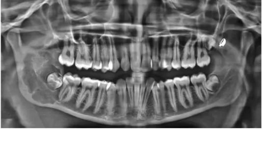
with exposure parameters of 73 kVp, 10 mA, and 15 s expo- sure time. A well-defined multiloculated radiolucent lesion was observed extending from the distal border and apex of the lower right second molar into the ramus to approxi- mately 3 mm away from the sigmoid notch enclosing the developing tooth bud of the right lower third molar (Fig.
1). There was no expansion of the posterior or inferior bor- ders of the ramus, as seen on the panoramic radiograph.
A cone-beam computed tomographic (CBCT) scan was obtained using a Kodak 9000 3D machine (Carestream Health Inc., Rochester, NY, USA), with a field-of-view (FOV) of 50 mm×37 mm, voxel size of 76.5μm×76.5 μm×76.5 μm, and exposure time of 10.8 s. The captured images were reconstructed using a high-spatial-frequency reconstruction algorithm, and these images revealed a well- defined multiloculated radiolucent lesion in the right man- dibular ramal region (Figs. 2A and B). This lesion extend- ed from the right lower second molar to the upper third of the ramus, with resorption of the apex of the right mandi-
Fig. 1. A panoramic radiograph shows a well-defined multiloculat- ed radiolucent lesion extending from the distal border and apex of lower right second molar into the ramus till approximately 3 mm away from the sigmoid notch enclosing the developing tooth bud of right lower third molar.
bular second molar having well-defined, sharp right-angl- ed septa (Fig. 2C). The radiolucency had scalloped borders and surrounded the developing third molar tooth bud (Figs.
2B and C). There was perforation of the lingual cortical plate and thinning of the buccal cortical plate, as seen on the CBCT coronal section of the lesion (Fig. 2D).
Based on the clinical and radiographic findings, a pro- visional diagnosis of keratocystic odontogenic tumor due to minimal expansion with considerable bony involvement was proposed with a differential diagnosis of odontogenic myxoma due to the presence of right-angled straight septa,
as seen on the reconstructed panoramic section in the CBCT image.
Thereafter, an incisional biopsy of the lesion along with enucleation of the third molar tooth bud was performed under antibiotic coverage and local anesthesia. The speci- men was submitted for histopathological evaluation, which revealed a pseudo-stratified, ciliated columnar epithelial cystic lining covering mature fibrous connective tissue.
Thus, the overall histopathological findings were sugges- tive of a GOC. Following these investigations, surgical exploration with curettage of the GOC of the hemimandi- ble was performed under general anesthesia, and the speci- men was sent for histopathological confirmation. The he- matoxylin and eosin stained section showed cystic lumen lined by pseudo-stratified columnar epithelium with fili- form extensions of the cytoplasm and mucous-secreting cells with intra-epithelial spherule formation, which is characteristic of GOCs, thus confirming the previous histo- pathological diagnosis of GOC (Fig. 3).
After a 3-month follow-up of the patient, no complica- tion or recurrence was reported (Fig. 4).
Discussion
As stated previously in the Introduction section, a case of GOC, an unusual odontogenic developmental cyst of the jaws, is presented herein. In accordance with previous studies and cases, our case demonstrated significant mandi- bular involvement.7In addition, the radiological features showed similarities with previous reports, including a well- defined radiolucency with distinct borders along with loss of cortical integrity and root resorption.
Fig. 4. A panoramic radiograph shows healing of the surgical defect by new bone formation 3 months post-surgery (white arrow heads).
Fig. 3.Histopathologic examination shows cystic lumen lined by pseudo-stratified columnar epithelium with filiform extensions of the cytoplasm and mucous-secreting cells with intra-epithelial spherule formation, characteristic of glandular odontogenic cyst (H&E stain, 400x).
The histopathological features were also suggestive of a cystic cavity lined with pseudo-stratified, ciliated colum- nar epithelial lining, and fibrous vascular connective tis- sue.8-12The disagreement was related to gender predilec- tion, age, and site prevalence: the literature showed a pre- dilection toward males and a mean age of 49.5 years with the anterior mandible being the most-commonly affected site,6,7,13,14 whereas the present case was reported in a young teenage girl with the involvement of the posterior mandible and the ramus region.
GOC recognition based on physical and radiological examinations alone is practically impossible, a fact that the authors of all previous studies harmoniously stress upon.
Only histopathological examinations allow for a certain diagnosis of the cyst.15
The GOC does not differ from other jawbone cysts in typical radiological projections. Thus, diagnoses of denti- gerous cysts, and botryoid, radicular, and keratocystic odontogenic tumors should be made on the basis of X-ray examinations. Furthermore, in the case of multilocular cysts, the differential diagnosis may shift toward amelo- blastoma, myxoma, central giant cell granuloma, and fibr- ous dysplasia.16 However, the most important diagnosis outcome worthy of consideration is central mucoepider- moid carcinoma due to the significant histopathological similarities.
Kaplan et al6,16 and Brannon et al12proposed a list of major and minor microscopic criteria for GOC on the basis of the frequency of each feature in the reported cases from the literature. On the basis of their analysis, it was suggest- ed that at least the focal presence of each of the following major criteria must be present for diagnosis: 1) squamous epithelial lining with a flat interface with the connective tissue wall, lacking basal palisading; 2) epithelium exhibit- ing variations in thickness along the cystic lining with or without epithelial “spheres,” “whorls,” or focal luminal proliferation; 3) cuboidal eosinophilic cells or “hobnail”
In addition, they listed the following minor criteria that supported the diagnosis but were not mandatory: 1) papil- lary proliferation of the lining epithelium, 2) ciliated cells, 3) multicystic or multiluminal architecture, and 4) clear or vacuolated cells in the basal or spinous layers.
The distinction between low-grade central mucoepider- moid carcinoma and GOC is difficult, if not impossible.
The only feature that has not been reported in low-grade central mucoepidermoid carcinoma and that may justify the existence of GOC as a separate entity is the occasional presence of epithelial plaques, similar to those seen in lat- eral periodontal cysts.17However, unlike lateral periodon- tal and botryoid odontogenic cysts, which are more inno- cuous, GOC is regarded as considerably aggressive.
Our case was considered to be GOC because it fulfilled all criteria specified by Gardner et al,2and unlike mucoepi- dermoid carcinoma, cellular atypia and solid and microcys- tic epithelial proliferation were not seen.
The reported treatment of GOC ranges from a conserva- tive approach (enucleation, marsupialization, curettage with or without peripheral ostectomy, curettage with adju- vant Carnoy’s solution, or cryotherapy) to marginal resec- tion and segmental resection. A few authors preferred mar- ginal and segmental resection due to the cyst’s tendency to recur after conservative treatment.14
However, it is unlikely that all cases and reports publish- ed thus far have used such strict histopathological criteria for GOC recognition. From this recommendation, it can be readily concluded that in a few cases, GOC histopathology can be non-specific to the extent that it is insufficient as the sole test for every suspected case of GOC.18Therefore, its criteria and clinical features should be identified and used to further refine the diagnosis of cases for which his- topathological diagnosis is ambiguous, as well as the dif- ferential diagnosis of this cyst (Table 1).18
In conclusion, GOC is a rare and aggressive lesion with a relatively high recurrence rate. Hence, a careful clinical
locularity, cortical integrity, expansion and extent of the lesion, and involvement of the contiguous soft tissue.
References
1. Padayachee A, Van Wyk CW. Two cystic lesions with features of both the botryoid odontogenic cyst and the central mucoepi- dermoid tumour: sialo-odontogenic cyst? J Oral Pathol 1987;
16: 499-504.
2. Gardner DG, Kessler HP, Morency R, Schaffner DL. The glan- dular odontogenic cyst: an apparent entity. J Oral Pathol 1988;
17: 359-66.
3. Kramer IR, Pindborg JJ, Shear M. The WHO histological typ- ing of odontogenic tumours: a commentary on the second edi- tion. Cancer 1992; 70: 2988-94.
4. Sadeghi EM, Weldon LL, Kwon PH, Sampson E. Mucoepi- dermoid odontogenic cyst. Int J Oral Maxillofac Surg 1991; 20:
142-3.
5. High AS, Main DM, Khoo SP, Pedlar J, Hume WJ. The poly- morphous odontogenic cyst. J Oral Pathol Med 1996; 25: 25- 31.
6. Kaplan I, Anavi Y, Hirshberg A. Glandular odontogenic cyst:
a challenge in diagnosis and treatment. Oral Dis 2008; 14: 575- 81.
7. Guruprasad Y, Chauhan DS. Glandular odontogenic cyst of maxilla. J Clin Imaging Sci 2011; 1: 54.
8. Nair RG, Varghese IV, Shameena PM, Sudha S. Glandular odontogenic cyst: report of a case and review of literature. J Oral Maxillofac Pathol 2006; 10: 20-3.
9. Gardner DG, Morency R. The glandular odontogenic cyst, a
rare lesion that tends to recur. J Can Dent Assoc 1993; 59: 929- 30.
10. Krishnamurthy A, Sherlin HJ, Ramalingam K, Natesan A, Premkumar P, Ramani P, et al. Glandular odontogenic cyst:
report of two cases and review of literature. Head Neck Pathol 2009; 3: 153-8.
11. Manor R, Anavi Y, Kaplan I, Calderon S. Radiological features of glandular odontogenic cyst. Dentomaxillofac Radiol 2003;
32:73-9.
12. Brannon RB, Kessler HP, Castle JT, Kahn MA. Glandular odontogenic cyst: analysis of 46 cases with special emphasis on microscopic criteria for diagnosis. Head Neck Pathol 2011;
5: 364-75.
13. Prabhu S, Rekha K, Kumar G. Glandular odontogenic cyst mimicking central mucoepidermoid carcinoma. J Oral Maxil- lofac Pathol 2010; 14: 12-5.
14. Salehinejad J, Saghafi S, Zare-Mahmoodabadi R, Ghazi N, Kermani H. Glandular odontogenic cyst of the posterior max- illa. Arch Iran Med 2011; 14: 416-8.
15. uczak K, Nowak R, Rzeszutko M. Glandular odontogenic cyst of the mandible associated with impacted tooth: report of a case and review of literature. Dent Med Probl 2007; 44: 403- 6.
16. Kaplan I, Anavi Y, Manor R, Sulkes J, Calderon S. The use of molecular markers as an aid in the diagnosis of glandular odon- togenic cyst. Oral Oncol 2005; 41: 895-902.
17. Noffke C, Raubenheimer EJ. The glandular odontogenic cyst:
clinical and radiological features; review of the literature and report of nine cases. Dentomaxillofac Radiol 2002; 31: 333-8.
18. MacDonald-Jankowski DS. Glandular odontogenic cyst: sys- tematic review. Dentomaxillofac Radiol 2010; 39: 127-39.