In 1923, the dentist Pierre Robin described a disorder characterized as a triad of micrognathia, glossoptosis, and airway obstruction.1 This disorder, now known as Pierre Robin sequence(PRS), has received much attention in den- tistry due to its relationship with anomalies in the maxillo- facial area. PRS does not always have a single pathogene- sis, but rather is often associated with various syndromes.2 Izumi el al.3 found that 60% of cases of PRS were associat- ed with another syndrome, while 40% were isolated.
Cerebro-costo-mandibular syndrome(CCMS) is a PRS- related syndrome associated with rib anomalies character- ized by posterior rib gap defects.4-6 Since the first report of CCMS by Smith et al.4 in 1966, over 80 cases have been reported to date.5 In addition to PRS and multiple rib gaps, scoliosis is a common feature of CCMS. The heart, the
kidneys, hearing ability, and intelligence are also affected in some patients with CCMS.7 Because the most consistent features of CCMS are associated with the hard tissues of the maxillofacial and thoracic vertebral area, radiographic evaluation, such as thoracic computed tomography, pos- teroanterior(PA) chest radiography, lateral cephalograms, and cone-beam computed tomography(CBCT), is required for the diagnosis and treatment planning of CCMS.
The patient in this report was suspected of having CCMS because she had PRS and severe scoliosis. However, no posterior rib gap defects were observed on PA chest radi- ography. Therefore, the authors could not make a definitive diagnosis of CCMS. Although many reports have been published on CCMS, few cases of PRS with scoliosis alone have been reported.8 This case report presents the clinical and radiological features of an adult patient with PRS and severe scoliosis, but without rib anomalies.
Case Report
A 35-year-old woman visited the Department of Ortho- dontics at Gangneung-Wonju National University Dental Hospital. Her chief complaints were a retruded mandible, Pierre Robin sequence(PRS) is characterized by the triad of micrognathia, glossoptosis, and airway obstruction.
PRS does not have a single pathogenesis, but rather is associated with multiple syndromes. This report presents the case of a 35-year-old woman with PRS and scoliosis. Among the syndromes related to PRS, cerebro-costo- mandibular syndrome(CCMS), which is characterized by posterior rib gap defects and vertebral anomalies, was suspected in this patient. However, no posterior rib gap defect was detected on radiological examinations. Although over 80 cases of CCMS have been reported to date, few cases of PRS with scoliosis alone have been reported.
Therefore, this report demonstrated the clinical, radiological, and cephalometric characteristics of an adult patient with PRS and scoliosis, but without rib anomalies.(Imaging Sci Dent 2019; 49: 323-9)
Key woRds: Pierre Robin Syndrome; Scoliosis; Micrognathism
Copyright ⓒ 2019 by Korean Academy of Oral and Maxillofacial Radiology
This is an Open Access article distributed under the terms of the Creative Commons Attribution Non-Commercial License(http://creativecommons.org/licenses/by-nc/3.0) which permits unrestricted non-commercial use, distribution, and reproduction in any medium, provided the original work is properly cited.
Imaging Science in Dentistry·pISSN 2233-7822 eISSN 2233-7830 Received September 19, 2019; Revised October 30, 2019; Accepted November 1, 2019
*Correspondence to: Prof. Bong-Keun Cha
Department of Orthodontics, College of Dentistry, Gangneung-Wonju National University, 120 Gangneung Daehangno, Gangneung, Gangwon Province 25457, Korea Tel) 82-33-640-3192, E-mail) [email protected]
Prof. In-Woo Park
Department of Oral and Maxillofacial Radiology, College of Dentistry, Gangneung- Wonju National University, 120 Gangneung Daehangno, Gangneung, Gangwon Province 25457, Korea
Tel) 82-33-640-3187, E-mail) [email protected]
a large overjet, and crowding on the upper incisors. She underwent soft palate surgery when she was an infant and spinal surgery for severe scoliosis at age 15. She had no familial history of any relevant syndromes.
Clinical examination revealed an anterior open bite, crowding on the upper incisors, a narrow maxillary arch,
and Class II canine relationship(Fig. 1). The bilateral lower second premolars and first molars were missing and replaced by fixed partial prostheses. Due to tongue- tie(Fig. 2A) and her anterior open bite, the patient had difficulty pronouncing sounds such as ‘s’ and ‘z’. Scar tis- sue was present on the soft palate, and mild rhinism was
Fig. 1. Intraoral photos of the patient show an anterior open bite and a Class II canine relationship.
Fig. 2. A. Tongue-tie restraining the forward and upward movement of the tongue. B. Soft palate scar caused by palate surgery.
A B
Fig. 3. A panoramic radiograph rev- eals deep antegonial notches, poste- rior bowing, and a small anteropos- terior width of the bilateral condyles.
ondary dental caries, and the left lower third molar had a periapical lesion. Deep antegonial notches and posterior bowing of the bilateral condyles were observed.
Cephalometric analysis showed a small and retruded mandible and a steep mandibular plane(Table 1). The A point-nasion-B point angle was 10.1°, indicating a severe skeletal Class II relationship. The mandibular plane angle was larger than normal, at 51.4°. Narrowing of the airway was also observed(Fig. 4). To compare the lateral cepha- lograms between the patient and normal control images,9 the profilograms were superimposed at the sella using the sella-nasion line as a horizontal reference line(Fig. 5).
Severe bimaxillary retrusion, particularly of the mandible, as well as a relatively small mandible and a large inclina- tion of the mandibular plane were found.
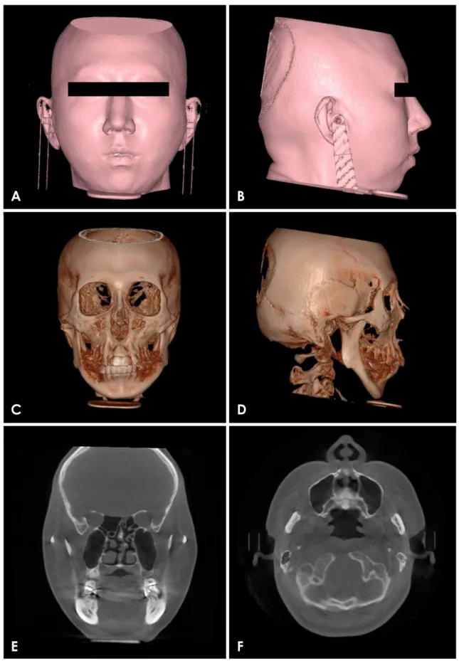
Mandibular asymmetry, with deviation of the chin to the left side and low-set ears(Figs. 6A-D), was exhibited on the CBCT images oriented according to the method described by Kim et al.10 A discontinuity of the nasal sep- tum extending from the posterior hard palate to the supe- rior nasal area was observed(Fig. 6E). A lack of soft tis- sue near the posterior nasal spine was also observed(Figs.
6E and F). The bilateral condyles were hypoplastic, but there were no signs of degenerative joint disease, such as
erosive changes of the condyle or articular eminence.11 Because PRS and scoliosis were observed during the clinical and radiological examinations, this patient was initially suspected to have CCMS. A PA chest radiograph was taken to identify rib defects and scoliosis(Fig. 7). Se-
Table 1. Comparison of cephalometric variables between the ref- erence values for Korean adult women and the patient in this study
Cephalometric variable Normal Patient
SNA(°) 81.6 77.6
SNB(°) 79.0 67.5
ANB(°) 2.6 10.1
FMA(°) 23.9 51.4
Gonion angle(°) 116.8 140.3
Lower anterior facial height(mm) 70.7 79.6
Anterior cranial base(mm) 72.0 66.0
Mandibular body length(mm) 80.6 65.3
Incisal overbite(mm) 1.7 -1.3
Incisal overjet(mm) 3.1 6.1
SNA: sella-nasion-A point angle, SNB: sella-nasion-B point angle, ANB:
A point-nasion-B point angle, FMA: Frankfort-mandibular plane angle The reference values for Korean adult women were obtained from [9].
Fig. 4. A lateral cephalogram shows a small and retruded mandible, a large inclination of the mandibular plane, and a narrow airway.
Fig. 5. Superimposition of the profilograms of the patient(dotted) and normal control(solid) shows severe micrognathia and a large mandibular plane angle.
A B
C D
E F
Fig. 6. Cone-beam computed tomography(CBCT) images. A-D. Three-dimensional reconstructed CBCT images demonstrate mandibular retrusion, asymmetry, and low-set ears. E and F. Coronal and axial CBCT images of the posterior soft palate area show discontinuity of the nasal septum. Note the lack of soft tissue in the posterior nasal spine area.
vere scoliosis was identified, but no signs of posterior rib gap defects were detected.
discussion
The patient in this report had PRS and severe scoliosis.
PRS is known to be associated with various syndromes and genetic anomalies.2 Among the syndromes accom- panied with PRS is CCMS, which is characterized by vertebral and rib anomalies. Campomelic dysplasia and diastrophic dysplasia also present with scoliosis,2 but no other symptoms of these syndromes-such as short stat- ure, hearing loss, or short limbs-matched the patient’s physical condition. Therefore, we suspected that this pa- tient had CCMS.
Antenatally, patients with CCMS usually exhibit raised nuchal translucency, intrauterine growth retardation, poly- hydramnios, and PRS,5,12 which are identified using ultra- sonography or magnetic resonance imaging.12 After birth, various radiographic techniques are available to detect the rib anomalies characteristic of CCMS. Because the major radiological findings are a narrow thorax, posterior rib gap defects, and defects of costo-transverse articulation,5 thoracic computed tomography or PA chest radiography is required to identify CCMS. Posterior rib gap defects in CCMS may resemble the costal fractures of the newborn that can occur when resuscitation is performed.13 Several researchers have reported that these rib gaps may become ossified with increasing age.5,14 However, it is uncertain
mortality. Therefore, it is thought that these severe rib anomalies may be rare in adult patients with CCMS. Re- cently, the presence of an accessory ossicle arising from the lesser cornua of the hyoid bone in CCMS was report- ed, although the mechanism behind this is uncertain.5 In our patient, an accessory ossicle of the hyoid bone was not observed.
The dentofacial anomalies of PRS, along with the rib and vertebral anomalies, are major features of CCMS.5,7 Micrognathia and glossoptosis can be detected by a phys- ical examination, but the skeletal anomalies in the maxil- lofacial area should be evaluated radiologically. The mor- phological features of the craniofacial area can be evalu- ated precisely by CBCT and lateral cephalograms(Figs.
4-7, Table 1).
The dentofacial traits of this patient, including mi- crognathia, cleft soft palate, and tongue-tie, are typical features of PRS. The presence of nasal septum deviation aligns with an earlier study that found that the nasal sep- tum deviation of the cleft group was more severe than that of the non-cleft group.17 On PA chest radiography, se- vere scoliosis was observed; however, there were no signs of posterior rib gap defects or other rib anomalies. A med- ical radiologist confirmed that posterior rib gap defects and traces of ossification were not observed upon further examination. Chest radiography at infancy or during the growth period could be useful to confirm the process of ossification, but the previous records of the patient could not be obtained.
Several researchers found that heterozygous mutations of the SNRPB gene are associated with CCMS.18,19 Ther- e fore, genetic tests, such as exome sequencing and Sanger sequencing, are recommended for confirmation of CCMS.19 However, our patient refused further genetic test- ing, so unfortunately, we could not confirm the presence of CCMS.
Maxillofacial radiologists, surgeons, and orthodontists play important roles in the skeletal and dental manage- ment of patients with PRS.20 The maxillofacial defor-
Fig. 7. A posteroanterior chest radiograph shows severe scoliosis and the presence of titanium rods without any signs of posterior rib gap defects.
mities associated with PRS are evaluated by an expert radiologist, and each evaluated deformity is managed by a maxillofacial surgeon. Generally, airway problems are the most critical. For those problems, noninvasive methods such as prone/lateral positioning, continuous positive airway pressure, or nasopharyngeal intubation are preferred. Invasive methods, including tracheostomy, tongue-lip adhesion, or mandibular distraction osteogene- sis, may be also used. Palate surgery is usually performed approximately 1 year after birth.21 From an orthodontic perspective, problems associated with PRS include Class II malocclusion, crowding in the narrow maxillary arch, and hypodontia. A phase I orthodontic treatment plan can include maxillary expansion and induce the normal erup- tion of teeth. Although a lack of sufficient space for prop- er alignment of the teeth is observed, it is recommended that the decision whether to perform tooth extraction be delayed until phase II of treatment. After growth is com- plete, a large majority of cases require orthodontic treat- ment with orthognathic surgery to improve the patients’
Angle Class II dentition and convex profiles due to an- teroposterior discrepancies between the maxilla and the mandible. Only patients with mild discrepancies are typi- cally treated using alternatives to orthognathic surgery.20
Because this patient showed a severe mandibular retru- sion with a Class II relationship, an asymmetric mandible, orthognathic surgery was recommended for correction of the skeletal discrepancy. However, the patient did not want to undergo orthognathic surgery for financial and psychological reasons. Therefore, camouflage treatment for the correction of Class II malocclusion and prostho- dontic rehabilitation for missing lower posterior teeth were planned. To advance the retruded chin, filler aug- mentation of the chin(or genioplasty) was also planned.
When treating a patient with PRS, clinicians should always suspect another underlying syndrome and, if nec- essary, identify the syndrome present. Radiological ex- aminations are often useful to identify the syndrome, and additional genetic testing can be performed as well. This report provides a better understanding of the clinical, ra- diological, and cephalometric features of an adult present- ing with PRS with scoliosis, but without rib anomalies.
Conflicts of Interest: None
References
1. Robin P. La chute de la base de la langue considérée comme une nouvelle cause de gêne dans la respiration nasopharyngi-
enne. Bull Acad Med(Paris) 1923; 89: 37-40.
2. Tan TY, Kilpatrick N, Farlie PG. Developmental and genetic perspectives on Pierre Robin sequence. Am J Med Genet C Se- min Med Genet 2013; 163C: 295-305.
3. Izumi K, Konczal LL, Mitchell AL, Jones MC. Underlying genetic diagnosis of Pierre Robin sequence: retrospective chart review at two children’s hospitals and a systematic literature review. J Pediatr 2012; 160: 645-50.e2.
4. Smith DW, Theiler K, Schachenmann G. Rib-gap defect with micrognathia, malformed tracheal cartilages, and redundant skin: a new pattern of defective development. J Pediatr 1966;
69: 799-803.
5. Tooley M, Lynch D, Bernier F, Parboosingh J, Bhoj E, Zackai E, et al. Cerebro-costo-mandibular syndrome: clinical, radiologi- cal, and genetic findings. Am J Med Genet A 2016; 170A: 1115- 6. Miller KE, Allen RP, Davis WS. Rib gap defects with microg-26.
nathia. The cerebro-costo-mandibular syndrome-a Pierre Rob- in-like syndrome with rib dysplasia. Am J Roentgenol Radium Ther Nucl Med 1972; 114: 253-6.
7. Smith KG, Sekar KC. Cerebrocostomandibular syndrome. Case report and literature review. Clin Pediatr(Phila) 1985; 24: 223- 8. Rymer AN, Porteous GH, Neal JM. Anesthetic challenges in an 5.
adult with Pierre Robin sequence, severe juvenile scoliosis, and respiratory failure. A A Case Rep 2015; 5: 95-8.
9. Kim JH, Gansukh O, Amarsaikhan B, Lee SJ, Kim TW. Com- parison of cephalometric norms between Mongolian and Ko- rean adults with normal occlusions and well-balanced profiles.
Korean J Orthod 2011; 41: 42-50.
10. Kim MS, Lee EJ, Song IJ, Lee JS, Kang BC, Yoon SJ. The location of midfacial landmarks according to the method of es- tablishing the midsagittal reference plane in three-dimensional computed tomography analysis of facial asymmetry. Imaging Sci Dent 2015; 45: 227-32.
11. Nah KS. Condylar bony changes in patients with temporoman- dibular disorders: a CBCT study. Imaging Sci Dent 2012; 42:
249-53.
12. Ogasawara K, Honda Y, Hosoya M. Ex utero intrapartum treat- ment for an infant with cerebro-costo-mandibular syndrome.
Pediatr Int 2014; 56: 613-5.
13. Matić A, Velisavljev-Filipović G, Lovrenski J, Gajdobranski D.
A case of severe type of cerebro-costo-mandibular syndrome.
Srp Arh Celok Lek 2016; 144: 431-5.
14. Leroy JG, Devos EA, Vanden Bulcke LJ, Robbe NS. Cere- bro-costo-mandibular syndrome with autosomal dominant in- heritance. J Pediatr 1981; 99: 441-3.
15. Merlob P, Schonfeld A, Grunebaum M, Mor N, Reisner SH.
Autosomal dominant cerebro-costo-mandibular syndrome:
ultrasonographic and clinical findings. Am J Med Genet 1987;
26: 195-202.
16. Silverman FN, Strefling AM, Stevenson DK, Lazarus J. Cere- bro-costo-mandibular syndrome. J Pediatr 1980; 97: 406-16.
17. Lee SS, You DS. Radiographic study on maxillary sinus de- velopment and nasal septum deviation in cleft palate patient. J Korean Acad Oral Maxillofac Radiol 1992; 22: 305-13.
18. Lynch DC, Revil T, Schwartzentruber J, Bhoj EJ, Innes AM, Lamont RE, et al. Disrupted auto-regulation of the spliceosomal