IMMUNE NETWORK http://www.ksimm.or.kr Volume 11 Number 4 August 2011 http://dx.doi.org/10.4110/in.2011.11.4.223
pISSN 1598-2629 eISSN 2092-6685
223
BRIEF COMMUNICATION
Received on June 1, 2011. Revised on July 18, 2011. Accepted on August 18, 2011.
CC This is an open access article distributed under the terms of the Creative Commons Attribution Non-Commercial License (http://creativecommons.org/licenses/by-nc/3.0) which permits unrestricted non-commercial use, distribu- tion, and reproduction in any medium, provided the original work is properly cited.
*Corresponding Author. Tel: 82-2-3277-2563; Fax: 82-2-3277-2846; E-mail: [email protected]
Keywords: Insulin-like growth factor-I, Toll-like receptor-4, Cytokines, Skeletal muscle cells, PI3K/Akt, MAPK
IGF-I Exerts an Anti-inflammatory Effect on Skeletal Muscle Cells through Down-regulation of TLR4 Signaling
Won Jun Lee*
Department of Exercise Science, College of Health Science, Ewha Womans University, Seoul 120-750, Korea
Although exercise-induced growth factors such as Insulin-like growth factor-I (IGF-I) are known to affect various aspects of physiology in skeletal muscle cells, the molecular mechanism by which IGF-I modulates anti-inflammatory effects in these cells is presently unknown. Here, we showed that IGF-I stim- ulation suppresses the expression of toll-like receptor 4 (TLR4), a key innate immune receptor. A pharmacological in- hibitor study further showed that PI3K/Akt signaling path- way is required for IGF-I-mediated negative regulation of TLR4 expression. Furthermore, IGF-I treatment reduced the expression of various NF-κB-target genes such as TNF-α and IL-6. Taken together, these findings indicate that the an- ti-inflammatory effect of exercise may be due, at least in part, to IGF-I-induced suppression of TLR4 and subsequent downregulation of the TLR4-dependent inflammatory signal- ing pathway.
[Immune Network 2011;11(4):223-226]
It is well known that the accumulation of low-grade chronic inflammation is common in individuals who live a sedentary lifestyle and this condition has been linked to the develop- ment of various diseases including diabetes and cardiovas- cular diseases (1). Increased expression of toll-like receptor 4 (TLR4), a central innate immune receptor, has been ob- served in humans suffering from chronic inflammatory dis- eases such as obesity and type II diabetes (2,3). TLR4 is re- sponsible for recognition of pathogens and plays an im- portant role in innate immune function and adaptive im- munity (4,5). It is well known that regular exercise is an ef- fective countermeasure against low-grade chronic inflam-
mation (6). It has recently been shown that exercise and reg- ular physical activity exert anti-inflammatory effects through downregulation of TLR4 in the immune cells (7-9).
Because skeletal muscle is the largest organ in the human body and considered to be an endocrine organ due to the production of growth hormones and cytokines in response to exercise stimuli, it is worth examining the modulating ef- fect of exercise on TLR4 in skeletal muscle cells. However, the molecular mechanisms by which exercise modulates TLR4 signaling pathways are currently unknown. Exercise induces various adaptations in skeletal muscle cells in terms of meta- bolic and hormonal aspects. Among them, increased levels of insulin-like growth factor-I (IGF-I) are one of the most well known adaptations induced by various exercise stimuli (10).
Therefore, we investigated IGF-I to determine if it would modulate TLR4 gene expression and its downstream signaling and subsequently exert an anti-inflammatory effect on skeletal muscle cells. To investigate the possible relationship between IGF-I and TLR4 expression, we examined the mRNA and pro- tein level of TLR4 in the presence or absence of IGF-I treatment. C2C12 cells were maintained in Dulbecco’s modi- fied Eagle’s medium (DMEM) (Welgene, Korea) supple- mented with 10% fetal bovine serum (FBS) (Hyclone, Logan, UT) and antibiotics (100 U/ml penicillin G and 100μg/ml streptomycin) (Welgene, Korea). For the experiments, C2C12 myoblasts were plated in six-well culture plates at a density of 5×10⁵ cells/well in growth medium (DMEM, 10% FBS).
For IGF-I treatment, cells at 90% confluence in DMEM supple- mented with 2% horse serum (Hyclone, Logan, UT) and anti- biotics (100 U/ml penicillin G and 100μg/ml streptomycin;
IGF-I-induced Down-regulation of TLR4 Signaling Won Jun Lee
224 IMMUNE NETWORK http://www.ksimm.or.kr Volume 11 Number 4 August 2011
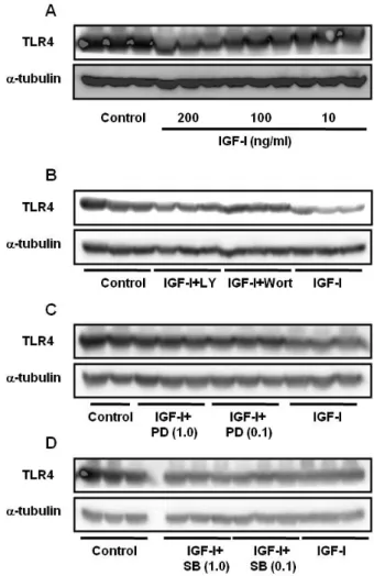
Figure 1. (A) Effect of different concentrations of IGF-I on TLR4 protein expression in differentiating C2C12 skeletal muscle cells. Cells were treated with IGF-I for 24 hr. (B) TLR4 protein expression in C2C12 cells treated with IGF-I (200 ng/ml) for 24 hr in the absence or presence of LY294002 (100μM) and Wortmannin (50 nM). (C) Cells were treated with IGF-I (200 ng/ml)+PD98059 (0.1 or 1.0μM) for 24 hr. (D) Cells were treated with IGF-I (200 ng/ml)+SB203580 (0.1 or 1.0μM) for 24 hr.
Welgene, Korea) were treated with IGF-I for 24 hr. The pro- tein expression levels were then examined by Western blot analysis using anti-TLR4 antibody (Santa Cruz Biotechnology, Santa Cruz, CA). Anti-α-tubulin was also used to normalize the amounts of loading proteins.
The results showed that IGF-I decreased TLR4 protein ex- pression in a dose-dependent manner, indicating that IGF-I has a modulating effect on TLR4 protein expression in C2C12 skeletal muscle cells (Fig. 1A). The effects of IGF-I are be- lieved to be mediated via signaling cascades including the PI3K/Akt and MAPK pathways (11). To determine if the PI3K/Akt pathway is involved in the IGF-I-mediated sup- pression of TLR4 protein expression, IGF-I-treated C2C12 my- otubes were treated with specific PI3K/Akt inhibitors (LY294002 or Wortmannin). As shown in Fig. 1B, IGF-I-in- duced TLR4 protein suppression was significantly attenuated by LY294002 or Wortmannin. These data indicate that IGF-I mediates the suppression of TLR4 through PI3K/Akt signaling.
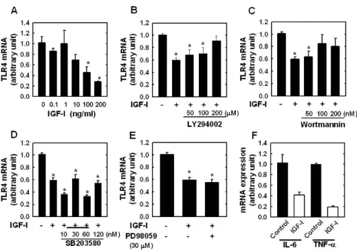
However, as shown in Fig. 1C and D, when we used PD98059 (a specific ERK1/2 inhibitor) or SB203580 (a specific inhibitor of the p38 MAPK), we found that TLR4 expression levels were not significantly affected, indicating that p38 MAPK or ERK1/2 pathways are not involved in IGF-I-induced suppression of TLR4 protein expression. To determine if the modulating effect of IGF-I on TLR4 protein expression was associated with TLR4 gene expression, TLR4 mRNA levels were determined by real-time PCR. The results showed de- creases in TLR4 mRNA in IGF-I-treated C2C12 cells of up to 73% with the maximum suppression occurring at an IGF-I concentration of 200 ng/ml (Fig. 2A). As shown in Fig. 2B and C, the suppression of TLR4 mRNA following IGF-I treat- ment in C2C12 cells was significantly attenuated by LY294002 (200μM) or Wortmannin (100 nM and 200 nM). However, the suppressive effect of IGF-I on TLR4 mRNA expression was not significantly blocked by SB203580 (Fig. 2D) or PD98059 (Fig. 2E). Taken together, these results indicate that the sup- pression of TLR4 expression of both mRNA and protein in skeletal muscle cells is regulated by IGF-I, and that the neg- ative-regulatory effect of IGF-I on TLR4 expression is regu- lated through activation of the PI3K/Akt pathways.
It is well known that TLR-mediated signaling activates NF- κB, which plays a critical role in regulation of the expression of pro-inflammatory genes such as tumor necrosis factor-al- pha (TNF-α) and interleukin-6 (IL-6) (12). Given that IGF-I treatment suppresses TLR4 expression, we investigated wheth- er IGF-I is also involved in the TLR4-mediated NF-κB-depend-
ent pro-inflammatory gene expression. As basal cytokine gene expression is chronically elevated in individuals who live a sedentary lifestyle and have many chronic diseases as- sociated with whole body chronic low-grade inflammation (1), we examined the basal expression level of IL-6 and TNF-α following IGF-I treatment. The results showed that IGF-I treat- ment greatly attenuated the endogenous expression of IL-6 and TNF-α, indicating that IGF-I exerts an anti-inflammatory effect on skeletal muscle cells by reducing the expression of pro-inflammatory cytokines under basal condition through
IGF-I-induced Down-regulation of TLR4 Signaling Won Jun Lee
IMMUNE NETWORK http://www.ksimm.or.kr Volume 11 Number 4 August 2011 225
Figure 2. (A) TLR4 mRNA expression determined by real-time PCR in C2C12 myotubes cultured for 24 hr with different concentration of IGF-I.
Effect of LY294002 (B), Wortmannin (C), SB203580 (D), or PD98059 (E) on IGF-I-mediated TLR4 expression. (F) The mRNA expression level of IL-6 and TNF-α in C2C12 cells cultured for 24 hr in the absence or presence of IGF-I (200 ng/ml). Target mRNA values are normalized to the GAPDH mRNA level for each sample. Samples were analyzed in duplicate in parallel with GAPDH. Values are menas±SE of three independent experiments. *p<0.05 vs. Control.
down-regulation of TLR4 expression. Although the exact mechanism remains to be elucidated, we can speculate that cells having low TLR4 expression are less sensitive to endoge- nous inflammation-stimulating ligands such as heat shock proteins, which contributes low basal cytokine expression.
In the present study, we demonstrated that IGF-I treatment causes suppression of TLR4 expression in differentiating C2C12 skeletal muscle cells. Our data provide the first evi- dence that growth hormone is a potent modulator of TLR4 expression in skeletal muscle cells. It has been suggested that normal inflammatory responses are the natural host responses to an acute infection, whereas chronic inflammation is linked to many chronic diseases such as heart disease, some cancers, and type II diabetes (2,3,13). Regular exercise has anti-in- flammatory effects and protects against diseases associated with chronic low-grade systemic inflammation. Skeletal mus- cle is now considered an endocrine organ and affects in- flammation throughout the body via the production of pro-in- flammatory cytokines (14). Therefore, it is possible that the IGF-I-induced suppression of TLR4 and cytokine expression in skeletal muscle cells observed in the present study may
provide a mechanistic basis for the anti-inflammatory effect of exercise.
ACKNOWLEDGEMENTS
The author acknowledges the technical assistance of Hey-Jin Kim.
CONFLICTS OF INTEREST
The author has no financial conflict of interest.
REFERENCES
1. Bruunsgaard H, Ladelund S, Pedersen AN, Schroll M, Jørgensen T, Pedersen BK: Predicting death from tumour ne- crosis factor-alpha and interleukin-6 in 80-year-old people.
Clin Exp Immunol 132;24-31, 2003.
2. Fessler MB, Rudel LL, Brown JM: Toll-like receptor signaling links dietary fatty acids to the metabolic syndrome. Curr Opin Lipidol 20;379-385, 2009.
3. Oliveira AG, Carvalho BM, Tobar N, Ropelle ER, Pauli JR,
IGF-I-induced Down-regulation of TLR4 Signaling Won Jun Lee
226 IMMUNE NETWORK http://www.ksimm.or.kr Volume 11 Number 4 August 2011
Bagarolli RA, Guadagnini D, Carvalheira JB, Saad MJ: Physical exercise reduces circulating lipopolysaccharide and TLR4 acti- vation and improves insulin signaling in tissues of DIO rats.
Diabetes 60;784-796, 2011.
4. Steinman RM, Hemmi H: Dendritic cells: translating innate to adaptive immunity. Curr Top Microbiol Immunol 311;17-58, 2006.
5. Cooper DM, Radom-Aizik S, Schwindt C, Zaldivar F Jr:
Dangerous exercise: lessons learned from dysregulated in- flammatory responses to physical activity. J Appl Physiol 103;700-709, 2007.
6. Walsh NP, Gleeson M, Shephard RJ, Gleeson M, Woods JA, Bishop NC, Fleshner M, Green C, Pedersen BK, Hoffman- Goetz L, Rogers CJ, Northoff H, Abbasi A, Simon P: Position statement. Part one: Immune function and exercise. Exerc Immunol Rev 17;6-63, 2011.
7. Petersen AM, Pedersen BK: The anti-inflammatory effect of exercise. J Appl Physiol 98;1154-1162, 2005.
8. Stewart LK, Flynn MG, Campbell WW, Craig BA, Robinson JP, McFarlin BK, Timmerman KL, Coen PM, Felker J, Talbert E: Influence of exercise training and age on CD14+ cell-sur-
face expression of toll-like receptor 2 and 4. Brain Behav Immun 19;389-397, 2005.
9. Gleeson M, McFarlin B, Flynn M: Exercise and Toll-like receptors. Exerc Immunol Rev 12;34-53, 2006.
10. Eliakim A, Nemet D: Exercise training, physical fitness and the growth hormone-insulin-like growth factor-1 axis and cy- tokine balance. Med Sport Sci 55;128-140, 2010.
11. Meng D, Shi X, Jiang BH, Fang J: Insulin-like growth factor-I (IGF-I) induces epidermal growth factor receptor trans- activation and cell proliferation through reactive oxygen species. Free Radic Biol Med 42;1651-1660, 2007.
12. Baeuerle PA, Baltimore D: NF-kappa B: ten years after. Cell 87;13-20, 1996.
13. Mathur N, Pedersen BK: Exercise as a mean to control low-grade systemic inflammation. Mediators Inflamm 2008;
109502, 2008.
14. Gomez-Merino D, Drogou C, Guezennec CY, Chennaoui M:
Effects of chronic exercise on cytokine production in white adipose tissue and skeletal muscle of rats. Cytokine 40;23-29, 2007.