J Korean Ophthalmol Soc 2014;55(9):1277-1283 pISSN: 0378-6471⋅eISSN: 2092-9374
http://dx.doi.org/10.3341/jkos.2014.55.9.1277
Original Article
굴절교정레이저각막절제술 후 트라닐라스트의 각막혼탁 억제에 대한 펜타캠을 이용한 분석
Effects of Topical Tranilast on Corneal Haze with the Pentacam
Ⓡafter Photorefractive Keratectomy
김성인⋅오태훈
Sung In Kim, MD, Tae Hoon Oh, MD
새빛안과병원
Department of Ophthalmology, Saevit Eye Hospital, Goyang, Korea
Purpose: To evaluate the inhibitory effect of tranilast on formation of corneal haze using the Pentacam® after photorefractive ker- atectomy (PRK).
Methods: A prospective, randomized, paired eye study was performed. A total of 60 eyes from 30 patients were enrolled in the present study. Eyes were categorized as myopic eyes <-5 D and ≥-5 D. Patients undergoing PRK were randomized to receive tranilast in one eye and no medication in the contralateral eye. Three months postoperatively, corneal haze was measured with the Pentacam® and compared between the 2 groups.
Results: Statistical differences were not found in preoperative data in the tranilast or control groups (all p > 0.05). There was a strong decreasing density trend from the apex to the 3 mm radius in both groups (p < 0.05). However, postoperative corneal haze in the tranilast group was similar to the control group (p > 0.05).
Conclusions: The use of tranilast after PRK did not inhibit corneal opacity. Additionally, Pentacam® can provide a useful ob- jective measure of corneal haze.
J Korean Ophthalmol Soc 2014;55(9):1277-1283 Key Words: Corneal haze, Pentacam®, PRK, Tranilast
■Received: 2014. 2. 7. ■ Revised: 2014. 5. 5.
■Accepted: 2014. 8. 1.
■Address reprint requests to Tae Hoon Oh, MD
Department of Ophthalmology, Saevit Eye Hospital, #1065 Jungang-ro, Ilsandong-gu, Goyang 410-817, Korea Tel: 82-31-900-7700, Fax: 82-31-900-7777 E-mail: [email protected]
* This study was presented as a narration at the 110th Annual Meeting of the Korean Ophthalmological Society 2013.
ⓒ2014 The Korean Ophthalmological Society
This is an Open Access article distributed under the terms of the Creative Commons Attribution Non-Commercial License (http://creativecommons.org/licenses/by-nc/3.0/) which permits unrestricted non-commercial use, distribution, and reproduction in any medium, provided the original work is properly cited.
각막혼탁은 굴절교정레이저각막절제술(Photorefractive ke- ratectomy) 후 발생할 수 있는 합병증으로 시력저하, 대비감 도의 감소, 눈부심, 달무리, 빛번짐을 유발하게 된다.1-3 엑 시머레이저를 이용한 각막굴절교정술을 시행하는 경우 각
막상피층의 손상과 더불어 각막기질부위에 존재하는 각막세 포가 소실되고, 이 과정에서 다양한 사이토카인 물질 및 성 장인자들이 분비되게 된다.4 특히 transforming growth factor (TGF)-β가 각막기질 치유 과정을 조절하는 데 중심적 역할 을 하는 것으로 보고되고 있다.4-6 TGF-β는 각막세포의 증 식 및 이동, 교원질, 세포외기질 합성을 유도하고, 각막세포 의 중요한 화학유인물질로 작용하고, 근섬유아세포의 강력 한 유도인자 역할을 한다.6-8 근섬유아세포는 각막기질에서 교원질과 세포외기질을 축적시켜 각막혼탁을 유발하게 된 다.9 이러한 TGF-β의 작용을 억제하여 굴절교정레이저각막 절제술 후 각막혼탁을 억제하려는 연구가 많이 진행되고 있으며, 점안 TGF-β 억제제를 사용한 동물 연구에서 그 효
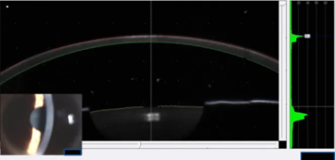
Figure 1. Scheimpflug image and slit-lamp photograph of a
cornea after PRK.과가 입증되었다.10,11
트라닐라스트는 임상적으로 항 알러지 제재로 혈관투과 성 억제 및 비만세포로부터의 histamine과 leukotriene의 길 항을 통한 항 염증작용 이외에도 TGF-β를 억제하여 각막실 질세포의 증식을 억제하고 교원질 같은 세포외기질의 분비 빛 축적을 억제하여 각막혼탁을 감소시킨다고 알려졌다.12-15 이와 같은 이유로 안과적으로는 굴절교정레이저각막절제 술 후 생기는 각막혼탁을 줄이기 위해 사용되고 있다.
굴절교정수술 후 각막혼탁의 평가는 주로 세극등현미경 을 이용한 관찰로 이루어지며, Fantes 분류법16 등을 사용하 여 그 정도에 따라 0-4단계로 분류된다. 0단계는 혼탁이 없 이 깨끗한 상태, 1단계는 홍채의 세밀한 관찰이 가능한 정 도의 혼탁을 나타낸다. 2단계는 홍채의 세밀한 관찰에 약간 의 지장이 있는 정도의 혼탁, 3단계는 홍채와 수정체의 관 찰에 상당한 제약이 되는 정도의 혼탁을 나타내며 4단계는 완전한 혼탁을 나타낸다. 하지만 이러한 방법은 관찰자의 주관적인 분류법으로 결과의 민감도, 신뢰도, 재현성이 결 여되어 있다. 또한 최근 레이저 장비와 수술 기법의 발달로 3단계와 4단계의 혼탁은 매우 드물게 되었다. 결과적으로 이러한 분류체계는 통계적인 비교 연구에서 유용성이 떨어 지게 되었다.17
Pentacam® (OCULUS Optikgerate GmbH, Wetzlar, Germany) 은 360°로 회전하는 Scheimpflug 카메라를 사용하여 각막 의 전면부터 수정체 후면까지 전안부 전체를 3차원적으로 분석할 수 있다. 또한 Pentacam®의 densitometry를 이용하 여 각막혼탁을 정량적으로 분석할 수 있고, 엑시머레이저 절삭 부위의 중심부터 주변부까지 전체 각막에 대해서 분 석이 가능하다.18
아직 국내외 보고에서 굴절교정레이저각막절제술 후 트 라닐라스트의 각막혼탁 억제에 대한 임상연구는 발표되지 않은 실정이다. 이에 본 연구에서는 트라닐라스트의 굴절 교정레이저각막절제술 후 발생할 수 있는 각막혼탁 억제효 과를 Pentacam®을 이용하여 알아보고자 하였다.
대상과 방법
2013년 3월부터 2013년 8월까지 본원에서 한 술자(OTH) 에 의해 굴절교정레이저각막절제술을 시행 받은 환자 30명 60안을 대상으로 전향적 연구를 시행하였다. 수술 전 설명 동의서(informed consent)를 받았으며, 이전에 안과적 질환 및 수술, 외상의 기왕력, 각막 상피 재생에 영향을 주는 전 신적인 질환이 있는 경우는 제외하였다. 술 전 구면대응치 에 따라 -5D 미만 근시 15명 30안, -5D 이상의 고도 근시 15명 30안으로 나누어 근시 정도에 따른 차이를 비교하고
자 하였다. 모든 환자는 수술 전 병력 문진, 나안 및 최대교 정시력, 조절 마비 및 현성굴절검사, 안압 측정, 각막형태검 사, 세극등검사, 안저검사, 동공크기 측정, 고위수차 검사를 시행 받았고 초음파 각막두께측정기(PacScan 300, Sonomed, Lake Success, NY)를 이용하여 중심각막두께를 측정하였다.
굴절교정레이저각막절제술은 먼저 환자의 각막을 0.5%
proparacaine hydrochloride (Alcaine®, Alcon, USA)를 사용 하여 점안마취 후 개검기를 사용해 각막을 노출시킨 다음 spatula로 8.0 mm 이내의 각막상피를 제거하였으며, 0.68 mm spot size의 Gaussian flying spot beam을 500 Hz의 속 도로 조사하는 WaveLight® EX500 excimer laser (Alcon Laboratories, Ft Worth, TX, USA)를 이용해 6.5 mm 광학 부로 각막을 절삭하였다. 각막 절삭 후 0.02% mitomycin C (MMC)를 절삭 부위에 국소적으로 30초간 접촉시킨 뒤 차 가운 평형염류용액(BSSⓇ, Alcon Laboratories, Inc.)으로 1 분간 각막표면과 결막낭을 충분히 세척한 후 치료용 콘택 트렌즈를 착용하였다. 수술 후에는 타리비드 점안액(0.5%
Ofloxacin drop, Tarivid®, Santen, Japan)과 스테로이드 점안 액(0.1% Fluorometholon drop, Ocumetholone®, Samil, Korea) 을 1일 4회 점안, 인공 눈물 점안액(0.1% Sodium hyaluro- nate drop, Unial®, Unimed, Korea)을 1일 6회 점안하였으 며, 수술 후 1개월부터 스테로이드 점안액을 회수를 점차 줄이면서 중단하였다. 수술 후 무작위로 배정한 한쪽 눈에 0.5% tranilast (KrixⓇ, JW pharmaceutical, Korea) 점안액을 1일 4회 3개월간 점안하였고, 대조군인 다른 쪽 눈에는 점 안하지 않았다.
술 후 3개월째에 두 군 간의 나안시력 및 최대교정시력, 현성굴절검사에 의한 구면대응치와 Pentacam®을 이용한 각막혼탁도(Fig. 1) 및 중심각막두께를 비교하였다. 각막의 혼탁도는 Pentacam®의 densitometry를 이용하여 절삭부위 를 각막 정점과 정점으로부터 1 mm, 2 mm, 3 mm 지점으 로 나누어 측정하였다. densitometry 프로그램을 이용하여 각막의 혼탁이 없는 상태를 0이라 하고 각막의 완전한 혼
Table 1. Preoperative data of myopic eyes <-5 D
Groupp-value Tranilast Control
Age (years) 26.63 ± 5.54
Sex (M/F) 15 (7/8)
CCT (μm) 524.63 ± 23.51 527.25 ± 24.31 0.31 Sphere (diopter) -3.26 ± 0.94 -3.25 ± 0.82 0.96 Cylinder (diopter) -0.66 ± 0.46 -0.60 ± 0.42 0.51 Values are presented as mean ± SD.
CCT = central corneal thickness.
Table 2. Postoperative data of myopic eyes <-5 D
Groupp-value Tranilast Control
UCVA (log MAR) 0.03 ± 0.07 0.04 ± 0.06 0.74 BCVA (log MAR) 0.00 ± 0.00 0.00 ± 0.00 1.00 MRSE (diopter) -0.18 ± 0.31 -0.19 ± 0.29 0.91 CCT (μm) 463.38 ± 30.00 465.56 ± 30.67 0.50 Values are presented as mean ± SD.
UCVA = uncorrected visual acuity; BCVA = best corrected visual acuity; MRSE = manifest refraction spherical equivalent; CCT = central corneal thickness.
Table 3. Mean maximum density values measured with the
PentacamⓇ for myopic eyes <-5 DDistance from
apex Tranilast Control p-value
0 mm 29.01 ± 2.82 29.50 ± 3.13 0.46
1 mm 27.94 ± 3.10 28.14 ± 2.94 0.69
2 mm 26.29 ± 2.75 26.20 ± 2.14 0.83
3 mm 22.78 ± 2.62 23.07 ± 2.33 0.39
Values are presented as mean ± SD.
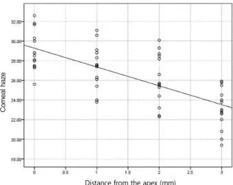
Figure 2. Correlation of corneal haze with distance from the
apex for myopic eyes <-5 D after photorefractive keratectomy (PRK) in tranilast (p < 0.05, r = -0.64).Figure 3. Correlation of corneal haze with distance from the
apex for myopic eyes <-5 D after photorefractive keratectomy (PRK) in control (p < 0.05, r = -0.66).탁이 있는 상태를 100으로 하여 그 사이의 값으로 각막혼 탁을 객관적으로 평가하였다.
표본 데이터에 대한 정규성 검정을 시행한 후 각막 정점 으로부터 거리에 따른 각막혼탁은 ANOVA를, 두 군 간 측 정치의 통계학적 비교는 Paired samples t-test를 이용하였 다. 통계처리는 SPSS 18.0을 이용하였으며, p-value의 유의 수준은 0.05 미만으로 하였다.
결 과
-5D 미만 근시 15명 30안에서 남자는 7명(47.7%), 여자 는 8명(52.3%)이었고, 수술 시 평균 연령은 26.63 ± 5.54세 였다. 수술 전 중심각막두께, 평균 구면 값과 난시 값은 트 라닐라스트군과 대조군에서 통계적으로 유의한 차이가 없 었다(p=0.31, 0.96, 0.51, Table 1). 수술 후 부작용은 발생하 지 않았으며, 술 후 3개월째 측정한 나안시력 및 최대교정 시력, 구면대응치, 중심각막두께는 두 군 간에 통계적으로
유의한 차이가 없었다(p>0.05, Table 2).
Pentacam®의 densitometry 프로그램을 이용한 수술 후 3 개월째 각막혼탁의 비교는 각막 정점에서 트라닐라스트군 이 29.01 ± 2.82, 대조군에서 29.50 ± 3.13으로 통계적으로 유의한 차이가 없었고(p=0.46), 정점으로부터 1 mm, 2 mm, 3 mm 지점에서 트라닐라스트군이 각각 27.94 ± 3.10, 26.29 ± 2.75, 22.78 ± 2.62였으며, 대조군에서 각각 28.14
± 2.94, 26.20 ± 2.14, 23.07 ± 2.33으로 각 지점 모두 통계 적으로 유의한 차이가 없었다(p=0.69, 0.83, 0.39 Table 3).
각막 정점으로부터 거리에 따른 각막혼탁은 트라닐라스트 군과 대조군 모두 정점으로부터 멀어질수록 통계적으로 유 의하게 각막혼탁도가 감소하였다(ANOVA, p<0.05, Fig. 2, 3).
-5D 이상 고도 근시 15명 30안에서 남자는 7명(47.7%),
Distance from the apex (mm) Distance from the apex (mm)
Table 4. Preoperative data of myopic eyes ≥-5 D
Groupp-value Tranilast Control
Age (years) 22.14 ± 8.47
Sex (M/F) 15 (7/8)
CCT (μm) 540.93 ± 22.43 543.71 ± 19.57 0.31 Sphere (diopter) -6.02 ± 0.21 -5.74 ± 0.24 0.26 Cylinder (diopter) -1.62 ± 0.56 -1.52 ± 0.68 0.46 Values are presented as mean ± SD.
CCT = central corneal thickness.
Table 5. Postoperative data of myopic eyes ≥-5 D
Group p-value
Tranilast Control
UCVA (log MAR) 0.05 ± 0.09 0.04 ± 0.08 0.75 BCVA (log MAR) 0.00 ± 0.00 0.00 ± 0.00 1.00 MRSE (diopter) -0.21 ± 0.27 -0.23 ± 0.32 0.90
CCT 427.86 ± 21.10 432.07 ± 22.20 0.15
Values are presented as mean ± SD.
UCVA = uncorrected visual acuity; BCVA = best corrected visual acuity; MRSE = manifest refraction spherical equivalent; CCT = central corneal thickness.
Table 6. Mean maximum density values measured with the
PentacamⓇ for myopic eyes ≥-5 DDistance from
apex Tranilast Control p-value
0 mm 29.16 ± 2.74 29.03 ± 2.00 0.85
1 mm 27.19 ± 4.17 27.39 ± 2.17 0.83
2 mm 25.75 ± 3.41 26.13 ± 2.55 0.75
3 mm 23.36 ± 1.64 23.06 ± 2.05 0.59
Values are presented as mean ± SD.
Figure 4. Correlation of corneal haze with distance from the
apex for myopic eyes ≥-5 D after photorefractive keratectomy (PRK) in tranilast (p < 0.05, r = -0.72).Figure 5. Correlation of corneal haze with distance from the
apex for myopic eyes ≥-5 D after photorefractive keratectomy (PRK) in control (p < 0.05, r = -0.69).여자는 8명(52.3%)이었고, 수술 시 평균 연령은 22.14 ± 8.47세였다. 수술 전 중심각막두께, 평균 구면 값과 난시 값
은 트라닐라스트군과 대조군에서 통계적으로 유의한 차이 가 없었다(p=0.31, 0.26, 0.46, Table 4). 수술 후 부작용은 발생하지 않았으며, 술 후 3개월째 측정한 나안시력 및 최 대교정시력, 구면대응치, 중심각막두께는 두 군 간에 통계 적으로 유의한 차이가 없었다(p>0.05, Table 5).
수술 후 3개월째 각막혼탁의 비교는 각막 정점에서 트라 닐라스트군이 29.16 ± 2.74, 대조군에서 29.03 ± 2.00으로 통계적으로 유의한 차이가 없었고(p=0.85), 정점으로부터 1 mm, 2 mm, 3 mm 지점에서 트라닐라스트군이 각각 27.19
± 4.17, 25.75 ± 3.41, 23.36 ± 1.64였으며, 대조군에서 각각 27.39 ± 2.17, 26.13 ± 2.55, 23.06 ± 2.05으로 각 지점 모두 통계적으로 유의한 차이가 없었다(p=0.83, 0.75, 0.59 Table 6). 또한, 각막 정점으로부터 거리에 따른 각막혼탁은 -5D 미만 근시 군들과 마찬가지로 트라닐라스트군과 대조군 모 두 정점으로부터 멀어질수록 통계적으로 유의하게 각막혼 탁도가 감소하였다(ANOVA, p<0.05, Fig. 4, 5).
고 찰
엑시머레이저를 이용한 굴절교정레이저각막절제술의 합 병증으로 생기는 각막혼탁은 시력저하, 대비감도의 감소를 유발하는 심각한 합병증이다. 이러한 술 후 각막혼탁의 위 험인자로는 근시가 심하여 많은 양의 절삭이 필요한 경우, 콘택트렌즈를 장기간 착용한 경우, 자가면역질환이 있는 경 우, 자외선 노출(UV-B)이 많은 경우 등으로 알려졌다.19-21 각막혼탁은 각막상피하에 새로운 교원질섬유의 재형성 과 기질화가 재배열되면서 발생하는데, 각막의 실질에 존 재하는 각막세포들이 중요한 역할을 한다.1 엑시머레이저에 의해 각막 실질의 각막세포가 손상되면 세포활성 물질이
Distance from the apex (mm)
Distance from the apex (mm)
섬유모세포의 증식을 촉진시키고 교원질과 세포외기질의 축적을 증가시키게 된다. 활성화된 각막세포에 의해 생성 된 교원질과 세포외기질은 정상적 각막에서보다 불규칙적 인 배열을 가지고 이것에 의해 각막혼탁이 생긴다.22,23 특히 TGF-β가 손상된 각막에서 섬유모세포의 증식을 촉진시키 고 세포간질의 축적을 증가시키는 데 가장 큰 역할을 수행하 고 있음이 밝혀졌으며,4-6 각막 창상 치유 과정에서도 TGF-β 가 세포 성장, 증식, 세포 외 기질 합성 등 반흔형성 과정을 조절하는 데 중심적 역할을 하는 것으로 확인되었다.24,25
각막혼탁을 예방하기 위한 여러 약제 중 스테로이드 제 재와 mitomycin C (MMC)가 많이 사용되고 있는데, 스테로 이드 제제는 활성화된 각막기질세포의 증식은 억제할 수 없지만, 교원질의 합성을 억제하여 각막혼탁을 예방하는 데 효과가 있다.26 MMC는 항암항생물질로 굴절교정레이저 각막절제술 후에 나타나는 각막세포의 증식을 억제하고, 근육섬유아세포의 분화과정을 억제하여 각막혼탁의 감소 를 가져오는 것으로 알려졌다.9,27,28
본 연구에서 사용한 트라닐라스트는 leukotriene 유사체 로서 트립토판이 indolamine 2,3-dioxygenase에 의해 대사 되어 발생되는 것으로 알려졌다.12 트라닐라스트의 효과를 살펴보면 비만세포와 호염구의 화학매개체의 분비를 억제 하는 항알러지 효과뿐만 아니라, TGF-β를 억제하여 조직 내 섬유모세포를 억제하여 교원질 분비를 억제하는 효과도 있다. 임상에서는 주로 기관지 천식, 아토피성 피부염, 알러 지 결막염에 사용되며, 교원질 합성을 억제하는 효과 때문 에 켈로이드, 비대흉터의 치료에도 쓰이고 있다.29-33
굴절교정레이저각막절제술 후 트라닐라스트가 사용된 연 구를 살펴보면, Song et al4은 토끼의 각막에 굴절교정레이 저각막절제술을 시행한 후 0.5% 트라닐라스트 점안액을 사 용하면 대조군에 비해 각막혼탁이 적게 발생한다고 보고하 였고, Okamoto et al34도 토끼에서 굴절교정레이저각막절제 술을 시행한 후 트라닐라스트를 사용하면 각막혼탁의 억제 에 효과가 있다고 보고하였다.
저자들은 현재까지 트라닐라스트의 각막혼탁 억제에 대 한 임상연구가 없기에 굴절교정레이저각막절제술을 시행 받은 환자 30명 60안을 대상으로 전향적 연구를 시행하였 다. 또한 -5D 미만 근시와 -5D 이상의 고도 근시로 나누어 근시 정도에 따른 차이를 비교하고자 하였다. 술 후 3개째 각막 정점과 정점으로부터 1 mm, 2 mm, 3 mm 구역의 각 막혼탁도를 분석하였다. 트라닐라스트를 점안한 군과 대조 군 모두 정점으로부터 멀어질수록 통계적으로 유의하게 각 막혼탁도가 감소하였다. 그 이유는 근시 굴절교정레이저각 막절제술에서 각막 정점이 가장 절삭 깊이가 깊고, 주변으 로 갈수록 얕아지기 때문이다. 트라닐라스트를 점안한 군
과 대조군을 비교한 연구에서는 -5D 미만 근시와 -5D 이상 의 고도 근시 모두 각막혼탁에서 두 군 간에 통계적으로 유 의한 차이가 없었다. 이는 각막혼탁을 줄이기 위해 수술 중 MMC를 사용하고, 술 후 3개월간 스테로이드 점안액을 사 용하기 때문에 트라닐라스트의 추가적인 사용이 술 후 3개 월째 각막혼탁을 더 억제하지는 못했다고 생각할 수 있다. 한편 본 연구에서는 각막혼탁을 정량적으로 분석하기 위 하여 Pentacam®을 이용하였다. 그동안의 연구에서 각막혼 탁에 대한 평가는 Fantes 분류법15 등을 이용하여 그 정도에 따라 0-4 단계로 분류하여 평가하였다. 이는 세극등현미경 관찰을 통한 주관적인 검사로 민감도, 신뢰도, 재현성이 떨 어지고, 최근 레이저 장비와 수술 기법의 발달로 각막혼탁 이 대부분 0-1 단계로 생기기 때문에 통계적인 비교 연구에 적합하지 않다. 그래서 각막 혼탁을 좀 더 객관적이고 세밀 하게 정량화하는 방법이 필요하게 되었다.15 이에 각막혼탁 을 객관적으로 평가하기 위한 방법으로 공초점 현미경이 사용되었다. 하지만 공초점 현미경의 광학슬라이스 두께는 약 11-30 µm로 각막의 전체 두께에서 평가하는 것이 어렵 고, 또한 주변부 각막까지 측정하기에는 어려움이 있다.18 초음파 생체현미경 검사도 사용되었는데 이 방법은 접촉식 방법이고, 대부분 중등도 이상의 혼탁을 발견하는 데 유용 하다.35
본 연구에서 저자들은 Pentacam®의 densitometry 프로그램 을 이용하여 각막혼탁을 정량적으로 분석하였다. Pentacam® 은 비접촉 장비로 360°로 회전하는 Scheimpflug 카메라를 사용하여 각막의 전면부터 수정체 후면까지 전안부 전체를 분석할 수 있다. 또한 densitometry 프로그램을 이용하면 수 정체와 각막을 포함하는 전안부 어느 곳이라도 혼탁도를 수치화하여 평가할 수 있다. 그래서 각막의 정점뿐만 아니 라 레이저 절삭부위 및 주변부 각막까지 혼탁도를 객관적 으로 분석할 수 있다.17
기존 연구에서 Pentacam®을 이용하여 수정체 및 후낭 혼 탁의 정량 분석에 대한 연구는 많이 있지만,36,37 각막 혼탁 에 대한 보고는 많지 않다. Matsuda et al38은 Pentacam®을 이용하여 라식과 에파라식 수술 전후 각막의 혼탁도 변화 에 대해 보고하였고, Takacs et al18은 굴절교정레이저각막 절제술 후 절삭 부위에 대해 각막의 정점과 정점으로부터 의 거리에 따른 각막 혼탁을 정량적으로 분석하였다.
본 연구는 현재까지 보고되지 않은 트라닐라스트의 각막 혼탁 억제에 대한 전향적인 임상연구를 시행하였고, 각막 혼탁을 Pentacam®을 이용하여 객관적으로 정량분석을 했 다는 데 의의가 있다고 하겠다. 하지만 굴절교정레이저각 막절제술에서 각막혼탁을 줄이기 위해 수술 중 MMC를 사 용하고, 술 후 3개월간 스테로이드 점안액을 사용했기 때문
에 트라닐라스트의 단독 효과를 보기에는 다소 제한점이 있다.
결론적으로 트라닐라스트의 추가적 사용이 근시에서 굴 절교정레이저각막절제술 후 생기는 각막혼탁의 억제에 효 과가 없었고, 각막혼탁을 정량적으로 평가하는 데 있어 Pentacam®을 이용하는 것이 유용하다고 하겠다.
REFERENCES
1) Caubet E. Course of subepithelial corneal haze over 18 months af- ter photorefractive keratectomy for myopia [corrected]. Refract Corneal Surg 1993;9:S65-70.
2) Gartry DS, Muir MG, Lohmann CP, Marshall J. The effect of top- ical corticosteroids on refractive outcome and corneal haze after photorefractive keratectomy. A prospective, randomized, dou- ble-blind trial. Arch Ophthalmol 1992;110:944-52.
3) Seiler T, Derse M, Pham T. Repeated excimer laser treatment after photorefractive keratectomy. Arch Ophthalmol 1992;110:1230-3.
4) Song JS, Jung HR, Kim HM. Effects of topical tranilast on corneal haze after photorefractive keratectomy. J Cataract Refract Surg 2005;31:1065-73.
5) Wilson SE, Walker JW, Chwang EL, He YG. Hepatocyte growth factor, keratinocyte growth factor, their receptors, fibroblast growth factor receptor-2, and the cells of the cornea. Invest Ophthalmol Vis Sci 1993;34:2544-61.
6) Grant MB, Khaw PT, Schultz GS, et al. Effects of epidermal growth factor, fibroblast growth factor, and transforming growth factor-beta on corneal cell chemotaxis. Invest Ophthalmol Vis Sci 1992;33:3292-301.
7) Ohji M, SundarRaj N, Thoft RA. Transforming growth factor-beta stimulates collagen and fibronectin synthesis by human corneal stromal fibroblasts in vitro. Curr Eye Res 1993;12:703-9.
8) Fini ME, Girard MT, Matsubara M, Bartlett JD. Unique regulation of the matrix metalloproteinase, gelatinase B. Invest Ophthalmol Vis Sci 1995;36:622-33.
9) Netto MV, Mohan RR, Ambrósio R Jr, et al. Wound healing in the cornea: a review of refractive surgery complications and new pros- pects for therapy. Cornea 2005;24:509-22.
10) Chen C, Michelini-Norris B, Stevens S, et al. Measurement of mRNAs for TGFss and extracellular matrix proteins in corneas of rats after PRK. Invest Ophthalmol Vis Sci 2000;41:4108-16.
11) Myers JS, Gomes JA, Siepser SB, et al. Effect of transforming growth factor beta 1 on stromal haze following excimer laser pho- torefractive keratectomy in rabbits. J Refract Surg 1997;13:
356-61.
12) Platten M, Ho PP, Youssef S, et al. Treatment of autoimmune neu- roinflammation with a synthetic tryptophan metabolite. Science 2005;310:850-5.
13) Tani E, Katakami C, Negi A. Effects of various eye drops on cor- neal wound healing after superficial keratectomy in rabbits. Jpn J Ophthalmol 2002;46:488-95.
14) Furukawa H, Nakayasu K, Gotoh T, et al. Effect of topical tranilast and corticosteroids on subepithelial haze after photorefractive ker- atectomy in rabbits. J Refract Surg 1997;13:S457-8.
15) Lee JE, Han HJ, Lee JS, Oum BS. Effect of tranilast on the pro-
liferation of human corneal keratocytes in vitro. J Korean Ophthalmol Soc 2005;46:510-20.
16) Fantes FE, Hanna KD, Waring GO 3rd, et al. Wound healing after excimer laser keratomileusis (photorefractive keratectomy) in monkeys. Arch Ophthalmol 1990;108:665-75.
17) van de Pol C, Soya K, Hwang DG. Objective assessment of tran- sient corneal haze and its relation to visual performance after pho- torefractive keratectomy. Am J Ophthalmol 2001;132:204-10.
18) Takacs AI, Mihaltz K, Nagy ZZ. Corneal density with the Pentacam after photorefractive keratectomy. J Refract Surg 2011;27:269-77.
19) Mirza MA, Qazi MA, Pepose JS. Treatment of dense subepithelial corneal haze after laser-assisted subepithelial keratectomy. J Cataract Refract Surg 2004;30:709-14.
20) Nagy ZZ, Tóth J, Nagymihály A, Süveges I. The role of ultra- violet-B in corneal healing following excimer laser in situ kerato- mileusis. Pathol Oncol Res 2002;8:41-6.
21) Nagy ZZ, Hiscott P, Seitz B, et al. Clinical and morphological re- sponse to UV-B irradiation after excimer laser photorefractive keratectomy. Surv Ophthalmol 1997;42:S64-76.
22) Jeong BJ, Kim HH, Park YJ, et al. Effect of mitomycin C to inhibit corneal haze formation after photorefractive keratectomy for high myopia. J Korean Ophthalmol Soc 2006;47:725-34.
23) Kim ES, Jin KH. Evaluation of the prophylactic use of mitomycin to inhibit haze formation after LASEK. J Korean Ophthalmol Soc 2007;48:623-9.
24) Mita T, Yamashita H, Kaji Y, et al. Effects of transforming growth factor beta on corneal epithelial and stromal cell function in a rat wound healing model after excimer laser keratectomy. Graefes Arch Clin Exp Ophthalmol 1998;236:834-43.
25) Lee JH, Kim ET, Oh JH. The inhibitory effect of TGF-β inhibitor on the corneal opacity after corneal laceration. J Korean Ophthalmol Soc 2009;50:450-61.
26) Vetrugno M, Maino A, Quaranta GM, Cardia L. The effect of early steroid treatment after PRK on clinical and refractive outcomes.
Acta Ophthalmol Scand 2001;79:23-7.
27) Kim TI, Tchah H, Lee SA, et al. Apoptosis in keratocytes caused by mitomycin C. Invest Ophthalmol Vis Sci 2003;44:1912-7.
28) Kim TI, Pak JH, Lee SY, Tchah H. Mitomycin C-induced reduction of keratocytes and fibroblasts after photorefractive keratectomy.
Invest Ophthalmol Vis Sci 2004;45:2978-84.
29) Kim HY, Lee HI, Chun YS, Kim JC. The effectiveness of tranilast in the prevention of posterior capsular opacity. J Korean Ophthalmol Soc 2008;49:1981-8.
30) Waseda T. [Concentrations of tranilast in keloid tissues]. Nihon Hifuka Gakkai Zasshi 1989;99:1159-65.
31) Suzawa H, Kikuchi S, Ichikawa K, Koda A. Inhibitory action of tranilast, an anti-allergic drug, on the release of cytokines and PGE2 from human monocytes-macrophages. Jpn J Pharmacol 1992;60:
85-90.
32) Suzawa H, Kikuchi S, Arai N, Koda A. The mechanism involved in the inhibitory action of tranilast on collagen biosynthesis of keloid fibroblasts. Jpn J Pharmacol 1992;60:91-6.
33) Isaji M, Aruga N, Naito J, Miyata H. Inhibition by tranilast of col- lagen accumulation in hypersensitive granulomatous inflammation in vivo and of morphological changes and functions of fibroblasts in vitro. Life Sci 1994;55:PL287-92.
34) Okamoto S, Sakai T, Iwaki Y, et al. Effects of tranilast on cultured rabbit corneal keratocytes and corneal haze after photorefractive
= 국문초록 =
굴절교정레이저각막절제술 후 트라닐라스트의 각막혼탁 억제에 대한 펜타캠을 이용한 분석
목적: 트라닐라스트의 굴절교정레이저각막절제술 후 발생할 수 있는 각막혼탁 억제효과를 팬타캠을 이용하여 알아보고자 한다.
대상과 방법: 굴절교정레이저각막절제술이 계획된 30명 60안을 각각 -5D 미만의 근시와 -5D 이상의 고도근시로 나누어 분석하였으 며, 술 후 무작위로 배정한 한쪽 눈에만 트라닐라스트 점안액을 사용하고, 다른 쪽 눈에는 사용하지 않았다. 술 후 3개월째 Pentacam® 을 이용하여 각막혼탁을 비교하였다.
결과: -5D 미만의 근시와 -5D 이상의 고도근시에서 트라닐라스트를 넣은 군과 넣지 않은 군 간의 술 전 구면 값과 난시 값은 통계적 으로 유의한 차이가 없었다(p>0.05). Pentacam®을 이용한 각막혼탁도에서는 -5D 미만의 근시와 -5D 이상의 고도근시 모두 각막정 점으로부터 멀어질수록 각막혼탁도는 감소하였지만(p<0.05), 트라닐라스트를 넣은 군과 대조군 간에 통계적으로 유의한 차이가 없 었다(p>0.05).
결론: 트라닐라스트의 사용이 굴절교정레이저각막절제술 후 생기는 각막혼탁의 억제에 효과가 없었고, 각막혼탁을 정량적으로 평가하 는 데 있어 Pentacam®을 이용하는 것이 유용하다.
<대한안과학회지 2014;55(9):1277-1283>
keratectomy. Jpn J Ophthalmol 1999;43:355-62.
35) Nagy ZZ, Németh J, Süveges I, Csákány B. [Examination of sub- epithelial scar formation after photorefractive keratectomy with the ultrasound biomicroscope]. Klin Monbl Augenheilkd 1996;
209:283-5.
36) Pei X, Bao Y, Chen Y, Li X. Correlation of lens density measured using the Pentacam Scheimpflug system with the Lens Opacities Classification System III grading score and visual acuity in age-re-
lated nuclear cataract. Br J Ophthalmol 2008;92:1471-5.
37) Grewal D, Jain R, Brar GS, Grewal SP. Pentacam tomograms: a novel method for quantification of posterior capsule opacification.
Invest Ophthalmol Vis Sci 2008;49:2004-8.
38) Matsuda J, Hieda O, Kinoshita S. [Quantification of corneal opac- ity after refractive corneal surgery using the anterior segment ana- lyzer]. Nihon Ganka Gakkai Zasshi 2007;111:447-53.