Copyright © 2018 The Korean Society for Bone and Mineral Research
This is an Open Access article distributed under the terms of the Creative Commons Attribution Non-Commercial Li- cense (http://creativecommons.org/licenses/by-nc/4.0/) which permits unrestricted non-commercial use, distribu- tion, and reproduction in any medium, provided the original work is properly cited.
α-Tocopheryl Succinate Inhibits Osteolytic Bone Metastasis of Breast Cancer by Suppressing
Migration of Cancer Cells and Receptor Activator of Nuclear Factor-κΒ Ligand Expression of Osteoblasts
Bongjun Kim, Hong-Hee Kim, Zang Hee Lee
Department of Cell and Developmental Biology, Dental Research Institute, School of Dentistry, Seoul National University College of Medicine, Seoul, Korea
Background: Breast cancer is one of the most common cancers affecting women and has a high incidence of bone metastasis, causing osteolytic lesions. The elevated expres- sion of receptor activator of nuclear factor-κB ligand (RANKL) in cancer activates osteo- clasts, leading to bone destruction. We previously reported that α-tocopheryl succinate (αTP-suc) inhibited interleukin-1-induced RANKL expression in osteoblasts. Here, we ex- amined the effect of αTP-suc on osteolytic bone metastasis in breast cancer. Methods:
To examine the effect of αTP-suc on the metastatic capacity of breast cancer, MDA-MB- 231-FL cells were injected into the left cardiac ventricle of BALB/c nude mice along with intraperitoneal injection of αTP-suc. The mice were then analyzed by bioluminescence imaging. To investigate the effect of αTP-suc on osteolysis, 4T1 cells were directly inject- ed into the femur of BALB/c mice along with intraperitoneal injection of αTP-suc. Micro- computed tomography analysis and histomorphometric analysis of the femora were performed. Results: αTP-suc inhibited cell migration and cell growth of 4T1 cells. In line with these results, bone metastasis of MDA-MB-231-FL cells was reduced in mice inject- ed with αTP-suc. In addition, αTP-suc decreased osteoclastogenesis by inhibiting 4T1-in- duced RANKL expression in osteoblasts. Consistent with these results, 4T1-induced bone destruction was ameliorated by αTP-suc, with in vivo analysis showing reduced tumor burden and osteoclast numbers. Conclusions: Our findings suggest that αTP-suc may be efficiently utilized to prevent and treat osteolytic bone metastasis of breast cancer with dual effects.
Key Words: Alpha-tocopheryl succinate, Breast neoplasms, Osteolysis
INTRODUCTION
Cancer is generally known to be incurable when the disease metastasizes into bone. Bone metastasis causes devastating clinical features, including excessive bone pain, pathological bone fractures, hypercalcemia, and spinal cord compres- sion.[1] Among the metastatic cancers, bone metastasis most commonly occurs in breast and prostate cancer, which account for 65% to 75% of patients with bone metastasis. Cancers of the lung, colon, stomach, bladder, uterus, rectum, thyroid, Corresponding author
Zang Hee Lee
Department of Cell and Developmental Biology, Dental Research Institute, School of Dentistry, Seoul National University College of Medicine, 103 Daehak-ro, Jongno-gu, Seoul 03080, Korea
Tel: +82-2-740-8672 Fax: +82-2-747-6589 E-mail: [email protected] Received: December 21, 2017 Revised: January 16, 2018 Accepted: January 23, 2018
No potential conflict of interest relevant to this article was reported.
and kidney cause up to 30% of bone metastasis.[2]
Breast cancer is one of the most common cancers in wom- en and has a high incidence of bone metastasis. The most common outcome of bone metastatic breast cancer is os- teolytic lesions due to increased osteoclast differentiation and activity.[3,4] Osteolytic lesions develop as a result of an interaction between the cancer and the bone marrow niche. Previous studies have shown that osteoblasts in the bone lining expressing C-X-C motif chemokine ligand 12 supply the metastatic niche to cancer cells expressing the C-X-C motif receptor 4.[5] Bone metastatic breast cancer migrates into the osteoblast-rich area and then interacts with cells in the bone marrow, including osteoblasts, fibro- blasts, and T cells, through the secretion of various inflam- matory factors, such as prostaglandin E2 (PGE2), interleukin 1 (IL-1), and IL-6. This inflammatory environment triggers cells including fibroblasts, T cells, and osteoblasts to increase the expression of receptor activator of nuclear factor (NF)- κB ligand (RANKL), a crucial cytokine for osteoclast differ- entiation. Elevated RANKL expression promotes osteoclast differentiation, leading to osteolysis. Osteolysis releases growth factors that have accumulated in the bone matrix, such as transforming growth factor-β (TGF-β), insulin-like growth factor (IGF), and platelet-derived growth factor (PDGF). The growth factors released from the bone matrix in turn stimulate more cancer growth and migration, lead- ing to accelerated bone destruction, forming a vicious cy- cle.[4,6]
Vitamin E consists of a group of eight structurally related compounds, four tocopherols (α, β, γ, and δ) and four toco- trienols (α, β, γ, and δ). Accumulating evidence suggests that the various vitamin E families have anti-cancer activi- ty. The vitamin E family inhibits cell growth and cell migra- tion and induces apoptosis of various cancer cells, such as colon cancer, lung cancer, prostate cancer, and breast can- cer.[7] For example, Husain et al.[8] showed that γ- and δ-tocotrienol inhibit cell growth and cell survival of pan- creatic cancers by inhibiting NF-κB, and Gysin et al. [9] showed that γ-tocopherol inhibits the cell cycle progression of os- teosarcoma cells by down-regulation of the cyclin family.
Moreover, various human clinical studies have shown that the vitamin E family is effective for cancer therapy.[7,10]
For bone metastatic osteolysis, elevated osteoclast activ- ity can be a therapeutic target to prevent bone destruction and tumor growth by this vicious cycle. We previously re-
ported that α-tocopheryl succinate (αTP-suc), an esterified compound of α-tocopherol (αTP), inhibits IL-1-induced RANKL expression in osteoblasts and thereby suppresses osteoclast differentiation.[11] Also, studies have shown that αTP-suc inhibits cell invasion and proliferation of cancers in vitro.[12] However, the effects of αTP-suc on the metastatic capacity of cancers in vivo and the ability of αTP-suc to pre- vent bone destruction by bone metastasis have not yet been elucidated.
In the present study, we investigated whether αTP-suc affects cancer migration in vivo. We also examined whether αTP-suc inhibits cancer-induced RANKL expression from osteoblasts and whether αTP-suc prevents osteolytic bone metastasis and bone destruction.
METHODS
1. Reagents and cells
Macrophage colony-stimulating factor (M-CSF) and RANKL were purchased from PeproTech (Rocky Hill, NJ, USA). αTP- suc was purchased from Sigma-Aldrich (St. Louis, MO, USA).
The 4T1 mouse breast cancer cell line and the MDA-MB-231 human breast cancer cell line were purchased from the American Type Culture Collection (Manassas, VA, USA) and cultured in Dulbecco’s modified Eagle’s medium (DMEM).
Primary bone marrow cells from 7-week-old BALB/c fe- male mice and primary osteoblasts from newborn BALB/c calvariae were prepared as described previously [13] and cultured in α-minimal essential medium (α-MEM). Cell cul- ture media and serum were purchased from Invitrogen (Carlsbad, CA, USA) and complete medium was supple- mented with 10% fetal bovine serum (FBS; Hyclone Labo- ratories, Logan, UT, USA) and 1% penicillin–streptomycin (Invitrogen). MDA-MB-231-FL cells were generated from MDA-MB-231 cells by stable transfection of the firefly lucif- erase gene as described previously.[14]
2. Animals
Six-week-old female BALB/c and BALB/c nude mice were purchased from Charles River Korea. All animal procedures were reviewed and approved by the animal care commit- tee of the Institute of Laboratory Animal Resources of Seoul National University.
3. Bone metastasis models
Starting 1 day before cancer injection, αTP-suc (0.1 mg/
head) or dimethyl sulfoxide (DMSO) was injected intraperi- toneally every 2 days for each model. For intracardiac in- jections (n=5), BALB/c nude mice were anesthetized and injected with 100 mL phosphate buffered saline (PBS) or 2
×105 MDA-MB-231-FL cells in 100 mL PBS into the left car- diac ventricle, a route of administration that allows for bone metastasis rather than lung infiltration of injected cells. Af- ter 7 days of injection, mice were subjected to biolumines- cence analysis. For intrafemoral injections (n=5), 5 mL PBS or 1×10⁴ 4T1 cells in 5 μL PBS was injected into the femo- ral marrow space of left femur through the femoral con- dyle using a 28-gauge Hamilton syringe as previous de- scribed.[15] After 7 days of injection, mice were sacrificed and femora were collected.
4. Bioluminescence imaging and analysis
The mice injected with MDA-MB-231-FL cells were intra- peritoneally injected with 150 mg/kg D-luciferin (Xenogen, Hopkinton, MA, USA) in PBS 12 min before bioluminescence imaging. Imaging was conducted by using a charge-cou- pled device camera (IVIS 100, Xenogen; exposure time of 3 min, binning of 8, field of view of 15 cm, f/stop of 1, and no filter). Mice were anesthetized with isoflurane (2% vapor- ized in O2). For analysis, total photon flux (photons per sec- ond) was measured from the whole body, and a fixed region of interest in the hind limbs and the mandible/maxilla by using Living Image software (Xenogen). Bioluminescent signals within the region were normalized to the background luminescence taken over the same region from animals not injected with D-luciferin.5. Quantitative micro-computed tomography analysis
Three-dimensional (3D) images of femora were recon- structed by microcomputed tomography (micro-CT) scan- ning (SMX-90CT system; 90 kVp, 109 mA, and 180-ms inte- gration time; Shimadzu, Kyoto, Japan). Trabecular bone parameters were calculated by using TRI 3D-BON (RATOC System Engineering Co., Tokyo, Japan). All micro-CT image acquisitions and analyses were carried out by an individual blinded to the composition of the experimental groups.
6. Histological analysis
Decalcified femoral bones were embedded in paraffin blocks. Histological sections (6 mm) were prepared and stained with hematoxylin and eosin (H & E) or with tartrate- resistant acid phosphatase (TRAP) to detect osteoclasts as described previously.[14] Histomorphometric analyses were conducted with the Osteomeasure analysis system (Osteo- Metrics, Decatur, GA, USA).
7. Cell migration assay
The 4T1 cells were harvested by using a cell dissociation solution (Sigma–Aldrich) and then resuspended in serum- free DMEM. A total of 1×10⁵ 4T1 cells in 100 mL of serum- free medium were seeded in the upper chamber of tran- swell chambers (8-mm pore membranes; Corning) with the indicated reagents. The lower chamber was filled with 900 mL of 10% FBS-containing medium. After 12 hr, cells on the upper surface of the filter were wiped off with cot- ton swabs. Cells on the lower surface of the filter were fixed with 3.7% formaldehyde and methanol and stained with H
& E. Stained cells were counted under a microscope.
8. Cell viability assay
Cell viability and proliferation were determined with the EZ-Cytox Cell Viability Assay Kit (Daeil Lab, Seoul, Korea) based on the cleavage of the tetrazolium salt to water-sol- uble formazan by succinate-tetrazolium reductase. Briefly, 2×10³ 4T1 cells were seeded on 96-well plates and cul- tured for 24 hr. Then, the culture medium was exchanged for media containing different concentrations of αTP-suc (0-10 μM). After 2 days of culture, cells were incubated with 20 μL of Ez-CyTox solution for 2 hr in a 37°C incubator. Ab- sorbance was measured at 450 nm.
9. Osteoclast differentiation assay
Primary bone marrow cells (1×10⁶ cells/well) and pri- mary osteoblasts (1×10⁵/well) were co-cultured with 4T1 cells (5×10² cells/ well) in 12-well (1.5 mL/well) tissue cul- ture plates for 8 days in α-MEM complete medium. The cul- tures were replenished with fresh medium containing the same supplements every 2 days. At the end of the culture period, cells were fixed in 10% formalin for 10 min, perme- abilized with 0.1% Triton X-100, and then stained for TRAP activity with the leukocyte acid phosphatase assay kit (Sig- ma–Aldrich).
10. Preparation of conditioned medium (CM)
The 4T1 cells were cultured in DMEM complete medium on 10-cm tissue culture dishes until optimal confluence (70%). Then the cells were cultured in fresh DMEM com- plete medium. CM was harvested after 24 hr of incubation, centrifuged at 2,000 rpm for 5 min, and stored at -80°C.11. Real-time polymerase chain reaction (PCR) analysis
Primary osteoblasts (4×10⁵/well) were cultured in 6-well tissue culture plates with or without 4T1-CM (30%) in the presence of DMSO or αTP-suc (10 μM). After 24 hr of cul- ture, cells were harvested for real-time PCR analysis and the culture medium was collected for enzyme-linked im- munosorbent assay (ELISA). Total RNA was prepared by us- ing the RNeasy mini kit (Qiagen, Valencia, CA, USA), and cDNA was synthesized from 2 mg of total RNA by use of re- verse transcriptase (Superscript II Preamplification System;
Invitrogen). Real-time PCR was performed as described pre- viously.[13] The primers were as follows: for mouse RANKL, (sense) TGGAAGGCTCATGGTTGGAT and (antisense) CATT- GATGGTGAGGTGTGCA; for mouse osteoprotegerin (OPG), (sense) TGGAACCCCA-GAGCGAAACA and (antisense) GCAG- GAGGCCAAATGTGCTG; for mouse b-actin, (sense) ATGTG- GATCAGCAAG-CAGGA and (antisense) AAGGGTGTAAAAC- GCAGCTC.
12. Enzyme-linked immunosorbent assay
The protein levels of mouse RANKL and mouse OPG in samples collected as indicated above were measured by using the corresponding ELISA kits (R & D Systems) accord- ing to the manufacturer’s instructions.13. Statistical analysis
Data are presented as the mean±standard deviation (SD; for in vitro data) or the mean±standard error of the
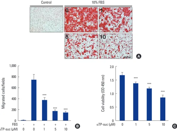
Fig. 1. Effect of α-tocopheryl succinate (αTP-suc) on the cell migration and cell viability of 4T1 cells. (A, B) Cell migration of serum-starved 4T1 cells in response to 10% fetal bovine serum in the presence of dimethyl sulfoxide (DMSO) or different concentrations of αTP-suc (1-10 μM) was assessed in transwell chambers for 12 hr. (A) Representative images of hematoxylin and eosin staining, ×100 magnification; and scale bar, 200 μm. (B) The number of migrated 4T1 cells per field (n=6 per each well). (C) The 4T1 cells were cultured with DMSO or different concentrations of αTP-suc (1-10 μM) for 2 days and then cell viability was measured. The results shown are representative of three independent experiments (n=3), and the values are expressed as mean±standard deviation. ***P<0.001 vs. vehicle treatment by unpaired Student’s t-test. FBS, fetal bovine serum.
Control 10% FBS
A 1,000
800
600
400
200
0
Migrated cells/fields
FBS - + + + + αTP-suc (μM) 0 0 1 5 10
***
*** ***
2.0
1.5
1.0
0.5
0
Cell viability (OD 450 nm)
αTP-suc (μM) 0 1 5 10
***
***
***
B C
mean (SEM; for in vivo data). Statistical analysis was per- formed by either unpaired, two-tailed Student’s t-test or one-way ANOVA followed by Dunnett’s test using Graph- Pad Prism 5.0 (GraphPad Software, San Diego, CA, USA). All P-values of less than 0.05 were considered to indicate sta- tistical significance.
RESULTS
1. αTP-suc inhibited cell migration and proliferation of cancer cells
Studies have shown that αTP-suc inhibits the invasive- ness and cell growth of 4T1 cells, a breast cancer cell line.
[12,16] Thus, we confirmed the effect of αTP-suc in our ex- perimental condition. As shown in Figure 1A and B, serum- induced cell migration of 4T1 cells was significantly inhib- ited by αTP-suc in a dose-dependent manner. Cell viability was also decreased by αTP-suc in a dose-dependent man- ner (Fig. 1C). The cell migration of 4T1 cells was inhibited
by αTP-suc at 1 μM by approximately 50% in 12 hr. Howev- er, αTP-suc inhibited 4T1 cell growth by about 50% at 10 μM for 48 hr suggesting that inhibition of cell migration by αTP-suc was not due to inhibited cell growth by αTP-suc.
These findings suggested that αTP-suc effectively inhibited cell mobility and proliferation in our laboratory conditions as reported previously.
2. αTP-suc inhibited cancer migration in vivo
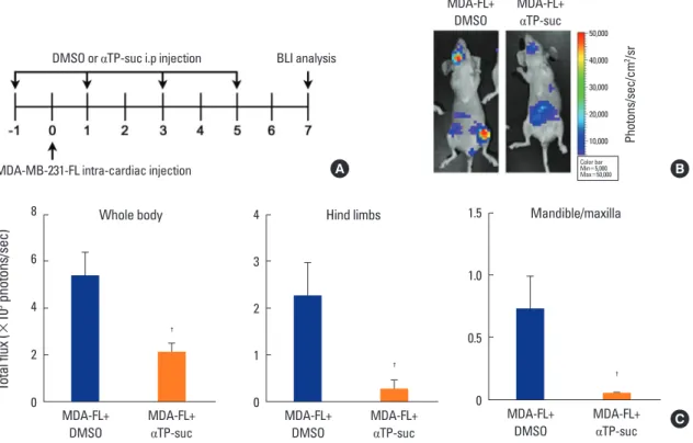
Next, to determine whether αTP-suc is effective in vivo as demonstrated by the in vitro experiments, we used a car- diac injection mouse model. MDA-MB-231 human breast cancer cells expressing firefly luciferase (MDA-MB-231-FL) were injected into the left ventricle, and starting 1 day be- fore cancer injection, αTP-suc was injected intraperitoneally every 2 days (Fig. 2A). After 7 days of cancer injection, bio- luminescence imaging analysis was performed to deter- mine the metastatic status of the cancer cells. In the bones, MDA-MB-231-FL cells mainly metastasized into hind limbsFig. 2. Effect of α-tocopheryl succinate (αTP-suc) on metastasis by intracardiac injection of MDA-MB-231-FL (MDA-FL) cells. MDA-MB-231-FL cells or phosphate-buffered saline was injected into the left cardiac ventricle and metastasis of cancer was analyzed on day 7 after injection of cancer. Starting 1 day before cancer injection, αTP-suc (0.1 mg/head) or dimethyl sulfoxide (DMSO) was injected intraperitoneally every 2 days (n
=5 each). (A) Scheme of the intracardiac injection mouse model. (B) Representative bioluminescence images. (C) Bioluminescence imaging anal- ysis of tumor burden in the whole body. The values are expressed as mean±standard error of the mean. †P<0.05 vs. DMSO-injected mice with injection of MDA-MB-231-FL cells by one-way repeated-measures analysis of variance followed by Dunnett’s test. i.p., intraperitoneally.
8
6
4
2
0 Total flux (×105 photons/sec)
MDA-FL+
DMSO
MDA-FL+
αTP-suc
†
Whole body 4
3
2
1
0 MDA-FL+
DMSO
MDA-FL+
αTP-suc
†
Hind limbs 1.5
1.0
0.5
0 MDA-FL+
DMSO
MDA-FL+
αTP-suc
†
Mandible/maxilla MDA-MB-231-FL intra-cardiac injection
DMSO or αTP-suc i.p injection BLI analysis
MDA-FL+
DMSO
MDA-FL+
αTP-suc
50,000 40,000 30,000 20,000
10,000 Color bar Min=5,000 Max=50,000
Photons/sec/cm2/sr
A B
C
and mandible/maxilla (Fig. 2B). However, the mice injected with αTP-suc showed significantly less tumor burden (Fig.
2B). Tumor-induced total flux of the whole body, hind limbs, and mandible/maxilla were also decreased in αTP-suc-inject- ed mice (Fig. 2C). These results showed that αTP-suc effec- tively inhibited cancer metastasis in vivo.
3. αTP-suc inhibited osteoclastogenesis by inhibiting 4T1-CM-induced RANKL expression in osteoblasts
We previously reported that αTP-suc inhibits IL-1-pro- moted RANKL expression in osteoblasts.[16] Breast cancer cells secrete various factors, including IL-1, IL-6, and PGE2,
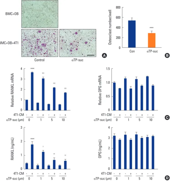
Fig. 3. Effect of α-tocopheryl succinate (αTP-suc) on osteoclast differentiation in triple co-culture and on 4T1 cell-conditioned medium (4T1-CM)- induced receptor activator of nuclear factor-κB ligand (RANKL) expression in osteoblasts (OBs). (A, B) Primary bone marrow cells and OBs were co-cultured with or without 4T1 cells in the presence of dimethyl sulfoxide (DMSO) or αTP-suc (10 μM) for 8 days. (A) Representative images of tartrate-resistant acid phosphatase staining, ×100 magnification; and scale bar, 200 μm. (B) The number of osteoclasts. ***P<0.001 vs. DMSO- treated control by unpaired Student’s t-test. (C, D) OBs were cultured with or without 4T1-CM (30%) in the presence of DMSO or different con- centrations of αTP-suc (1-10 μM) for 24 hr. (C) Levels of RANKL messenger RNA (mRNA) (left panel) and osteoprotegerin (OPG) mRNA (right pan- el) were measured by real-time polymerase chain reaction analysis. (D) Levels of RANKL protein (left panel) and OPG protein (right panel) in the culture media were measured by enzyme-linked immunosorbent assay. The results shown are representative of three independent experiments (n
=3), and the values are expressed as mean±standard deviation. ***P<0.001 vs. untreated control and †††P<0.001; ††P<0.01; †P<0.05 vs. DM- SO-treated cells in response to 4T1-CM by unpaired Student’s t-test. BMC, bone marrow cell.
BMC+OB
BMC+OB+4T1
Control αTP-suc
800 600
400
200
0 Con αTP-suc
***
Osteoclast number/well
A B
4
3
2
1
0
4T1-CM - + - + - + - + αTP-suc (μm) 0 1 5 10
***
†††
†††
†††
Relative RANKL mRNA
1.5
1.0
0.5
0
4T1-CM - + - + - + - + αTP-suc (μm) 0 1 5 10
Relative OPG mRNA
C 3
2
1
0
4T1-CM - + - + - + - + αTP-suc (μm) 0 1 5 10
***
†
†† ††
RANKL (ng/mL)
4
3
2
1
0
4T1-CM - + - + - + - + αTP-suc (μm) 0 1 5 10
OPG (ng/mL)
D
that increase RANKL expression in osteoblasts, leading to enhanced osteoclastogenesis. Therefore, we investigated whether αTP-suc inhibited cancer-induced osteoclast dif- ferentiation in a triple co-culture of mouse primary osteo-
blasts, mouse primary bone marrow cells, and 4T1 cells. As shown in Figure 3A and B, 4T1-induced osteoclast differen- tiation was greatly reduced by αTP-suc (Fig. 3A, B). Next, we performed real-time PCR analysis and enzyme immu-
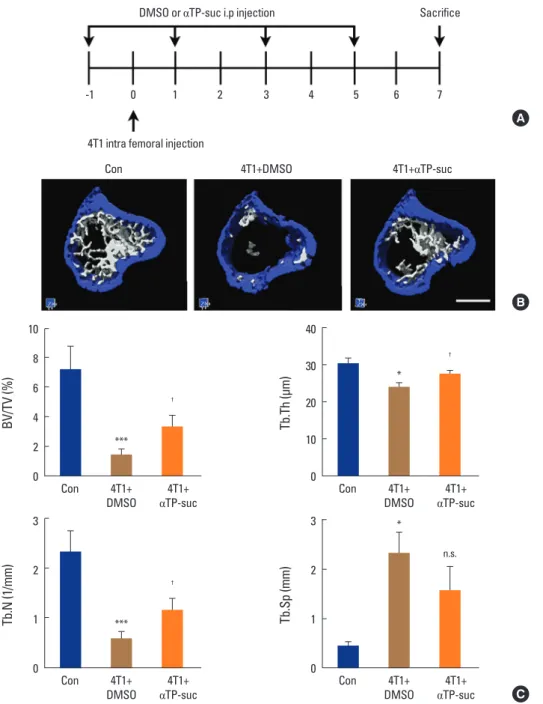
Fig. 4. Effect of α-tocopheryl succinate (αTP-suc) on the bone destruction by 4T1 cells in the mouse model of intrafemoral injection. The 4T1 cells were directly injected into the left femur. Starting 1 day before cancer injection, αTP-suc (0.1 mg/head) or dimethyl sulfoxide (DMSO) was inject- ed intraperitoneally every 2 days (n=5 each). After 7 days of 4T1 injection, left femora were collected and analyzed by microcomputed tomogra- phy (micro-CT) scanning. (A) Scheme of the intrafemoral injection mouse model. (B) Representative reconstructed images by micro-CT (scale bar, 0.7 mm). (C) Quantification of trabecular bone parameters: bone volume per total volume (BV/TV, left upper panel), trabecular thickness (Tb.Th, right upper panel), trabecular number (Tb.N, left bottom panel), and trabecular separation (Tb.Sp, right bottom panel). The values are expressed as mean±standard error of the mean. ***P<0.001; *P<0.05 vs. phosphate-buffered saline-injected mice and †P<0.05 vs. DMSO-injected mice with injection of 4T1 cells by one-way repeated-measures analysis of variance followed by Dunnett’s test. n.s., not significant; i.p., intraperitoneally.
4T1 intra femoral injection
-1 0 1 2 3 4 5 6 7 DMSO or αTP-suc i.p injection Sacrifice
A
B 10
8 6 4 2 0
BV/TV (%)
Con 4T1+
DMSO 4T1+
αTP-suc
†
***
3
2
1
0
Tb.N (1/mm)
Con 4T1+
DMSO 4T1+
αTP-suc
†
***
40
30
20
10
0
Tb.Th (μm)
Con 4T1+
DMSO 4T1+
αTP-suc
†
*
3
2
1
0
Tb.Sp (mm)
Con 4T1+
DMSO 4T1+
αTP-suc n.s.
*
C
Con 4T1+DMSO 4T1+αTP-suc
noassay to investigate the effect of αTP-suc on RANKL ex- pression in osteoblasts induced by 4T1-CM. Osteoblasts treated with 4T1-CM showed elevated expression of RANKL, which was inhibited by αTP-suc in a dose-dependent man- ner (Fig. 3C). However, expression of OPG, a decoy receptor of RANKL, was not affected by αTP-suc (Fig. 3C). To confirm these results, we examined protein levels in the culture me- dia. As shown in Figure 3D, the 4T1-CM-promoted expres- sion of RANKL protein in the culture media of osteoblasts was also decreased by αTP-suc (Fig. 3D). These data sug- gested that αTP-suc inhibited the usual increase in RANKL expression in response to the secretion of factors from 4T1 cells in osteoblasts, leading to reduced osteoclastogenesis.
4. αTP-suc decreased bone destruction by 4T1 cells by inhibiting osteoclastogenesis in vivo
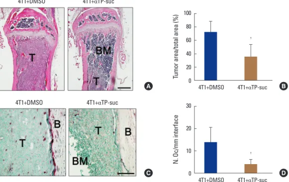
When cancer migrates into bones, communication be- tween the bone marrow niche and the cancer cells forms a vicious cycle, accelerating bone destruction. Thus, to inves- tigate whether αTP-suc affects bone destruction by this vi- cious cycle, 4T1 cells were directly injected into femora,Fig. 5. Effect of α-tocopheryl succinate on tumor burden and osteoclast number by 4T1 cells. Histological analysis of bone metastasis from the experimental groups in Figure 4 (n=5). (A) Representative images of hematoxylin and eosin (H & E)-stained femur sections, ×100 magnification;
and scale bar, 500 μm. (B) Histomorphometric quantification of tumor burden from H & E sections. (C) Representative images of tartrate-resistant acid phosphatase (TRAP)-stained femur sections, ×400 magnification; and scale bar, 200 μm. (D) Histomorphometric quantification of osteoclast numbers from TRAP-stained sections. †P<0.05 vs. dimethyl sulfoxide (DMSO)-injected mice with injection of 4T1 cells by one-way repeated- measures analysis of variance followed by Dunnett’s test. T, tumor; BM, bone marrow; B, bone; αTP-suc, α-tocopheryl succinate.
100 80 60 40 20 Tumor area/total area (%) 0
4T1+DMSO 4T1+αTP-suc
†
30
20
10
0
N. Oc/mm interface
4T1+DMSO 4T1+αTP-suc
†
4T1+DMSO 4T1+αTP-suc
4T1+DMSO 4T1+αTP-suc
A B
C D
and starting 1 day before cancer injection, αTP-suc was in- jected intraperitoneally every 2 days (Fig. 4A). Reconstruct- ed images by micro-CT showed that the significant reduc- tion in trabecular bones resulting from injection of 4T1 cells was greatly rescued in mice co-injected with αTP-suc (Fig.
4B). The trabecular bone parameters analyzed by micro-CT showed the same tendencies as the reconstructed images.
The reductions in bone volume, trabecular thickness, and trabecular number induced by 4T1 cells were relieved in mice co-injected with αTP-suc (Fig. 4C). Histomorphomet- ric analysis showed that tumor burden and the number of osteoclasts at the tumor-bone interface were significantly reduced in the αTP-suc-injected mice (Fig. 5A-D). These data demonstrated that αTP-suc suppressed the osteoclast formation and tumor growth induced by metastatic can- cer, ameliorating the bone destruction.
DISCUSSION
Once bone metastasis of cancer occurs, it has historically been incurable, and patients with bone metastasis of breast
cancer have a reported 5-year survival rate of only 20%.[1]
Thus, it is urgent to develop therapeutic approaches to pre- vent and treat bone metastasis of breast cancer. In the pres- ent study, we found that αTP-suc (4 mg/kg every other day, delivered intraperitoneally) not only inhibited metastasis into bone but also decreased the tumor burden in bone and bone destruction by bone metastatic breast cancer cells.
Vitamins are organic compounds abundant in nature.
Many studies have shown that various vitamins have anti- cancer effects. Bioactive vitamins including vitamins A, C, D, E, and K have anti-cancer potency such as inhibiting cell growth, differentiation, and migration and promoting apop- tosis of cancer cells.[10,17-21] The vitamin E family exhibits various functions including not only anti-cancer but also antioxidant, anti-inflammatory, and anti-thrombolytic ac- tivities.[21-23] Among the vitamin E family, αTP is the most abundant in nature and in the human body and is expect- ed to be useful as a supplement to prevent chronic diseas- es associated with oxidative stress.[24] In addition, αTP has the highest antioxidant capacity among the tocopherols in the following order: αTP >βTP >γTP >δTP.[25] αTP-suc is the most effective form of αTP for cancer therapy.[26] In vi- tro studies have shown the anti-cancer effect of αTP-suc.
[12,16] In addition, it was reported that cancer treatment is improved by using αTP-suc as an adjunct to radiation and chemotherapy.[26] In the present study, we demon- strated the in vivo effect of αTP-suc on cancer migration. As shown in Figure 2, metastasis of MDA-MB-231 cells into the whole body was greatly reduced in mice injected with αTP-suc (Fig. 2B, C). In addition, mice injected with αTP-suc showed minimal metastasis into the mandible/maxilla and limbs (Fig. 2B, C). Taken together, these results demonstrat- ed that αTP-suc is also effective for preventing metastasis of breast cancer cells, especially metastasis into bone, in vivo. However, additional studies are required to compare the anti-cancer effect of the different αTP derivatives in vivo.
The mechanism of bone metastasis is complex and in- volves cooperative, reciprocal interactions among cancer cells, bone marrow cells, and the mineralized bone matrix.
The excess of soluble and cellular components, the signal- ing network, and coordinated gene expression have been shown to contribute to the interplay among bone degra- dation, bone formation, and tumor growth. The interaction between the metastatic tumor and the bone marrow has
been commonly referred as the “vicious cycle”.[27] This vi- cious cycle leads to two separated physiological phenom- enon: osteolytic or osteoblastic bone metastasis, which depends on the type of cancer.[27,28] Among the cancers, breast cancer undergoes osteolytic bone metastasis, which leads to overall bone loss.[4] The molecular mechanisms of bone destruction by metastatic breast cancer are well es- tablished. The migrated cancer cell initiates a vicious cycle by secreting inflammatory factors including parathyroid hormone-related protein, IL-1, IL-6, and PGE2. These inflam- matory factors work on the osteoblasts, leading to increased expression of RANKL. The RANKL expressed from osteo- blasts promotes osteoclast differentiation and activation, and the activated osteoclasts destroy bones. The destruct- ed bone matrix releases growth factors that have accumu- lated in the bone such as TGF-β, IGF-1, and PDGF. These growth factors promote cell growth of metastatic tumors, promot- ing the release of more inflammatory factors.[4,6,29] In ad- dition, cancer-induced factors also stimulate RANKL ex- pression in CD4+ T cells, contributing to bone destruction.
[30] Moreover, RANKL+ regulatory T cells express more RA- NKL by breast cancer cells and then stimulate cancer me- tastasis.[31] The vicious cycle of bone metastasis also causes systemic inflammation, which promotes severe metastasis and low bone mineral density.[32,33] Therefore, communi- cation between cancer cells and bone marrow niches can be a therapeutic target for preventing osteolytic bone de- struction. We previously reported that Trolox, a hydrophilic derivative of αTP, inhibits osteolytic bone metastasis by in- hibiting cancer-induced RANKL expression from osteoblasts, but that αTP had no such effect.[13,34] In addition, our pre- vious report showed that αTP-suc inhibits IL-1-induced RANKL expression in osteoblasts and prevents osteoclast differentiation and bone resorption.[11] In the present study, we showed that αTP-suc also strongly inhibits can- cer-induced RANKL expression in osteoblasts, leading to the inhibition of osteoclast differentiation (Fig. 3). These results indicate that αTP-suc can effectively inhibit cancer- induced RANKL expression, leading to the prevention of osteoclastogenesis.
Various animal models are established for the study of bone metastasis. The ideal model should be clinically rele- vant, recapitulate the human disease, and be reproducible.
Thus, the best method is to inject cancer cells directly into the primary site and study the individual cells that have
metastasized to the bone over time. However, the incidence of bone metastasis is very low and animals usually cannot survive until bone metastasis occurs. Therefore, a method of injecting cancer cells into blood vesicles, such as the left cardiac ventricle or the tail vein, is used to study bone me- tastasis. Of these methods, cardiac injection is considered a better method because the intravenous injection of can- cer cells generally results in the accumulation of cancer cells in the lungs.[35] In the present study, we used intra- cardiac injection of MDA-MB-231-FL cells (Fig. 2). Metasta- sis of MDA-MB-231-FL cells into the whole body, including bones, was greatly reduced in αTP-suc-injected mice (Fig.
2). Because αTP-suc inhibited cell migration in the intracar- diac injection model, we next used intrafemoral injection of 4T1 cells to study the effect of αTP-suc on the interac- tion between cancers and bone marrow cells after metas- tasis of cancer cells into bone (Fig. 4). As shown in Figure 4 and 5, the 4T1-induced bone destruction, tumor burden, and number of osteoclasts were greatly reduced by αTP- suc injection (Fig. 4, 5). These results indicated that αTP-suc was effective for inhibiting bone destruction and tumor growth.
CONCLUSIONS
In the present study, we showed that αTP-suc has dual effects on bone metastatic breast cancers: inhibition of cell growth and migration of cancer, and suppression of the vi- cious cycle by decreasing cancer-derived-factor-induced RANKL expression in osteoblasts. Therefore, the results of the present study indicate that αTP-suc can be efficiently utilized to prevent and treat osteolytic bone metastasis of breast cancer with dual effects.
ACKNOWLEDGMENTS
This study was supported by the Basic Science Research Program through the National Research Foundation of Ko- rea funded by the Ministry of Science, ICT & Future Plan- ning (NRF-2017R1A2B2002312) and the research funding from Korean Society for Bone and Mineral Research.
REFERENCES
1. Roodman GD. Mechanisms of bone metastasis. N Engl J
Med 2004;350:1655-64.
2. Coleman RE. Metastatic bone disease: clinical features, pathophysiology and treatment strategies. Cancer Treat Rev 2001;27:165-76.
3. Mundy GR. Metastasis to bone: causes, consequences and therapeutic opportunities. Nat Rev Cancer 2002;2:584-93.
4. Chen YC, Sosnoski DM, Mastro AM. Breast cancer metasta- sis to the bone: mechanisms of bone loss. Breast Cancer Res 2010;12:215.
5. Wang N, Docherty FE, Brown HK, et al. Prostate cancer cells preferentially home to osteoblast-rich areas in the early stages of bone metastasis: evidence from in vivo models. J Bone Miner Res 2014;29:2688-96.
6. Croucher PI, McDonald MM, Martin TJ. Bone metastasis:
the importance of the neighbourhood. Nat Rev Cancer 2016;16:373-86.
7. Ju J, Picinich SC, Yang Z, et al. Cancer-preventive activities of tocopherols and tocotrienols. Carcinogenesis 2010;31:
533-42.
8. Husain K, Francois RA, Yamauchi T, et al. Vitamin E delta-to- cotrienol augments the antitumor activity of gemcitabine and suppresses constitutive NF-kappaB activation in pan- creatic cancer. Mol Cancer Ther 2011;10:2363-72.
9. Gysin R, Azzi A, Visarius T. Gamma-tocopherol inhibits hu- man cancer cell cycle progression and cell proliferation by down-regulation of cyclins. FASEB J 2002;16:1952-4.
10. Alqahtani S, Kaddoumi A. Vitamin E transporters in cancer therapy. Aaps j 2015;17:313-22.
11. Kim HN, Lee JH, Jin WJ, et al. alpha-Tocopheryl succinate inhibits osteoclast formation by suppressing receptor ac- tivator of nuclear factor-kappaB ligand (RANKL) expres- sion and bone resorption. J Bone Metab 2012;19:111-20.
12. Savitskaya MA, Onischenko GE. alpha-Tocopheryl succi- nate affects malignant cell viability, proliferation, and dif- ferentiation. Biochemistry (Mosc) 2016;81:806-18.
13. Lee JH, Kim HN, Yang D, et al. Trolox prevents osteoclasto- genesis by suppressing RANKL expression and signaling. J Biol Chem 2009;284:13725-34.
14. Lee JH, Kim HN, Kim KO, et al. CXCL10 promotes osteolytic bone metastasis by enhancing cancer outgrowth and os- teoclastogenesis. Cancer Res 2012;72:3175-86.
15. Jin WJ, Kim B, Kim D, et al. NF-kappaB signaling regulates cell-autonomous regulation of CXCL10 in breast cancer 4T1 cells. Exp Mol Med 2017;49:e295.
16. Wang D, Chuang HC, Weng SC, et al. alpha-Tocopheryl
succinate as a scaffold to develop potent inhibitors of breast cancer cell adhesion. J Med Chem 2009;52:5642-8.
17. Davis-Yadley AH, Malafa MP. Vitamins in pancreatic cancer:
a review of underlying mechanisms and future applica- tions. Adv Nutr 2015;6:774-802.
18. Doldo E, Costanza G, Agostinelli S, et al. Vitamin A, cancer treatment and prevention: the new role of cellular retinol binding proteins. Biomed Res Int 2015;2015:624627.
19. Fritz H, Flower G, Weeks L, et al. Intravenous vitamin C and cancer: A systematic review. Integr Cancer Ther 2014;13:
280-300.
20. Garland CF, Garland FC, Gorham ED, et al. The role of vita- min D in cancer prevention. Am J Public Health 2006;96:
252-61.
21. Lamson DW, Plaza SM. The anticancer effects of vitamin K.
Altern Med Rev 2003;8:303-18.
22. Frank J. Beyond vitamin E supplementation: an alternative strategy to improve vitamin E status. J Plant Physiol 2005;
162:834-43.
23. Azzi A, Ricciarelli R, Zingg JM. Non-antioxidant molecular functions of alpha-tocopherol (vitamin E). FEBS Lett 2002;
519:8-10.
24. Diplock AT. Safety of antioxidant vitamins and beta-caro- tene. Am J Clin Nutr 1995;62:1510s-6s.
25. Müller L, Theile K, Böhm V. In vitro antioxidant activity of tocopherols and tocotrienols and comparison of vitamin E concentration and lipophilic antioxidant capacity in hu- man plasma. Mol Nutr Food Res 2010;54:731-42.
26. Prasad KN, Kumar B, Yan XD, et al. Alpha-tocopheryl succi-
nate, the most effective form of vitamin E for adjuvant can- cer treatment: a review. J Am Coll Nutr 2003;22:108-17.
27. Futakuchi M, Fukamachi K, Suzui M. Heterogeneity of tu- mor cells in the bone microenvironment: mechanisms and therapeutic targets for bone metastasis of prostate or breast cancer. Adv Drug Deliv Rev 2016;99:206-11.
28. Clezardin P, Teti A. Bone metastasis: pathogenesis and ther- apeutic implications. Clin Exp Metastasis 2007;24:599-608.
29. Hauschka PV, Chen TL, Mavrakos AE. Polypeptide growth factors in bone matrix. Ciba Found Symp 1988;136:207-25.
30. Monteiro AC, Leal AC, Gonçalves-Silva T, et al. T cells induce pre-metastatic osteolytic disease and help bone metasta- ses establishment in a mouse model of metastatic breast cancer. PLoS One 2013;8:e68171.
31. Tan W, Zhang W, Strasner A, et al. Tumour-infiltrating reg- ulatory T cells stimulate mammary cancer metastasis through RANKL-RANK signalling. Nature 2011;470:548-53.
32. Chechlinska M, Kowalewska M, Nowak R. Systemic inflam- mation as a confounding factor in cancer biomarker dis- covery and validation. Nat Rev Cancer 2010;10:2-3.
33. Okazaki R, Watanabe R, Inoue D. Osteoporosis associated with chronic obstructive pulmonary disease. J Bone Metab 2016;23:111-20.
34. Ha H, Lee JH, Kim HN, et al. alpha-Tocotrienol inhibits os- teoclastic bone resorption by suppressing RANKL expres- sion and signaling and bone resorbing activity. Biochem Biophys Res Commun 2011;406:546-51.
35. Simmons JK, Hildreth BE 3rd, Supsavhad W, et al. Animal models of bone metastasis. Vet Pathol 2015;52:827-41.