http://dx.doi.org/10.3988/jcn.2013.9.4.231 J Clin Neurol 2013;9:231-236
Introduction
Intracranial atherosclerotic stenosis (ICAS) is one of the ma-
jor causes of ischemic stroke. Although the progression of ICAS is a strong predictor for stroke recurrence,1 the nature and risk factors of ICAS progression have yet to be clarified.
In particular, the association between the natural course of ICAS and systemic atherosclerosis has never been investi- gated.
Carotid intima-media thickness (CIMT) is a well-estab- lished marker of generalized atherosclerosis. Its relationship
The Response of Carotid Intima-Media Thickness to Medical Treatment Is Correlated with That of Intracranial Atherosclerosis
Sun U. Kwon,a Bum Joon Kim,a Seong Rae Kim,b Dong-Eog Kim,c Hahn Young Kim,d Ju-Hun Lee,e Hee-Joon Bae,f Moon-Ku Han,f Dong-Wha Kang,a Jong S. Kim,a Joung-Ho Rhab
aDepartment of Neurology, Asan Medical Center, University of Ulsan College of Medicine, Seoul, Korea
bDepartment of Neurology, Inha University Hospital, Incheon, Korea
cDepartment of Neurology, Dongguk University Hospital, Goyang, Korea
dDepartment of Neurology, Konkuk University Hospital, Seoul, Korea
eDepartment of Neurology, Kangdong Sacred Heart Hospital, Seoul, Korea
fDepartment of Neurology, Seoul National University Bundang Hospital, Seongnam, Korea
Received May 6, 2013 Revised July 6, 2013 Accepted July 6, 2013 Correspondence Joung-Ho Rha, MD Department of Neurology, Inha University Hospital, 27 Inhang-ro, Jung-gu, Incheon 400-711, Korea Tel +82-32-890-3418 Fax +82-32-890-3864 E-mail [email protected]
Background and PurposezzIntracranial atherosclerotic stenosis (ICAS) is considered as a major cause of stroke. The carotid intima-media thickness (CIMT), which accurately reflects the burden of generalized atherosclerosis, is also associated with stroke. The aim of this study was to determine the association between the CIMT and ICAS responses to medical treatment.
MethodszzThis study constituted part of the “Trial of cilostazol in symptomatic intracranial arterial stenosis”-2 that evaluated the ICAS response after randomized antiplatelet treatment.
Magnetic resonance angiography and CIMT measurement were performed at baseline and after 7 months of treatment. CIMT was measured using semiautomated software, and was presented as maximum (CIMT-max) and average (CIMT-ave) values. The change in CIMT was compared relative to the ICAS response (i.e., progression, no-change, and regression). Ordinal logistic re- gression and analysis of covariance (ANCOVA) were used to analyze the association between the responses.
ResultszzAmong the 101 enrolled patients, 85 underwent follow-up CIMT measurement. CIMT increased most in the ICAS progression group (CIMT-max: 0.09±0.23, CIMT-ave: 0.04±0.12), and to a lesser degree in the no-change group (CIMT-max: 0.02±0.16, CIMT-ave: 0.02±0.11), but decreased in patients with ICAS regression (CIMT-max: -0.04±0.11, CIMT-ave: -0.03±0.07;
CIMT-max: p=0.010, CIMT-ave: p=0.015). Ordinal logistic regression analysis demonstrated that the change in CIMT-max was independently associated with the ICAS response (p=0.032).
However, the ANCOVA revealed that the reverse was not true, in that the ICAS response was not independently associated with the change in CIMT after adjusting for confounding factors.
ConclusionszzThe ICAS response may be associated with the CIMT response to medical
treatment. J Clin Neurol 2013;9:231-236
Key Wordszz carotid intima media thickness, intracranial atherosclerosis, antiplatelet.
Open Access
cc This is an Open Access article distributed under the terms of the Cre- ative Commons Attribution Non-Commercial License (http://creative- commons.org/licenses/by-nc/3.0) which permits unrestricted non-com- mercial use, distribution, and reproduction in any medium, provided the ori- ginal work is properly cited.
with atherosclerosis in arteries at various sites has been re- ported previously. Increased CIMT was associated with the presence and extent of atherosclerosis in the coronary arter- ies,2 peripheral arteries,3 and the carotid bifurcation or internal carotid artery.4 A recent community-based cross-sectional study performed in an Asian country found that CIMT was increased in subjects with ICAS. However, the increased CIMT was not independently associated with ICAS after ad- justing for confounding factors.5
There are pathophysiologic differences in the occurrence and development of atherosclerosis between the intra- and ex- tracranial arteries, and these differences may have influenced the previous results.5 However, the correlation between the responses of atherosclerosis to medical treatment in the intra- and extracranial arteries has not yet been elucidated. There- fore, the present study investigated the change in CIMT rela- tive to the degree of ICAS response to medical treatment in ischemic stroke patients with symptomatic ICAS.
Methods
Participants and study design
This study was performed as a substudy of the “Trial of cilo- stazol in symptomatic intracranial arterial stenosis” (TOSS)- 2 (unique identifier: NCT00130039),6 which is a randomized controlled multicenter trial that enrolled 457 acute (≤2 weeks of onset) ischemic stroke patients with symptomatic ICAS in the middle cerebral or basilar arteries, as assessed by mag- netic resonance angiography (MRA). To ensure that only pa- tients with intracranial stenosis due to atherosclerosis were included, patients with 1) other vasculopathy such as arterial dissection or moyamoya disease, 2) embolic heart disease, or 3) significant stenosis of arteries proximal to the symptomatic stenosis were excluded. After obtaining their written in- formed consent to participate, subjects who were enrolled in TOSS-2 were randomly assigned to either the cilostazol (100-mg aspirin plus 200-mg cilostazol daily) or clopidogrel (100-mg aspirin plus 75-mg clopidogrel daily) groups. Ag- gressive control of atherosclerosis risk factors including statin therapy was strongly recommended in the protocol. The pro- gression of ICAS between the two antiplatelet regimens was compared after 7 months. The TOSS-2 study is described in detail elsewhere.6
In the present substudy, demographic characteristics and clinical data including vascular risk factors were obtained at baseline. The results of various blood tests including lipid pro- files, fasting glucose, and C-reactive protein (CRP) were also obtained. Three-dimensional time-of-flight MRA and carotid ultrasonography were used to evaluate the baseline ICAS and CIMT, respectively. After 7 months of treatment, the details
of any concomitant medications used during the observation period were obtained, and the blood tests, MRA, and carotid ultrasonography were followed-up so that the responses to treatment could be evaluated. The protocol was approved by the ethics committee of each participating center.
Evaluation of ICAS and CIMT
MRA was performed twice: first at baseline and then at the 7 months follow-up. According to the TOSS-2 MRA grading system, the severity of ICAS was classified into normal (0), mild (1; signal reduction <50%), moderate (2; signal reduction
>50%), severe (3; focal signal loss with distal flow), or occlu- sion (4; sudden cutoff without distal flow void). The change in ICAS after 7 months of treatment was categorized into three groups: progression, regression, and no-change. The severity of symptomatic ICAS was evaluated independently by two investigators who were blinded to all of the clinical in- formation. If any discrepancies arose, a third investigator re- solved the discrepancy by consensus with the other two in- vestigators.
Six of the 20 centers that participated in TOSS-2 were ca- pable of assessing the CIMT in acute ischemic stroke pa- tients, and could therefore participate in this substudy. Before the data acquisition began, a sample carotid ultrasonography image was assessed from each center to evaluate the appropri- ateness of image quality. Carotid ultrasonography was per- formed according to a standard protocol by skilled sonogra- phers at each center, and using the same schedule as for MRA.
CIMT was measured from each right and left carotid artery with a 10-MHz linear vascular probe. The insonation angle was 120-150° for the left side and 210-240° for the right side;
the variation among patients meant that the best angle for measurement was set by the examiner. The depth of focus was 30-40 mm and the log gain for measurement was approxi- mately 60 dB. Standardized longitudinal B-mode images were obtained from the far wall of the common carotid ar- tery, which was defined as the segment extending from 10 to 20 mm proximal to the tip of the bifurcation site according to the Mannheim carotid intima-media thickness consensus.7 All of the DICOM images that contained the proper CIMT for each side were archived electronically from each center to the central laboratory for CIMT measurements.
Carotid intima-media thickness was measured using dedi- cated semiautomated software (Intimascope, Media Cross, Tokyo, Japan) by a single reader (S.R.K.) who was blinded to all clinical information to avoid interrater disagreement.8 The resulting data are presented as the maximum (CIMT- max) and average (CIMT-ave) values. CIMT-max was calcu- lated as the mean of the maximum points in both common ca- rotid arteries, while CIMT-ave was the mean of the average
value obtained from 250 computer-based points for each com- mon carotid artery.8
Statistical analysis
General characteristics including demographic characteristics, vascular risk factors, and concomitant medications were com- pared relative to the ICAS response to treatment. Changes in lipid profiles, fasting glucose, CRP, and CIMT after 7 months of medical treatment were also compared. The ICAS response was used as a categorical variable with an ordinal nature. The analysis was performed using chi-square statistics for a linear- by-linear association model and appropriate contrast weights from ANOVA. Bonferroni’s post-hoc analysis was also per- formed in order to compare the change in CIMT between the progression and regression groups.
Ordinal logistic regression models were used to test the presence of an independent association between the change in CIMT and the ICAS response after adjusting for potential confounding factors. All of the variables that exhibited a po- tential association (i.e., p<0.15) with the ICAS response in bi- variate analysis were selected. Any suspicion of multicol- linearity between CIMT-max and CIMT-ave was prevented by using two models. Models 1 and 2 both contained sex, smoking, and the type of antiplatelet agent, with model 1 containing CIMT-max whereas, model 2 containing CIMT- ave. In addition, a potential independent association between the ICAS response and the follow-up CIMT-max or CIMT- ave was evaluated using separate models of analysis of covari- ance (ANCOVA) after adjusting for age, sex, hypertension, diabetes, hyperlipidemia, smoking, type of antiplatelet, and baseline CIMT. Statistical analyses were performed using
SPSS 16.0 (SPSS Inc., Chicago, IL, USA). Except where stated otherwise, the data are presented as mean±SD values, and the level of statistical significance was set at p<0.05.
Results
Among the 457 patients who were enrolled into TOSS-2 from the 20 centers, 223 patients were registered from the 6 cen- ters that were able to offer carotid ultrasonography. This sub- study was proposed after the initiation of TOSS-2; therefore, the 122 patients who had already been registered to TOSS-2 before the commencement of this substudy could not receive carotid ultrasonography and were unable to participate in this substudy. Among the remaining 101 patients, follow-up ca- rotid ultrasonography was not performed in 16 patients due to various causes, and hence 85 patients were ultimately in- cluded in the present study. Due to technical problems, ultra- sonography was performed on one side only in two patients.
ICAS progressed in 13 (15.3%) patients, regressed in 29 (34.1%) patients, and remained unchanged in 43 (50.6%) pa- tients.
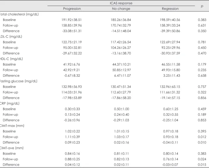
There were no significant differences among the groups with respect to demographic characteristics, risk factors, and concomitant medications relative to the ICAS response (Table 1), nor in lipid profiles, fasting glucose, and CRP between baseline and follow-up (Table 2). However, there was a signif- icant change in CIMT according to the relative progression of ICAS. The CIMT increased in patients with ICAS progres- sion (CIMT-max: 0.09±0.23 mm and CIMT-ave: 0.04±0.12 mm) or with no change in ICAS status (CIMT-max: 0.02±
0.16 mm and CIMT-ave: 0.02±0.11 mm). Conversely, CIMT Table 1. General characteristic according to the ICAS response
ICAS response
Progression (n=13) No change (n=43) Regression (n=29) p
Age (years) 60.9±13.0 68.6±9.4 65.5±12.8 0.509
Male 6 (46.2%) 18 (41.9%) 20 (69.0%) 0.068
Hypertension 10 (76.9%) 28 (65.1%) 17 (58.6%) 0.264
Diabetes 6 (46.2%) 14 (32.6%) 8 (27.6%) 0.268
Hyperlipidemia 8 (61.5%) 13 (30.2%) 14 (48.2%) 0.849
Smoking 4 (30.8%) 19 (44.2%) 16 (55.2%) 0.137
Cardiac disease 1 (7.7%) 1 (2.3%) 2 (6.9%) 0.853
Systolic pressure (mm Hg) 131.6±15.3 139.6±19.8 134.3±19.0 0.994
Diastolic pressure (mm Hg) 79.8±9.6 80.7±11.3 78.7±11.5 0.647
Concurrent medications
Cilostazol 4 (30.8%) 19 (44.2%) 16 (55.2%) 0.137
ARB 4 (30.8%) 18 (41.9%) 8 (27.6%) 0.583
ACEI 1 (7.7%) 1 (2.3%) 2 (6.9%) 0.853
Statin 10 (76.9%) 21 (48.8%) 17 (58.6%) 0.513
Data are mean±SD or number (percentage) values.
ACEI: angiotensin converting enzyme inhibitor, ARB: angiotensin-II receptor blocker, ICAS: intracranial atherosclerotic stenosis.
decreased in patients with ICAS regression (CIMT-max:
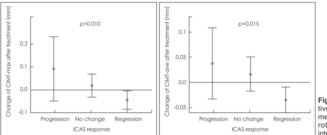
-0.04±0.11 mm and CIMT-ave: -0.03±0.07 mm). The trend analysis demonstrated a significant linear decrease in CIMT among those with a favorable ICAS response to treatment (CIMT-max: p=0.010; CIMT-ave: p=0.015) (Fig. 1).
Post-hoc analysis between patients with ICAS regression and progression revealed that the change in CIMT-max de- creased significantly in the ICAS regression group (mean dif- ference=0.14; p=0.037). The results of ordinal logistic regres- sion demonstrated that the change in CIMT-max was independently associated with the ICAS response after ad- justing for potential confounding factors (p=0.032) (Table 3).
However, the ICAS response was not independently associ- ated with the follow-up CIMT after adjusting for age, sex, conventional risk factors, type of antiplatelet, and baseline CIMT (CIMT-max, ANCOVA F=2.677, ANCOVA p=0.075;
CIMT-ave, ANCOVA F=1.508, ANCOVA p=0.228).
Discussion
The objective of this study was to determine the association between the ICAS and CIMT responses to medical treat- ment. The results indicated that CIMT decreased in patients with ICAS regression and increased both in patients with no change in ICAS status and in those with ICAS progression.
This illustrates a similarity in the CIMT and ICAS responses.
The change in CIMT-max differed significantly between the ICAS progression and regression groups, and was also inde- pendently associated with the ICAS response. However, there was no statistically significant independent association be- tween the ICAS response and the follow-up CIMT after ad- justing for confounding factors.
Table 2. Difference in lipid profiles and changes in CIMT after antiplatelet therapy according to the ICAS response ICAS response
Progression No change Regression p
Total cholesterol (mg/dL)
Baseline 191.92±38.51 185.26±36.84 198.59±40.56 0.383
Follow-up 158.85±39.96 170.74±32.79 158.39±33.24 0.631
Difference -33.08±51.31 -14.51±48.04 -39.39±50.86 0.350
LDL-C (mg/dL)
Baseline 122.75±21.19 117.42±26.54 122.69±27.94 0.781
Follow-up 95.00±32.81 104.26±26.27 92.25±29.96 0.450
Difference -29.67±32.22 -13.16±38.70 -30.93±37.59 0.470
HDL-C (mg/dL)
Baseline 41.92±6.76 44.37±10.21 46.55±11.58 0.179
Follow-up 42.92±9.21 50.85±12.97 49.93±15.80 0.235
Difference -0.67±8.32 6.47±11.07 3.25±11.43 0.658
Fasting glucose (mg/dL)
Baseline 132.98±56.93 130.47±51.34 132.96±65.15 0.757
Follow-up 114.03±31.96 112.60±27.79 111.66±31.32 0.322
Difference -17.98±53.89 -17.86±58.20 -19.14±57.15 0.856
CRP (mg/dL)
Baseline 0.30±0.33 0.50±1.00 0.60±1.25 0.459
Follow-up 0.13±0.24 0.24±0.40 0.32±0.55 0.189
Difference -0.26±0.96 -0.29±1.03 -0.25±1.04 0.853
CIMT-max (mm)
Baseline 1.02±0.22 1.01±0.15 0.97±0.18 0.395
Follow-up 1.11±0.39 1.03±0.17 0.93±0.18 0.012
Difference 0.09±0.23 0.02±0.16 -0.04±0.11 0.010
CIMT-ave (mm)
Baseline 0.84±0.16 0.81±0.11 0.80±0.14 0.383
Follow-up 0.88±0.25 0.82±0.13 0.76±0.14 0.024
Difference 0.04±0.12 0.02±0.11 -0.03±0.07 0.015
Data are mean±SD values.
CIMT: carotid intima-media thickness, CRP: C-reactive protein, HDL-C: high-density lipoprotein cholesterol, ICAS: intracranial athero- sclerotic stenosis, LDL-C: low-density lipoprotein cholesterol.
Various factors may influence the occurrence and progres- sion of atherosclerosis in the intra- and extracranial arteries.
ICAS is more closely associated with hypertension, diabetes, and metabolic syndrome than is atherosclerosis in the carotid arteries.9 Patients with ICAS exhibited higher CIMTs, whereas the association disappeared after adjusting for risk factors from a previous study.5 Focusing on the progression of ath- erosclerosis, systemic endothelial dysfunction was associated with the progression of CIMT,10 whereas low high-density li- poprotein cholesterol levels were associated with symptom- atic ICAS progression.11 Although the risk factors of athero- sclerosis occurrence and progression differ according to the site, the present results demonstrate that the responses of ath- erosclerosis in distinct sites may actually be associated with each other.
Increased CIMT is frequently observed in ICAS patients,12 and medical treatment usually focuses on the target site. How- ever, effective antiatherosclerotic treatments at one site may also be effective for other vascular beds. It has been shown previously that lipid-lowering agents prevent the progression of symptomatic ICAS,13 and are significantly associated with a favorable decrease in CIMT.14 Cilostazol, an antiplatelet agent with an antiatherogenic effect, has been shown to pre- vent the progression of both ICAS6 and CIMT.15
Only subjects with symptomatic ICAS participated in the present study. The change in CIMT-max was independently associated with the ICAS response. Therefore, during the medical treatment for ICAS, subjects exhibiting a favorable CIMT-max response may also exhibit a favorable ICAS re-
sponse. However, the converse does not appear to be true, in that ICAS change was not associated with the follow-up CIMT (as indicated by ANCOVA). Therefore, subjects with a favorable ICAS outcome may not always demonstrate a fa- vorable CIMT response. However, the small sample may have been at least partially responsible for the failure to dem- onstrate an independent association in both directions. More- over, the results should be interpreted with further caution since the main trial included symptomatic ICAS patients and its focus was the treatment of ICAS and its response.
This substudy of TOSS-2 was subject to some limitations, the first of which stems from its small sample. Since the par- ticipation of each center was determined according to the fea- sibility, and the substudy was proposed during the main trial, the number of subjects available with a baseline CIMT was small. Second, volumetric evaluation of the change in plaque size was not included as an endpoint, and the measurement of plaque size by ultrasonography in multiple centers may have increased differences between the centers. Third, the measurement of CIMT may also vary considerably with the ultrasound setting, the study site, and the angle at which it is assessed.16 To minimize the possible effects of these factors, CIMT was measured according to a fixed protocol based on the Mannheim carotid intima-media thickness consensus,7 and the data were analyzed using semiautomated software.
Although there were some intercenter differences in the baseline and follow-up CIMT values, the change in CIMT, which was the main outcome of this substudy, did not differ significantly among the centers.
Table 3. Independent factors associated with the ICAS response
Variable Exp (b) SE 95% CI p
Model 1
CIMT-max 3.065 1.426 0.270-5.860 0.032
Model 2
CIMT-ave 4.308 2.353 -0.304-8.920 0.067
Variables entered to each model of ordinal logistic regression: Model 1: sex, smoking, type of antiplatelet agent, and CIMT-max, Model 2: sex, smoking, type of antiplatelet agent, and CIMT-ave.
CI: confidential interval, CIMT: carotid intima-media thickness, ICAS: intracranial atherosclerotic stenosis, SE: standard error.
Fig. 1. Difference in CIMT change rela- tive to the ICAS response after treat- ment of symptomatic ICAS. CIMT: ca- rotid intima-media thickness, ICAS:
intracranial atherosclerotic stenosis.
0.2 0.1 0.0
) trmmt (enmeaterC aftT-maxf CIM ogehan -0.1
Progression No change Regression ICAS response
p=0.010
0.1
0.05
0.0
-0.05
Change of CIMT-ave after treatment
(mm
)
Progression No change Regression ICAS response
p=0.015
In conclusion, there is a correlation between the ICAS re- sponse and the CIMT response to treatment in symptomatic ICAS patients. Therefore, the CIMT response, which is a sur- rogate marker of generalized atherosclerosis, may reflect the ICAS response to treatment.
Conflicts of Interest
The authors have no financial conflicts of interest.
Acknowledgements
This study was supported by a grant of the Korea Healthcare Technology R&D Project, Ministry of Health and Welfare Republic of Korea (HI10C2020).
Korea Otsuka Pharmaceutical, Korea Otsuka International Asia, and Korea Otsuka International Arab provided financial support for TOSS-2 and the CIMT substudy. However, these institutions played no role in the protocol development, data collection, or data analysis for the current study, or in the manuscript preparation.
REFERENCES
1. Arenillas JF, Molina CA, Montaner J, Abilleira S, González-Sánchez MA, Alvarez-Sabín J. Progression and clinical recurrence of symptom- atic middle cerebral artery stenosis: a long-term follow-up transcra- nial Doppler ultrasound study. Stroke 2001;32:2898-2904.
2. Coskun U, Yildiz A, Esen OB, Baskurt M, Cakar MA, Kilickesmez KO, et al. Relationship between carotid intima-media thickness and coronary angiographic findings: a prospective study. Cardiovasc Ul- trasound 2009;7:59.
3. Allan PL, Mowbray PI, Lee AJ, Fowkes FG. Relationship between ca- rotid intima-media thickness and symptomatic and asymptomatic pe- ripheral arterial disease. The Edinburgh Artery Study. Stroke 1997;28:
348-353.
4. Bots ML, Hofman A, De Jong PT, Grobbee DE. Common carotid inti- ma-media thickness as an indicator of atherosclerosis at other sites of the carotid artery. The Rotterdam Study. Ann Epidemiol 1996;6:147- 5. Leng XY, Chen XY, Chook P, Xiong L, Lin WH, Liu JY, et al. Corre-153.
lation of large artery intracranial occlusive disease with carotid inti- ma-media thickness and presence of carotid plaque. Stroke 2013;44:
68-72.
6. Kwon SU, Hong KS, Kang DW, Park JM, Lee JH, Cho YJ, et al. Effi- cacy and safety of combination antiplatelet therapies in patients with symptomatic intracranial atherosclerotic stenosis. Stroke 2011;42:
2883-2890.
7. Touboul PJ, Hennerici MG, Meairs S, Adams H, Amarenco P, Born- stein N, et al. Mannheim carotid intima-media thickness and plaque consensus (2004-2006-2011). An update on behalf of the advisory board of the 3rd, 4th and 5th watching the risk symposia, at the 13th, 15th and 20th European Stroke Conferences, Mannheim, Germany, 2004, Brussels, Belgium, 2006, and Hamburg, Germany, 2011. Cere- brovasc Dis 2012;34:290-296.
8. Yanase T, Nasu S, Mukuta Y, Shimizu Y, Nishihara T, Okabe T, et al.
Evaluation of a new carotid intima-media thickness measurement by B-mode ultrasonography using an innovative measurement software, intimascope. Am J Hypertens 2006;19:1206-1212.
9. Bang OY, Kim JW, Lee JH, Lee MA, Lee PH, Joo IS, et al. Associa- tion of the metabolic syndrome with intracranial atherosclerotic stroke.
Neurology 2005;65:296-298.
10. Halcox JP, Donald AE, Ellins E, Witte DR, Shipley MJ, Brunner EJ, et al. Endothelial function predicts progression of carotid intima-media thickness. Circulation 2009;119:1005-1012.
11. Kim DE, Kim JY, Jeong SW, Cho YJ, Park JM, Lee JH, et al. Associa- tion between changes in lipid profiles and progression of symptomat- ic intracranial atherosclerotic stenosis: a prospective multicenter study.
Stroke 2012;43:1824-1830.
12. Jung KW, Shon YM, Yang DW, Kim BS, Cho AH. Coexisting carotid atherosclerosis in patients with intracranial small- or large-vessel dis- ease. J Clin Neurol 2012;8:104-108.
13. Kim HJ, Kim EK, Kwon SU, Kim JS, Kang DW. Effect of statin on progression of symptomatic intracranial atherosclerosis. Can J Neurol Sci 2012;39:801-806.
14. Huang Y, Li W, Dong L, Li R, Wu Y. Effect of statin therapy on the progression of common carotid artery intima-media thickness: an up- dated systematic review and meta-analysis of randomized controlled trials. J Atheroscler Thromb 2013;20:108-121.
15. Heo SH, Lee JS, Kim BJ, Hwang KJ, Kim JH, Chang DI. Effects of cilostazol against the progression of carotid IMT in symptomatic ischemic stroke patients. J Neurol 2013;260:122-130.
16. Peters SA, den Ruijter HM, Palmer MK, Grobbee DE, Crouse JR 3rd, O’Leary DH, et al. Extensive or restricted ultrasound protocols to measure carotid intima-media thickness: analysis of completeness rates and impact on observed rates of change over time. J Am Soc Echocardiogr 2012;25:91-100.