CASE REPORT
만성적인 소화불량증을 주소로 내원한 고령의 환자에서 우연히 발견된 모르가니 헤르니아
김덕기, 문희석, 정현용, 성재규, 강선형, 김명희
충남대학교 의과대학·의학전문대학원 소화기내과학교실
An Incidental Discovery of Morgagni Hernia in an Elderly Patient Presented with Chronic Dyspepsia
Duk Ki Kim, Hee Seok Moon, Hyeon Yong Jung, Jae Kyu Sung, Sun Hyeong Gang, and Myeong Hee Kim
Division of Gastroenterology and Hepatology, Department of Internal Medicine, Chungnam National University of Medicine, Daejeon, Korea
A Morgagni hernia was first described in 1761 by Giovanni Morgagni. In adults, it is accompanied by gastrointestinal- or respiratory-type symptoms. Herein, we report an 84-year-old woman presented to our hospital with nausea and vomiting. After hospitalization, an X-ray revealed a right diaphragmatic hernia. Based on the results of abdominal computed tomography, duodenoscopy, and upper gastro- intestinography (gastrografin), we concluded that her symptoms were caused by Morgagni hernia. Our patient underwent laparoscopic surgery, and shortly thereafter, her symptoms resolved. (Korean J Gastroenterol 2017;69:68-73)
Key Words: Hernia, diaphragmatic; Dyspepsia; Laparoscopy
Received October 14, 2016. Revised November 13, 2016. Accepted December 5, 2016.
CC This is an open access article distributed under the terms of the Creative Commons Attribution Non-Commercial License (http://creativecommons.org/licenses/
by-nc/4.0) which permits unrestricted non-commercial use, distribution, and reproduction in any medium, provided the original work is properly cited.
Copyright © 2017. Korean Society of Gastroenterology.
교신저자: 문희석, 35015, 대전시 중구 문화로 266, 충남대학교 의과대학 소화기내과학교실
Correspondence to: Hee Seok Moon, Division of Gastroenterology and Hepatology, Department of Internal Medicine, Chungnam National University of Medicine, 266 Munhwa-ro, Jung-gu, Daejeon 35015, Korea. Tel: +82-42-280-7143, Fax: +82-42-257-5753, E-mail: [email protected]
Financial support: None. Conflict of interest: None.
INTRODUCTION
In 1761, Giovanni Morgagni described a type of hernia that today bears his name.1 Morgagni hernias account for only 3%
of all diaphragmatic hernias and are extremely rare in adults.
They are more common in the pediatric population and usu- ally occur on the right side or in the anterior mediastinum due to the retrosternal location of the foramen of Morgagni.2 According to one previously reported study, they occur on the right side in about 90% of cases, with the remainder occur- ring on the left side and on both sides (2% and 8% of cases, respectively).3 Elderly people rarely present with a dia- phragmatic hernia, and late presentations are often accom- panied by a range of symptoms, such as abdominal dis-
comfort, abdominal pain, constipation, loss of appetite, and chronic respiratory symptoms (e.g., Dyspnea on exertion [DOE]
and dyspnea). Since many of these symptoms are common, Morgagni hernias are often missed or attributed to other fac- tors (e.g. dyspepsia or insufficient exercise). Furthermore, some cases of Morgagni hernias can completely be asympto- matic, only being detected accidentally.
CASE REPORT
An 84-year-old woman visited the gastroenterology de- partment in our hospital with a 6-month history of post- prandial nausea and vomiting. Her medical history was un- remarkable (e.g., no trauma or surgery), and she had no other
Fig. 1. A chest X-ray displaying air and gas above the right diaphragm, indicating a diaphragmatic hernia.
Fig. 2. A CT scan showing a right diaphragmatic hernia, with small and large bowel contents in the right hemithorax.
Fig. 3. (A, B) Duodenoscopy showing a twisted stomach and duodenal bulb.
symptoms. Prior to her visit, she was prescribed prokinetics in a private hospital, but these were not effective. Recently, the discomfort had worsened, and she reported that she was unable to consume any food.
The patient was scheduled to undergo a colonoscopy.
However, after taking Coolprep powder, she was unable to defecate; she retched and complained of abdominal dis- comfort and pain. The patient was subsequently hospitalized.
At the time of the visit, a check of the patient’s vital signs revealed the following: systolic/diastolic blood pressure of 90/60 mmHg, heart rate of 64/min, respiratory rate of
18/min, and body temperature of 36.1℃. Auscultation on both lungs was clear. Her abdomen was soft but distended, especially in the right upper and left upper quadrant areas.
According to her abdominal examination, she had epigastric tenderness, without rebound tenderness.
The initial laboratory data, including electrolyte, blood urea nitrogen, Creatinine, cardiac enzyme, amylase, and li- pase findings, and an electrocardiography were all normal.
After admission, a plain X-ray revealed intestinal gas above the right diaphragm (Fig. 1), indicating a diaphr-agmatic hernia. The patient underwent abdominal and pelvic com- puted tomography (CT), as shown in Fig. 2. The abdominal pelvic CT also showed a diaphragmatic hernia, appearing to be a Morgagni hernia. Duodenoscopy (Fig. 3) and upper gas- trointestinography (UGI) using gastrografin were performed to detect any functional impairment (Fig. 4). Duodenoscopy showed stomach herniation and torsion of the duodenal bulb. UGI revealed a herniation in the antrum of the stomach and duodenum that extended into the mediastinum and
A
B
Fig. 4. (A, B) UGI (gastrografin) showing the bowel in an anterior area of the chest wall. UGI, upper gastrointestinal series.
beak-like luminal narrowing in the second portion of the duodenum. The gastrografin rarely passed the duodenal bulb. We concluded that the symptoms were caused by Morgagni hernia, and the patient was scheduled for surgery.
Diaphragmatic hernia was repaired with a mesh, using a laparoscopic approach. A crescent-shaped defect (5-7 cm) was located on the right retrosternal area of the diaphragm.
A transverse colon and omentum herniation were also ob- served, in addition to midgut malrotation, a hernia sac, and partial rotation of the duodenojejunal junction. First, the sur- geon repaired the diaphragm defect in the abdominal wall us- ing a dual mesh (10-15 cm) and transfascial fixation. Next, to address the laxity of the perigastric ligament and recurrent mesenteroaxial volvulus, gastropexy of the fundus and mid body was performed. After the surgery, the patient ate well
and experienced no abdominal discomfort or pain. During a 6-month follow-up period, she had no complaints or symp- toms, and an abdominal X-ray revealed no abnormalities.
DISCUSSION
Congenital diaphragmatic hernia (CDH) is a birth defect of the diaphragm. It occurs in one in every 2,000-3,000 live births and accounts for 8% of all major congenital anomalies.
The three basic types of CDHs are Bochdalek hernias, ante- rior Morgagni hernias, and hiatus hernias. All three types of hernias are associated with high mortality rates, approx- imately 40-62%.4
Morgagni hernias account for approximately 2% of all CDH cases. They refer to herniation through the foramina of Morgagni (anterior defect of the diaphragm), which is located beside the xiphoid process of the sternum. Newborns with Morgagni hernias may present with respiratory distress or acute abdomen, similar to other CDHs. In contrast, the only symptom of Morgagni hernias in adults is thought to be discomfort. However, recent data suggest that this is not the case, and about 70% of patients with a Morgagni hernia pres- ent with significant symptoms.
Hernias frequently contain omental fat and are accom- panied by a transverse colon. Sometimes, they contain the liver, stomach, biliary system, and right side heart, causing cardiomegaly.5
The pathophysiology of Morgagni hernias in adults is not well known due to their rarity; however, they are frequently as- sociated with minimal symptoms and are found accidentally.
It is unclear whether they truly have a congenital origin or are the result of acquired disease. We presume that the develop- ment of Morgagni hernias among elderly individuals is due to hereditary factors and predisposing conditions, with hernia being the result of a pre-existing diaphragmatic defect.
Subsequent detection of Morgagni hernias among patients who initially had normal radiographs suggests that these her- nias may be acquired.6 The rarity of congenital Morgagni her- nias indicates that other factors, such as pregnancy, trauma, obesity, chronic constipation, and chronic cough, are likely the common predisposing conditions for these hernias among adults.5 All aforementioned predisposing factors have one thing in common: An increased intra-abdominal pressure.
A
B
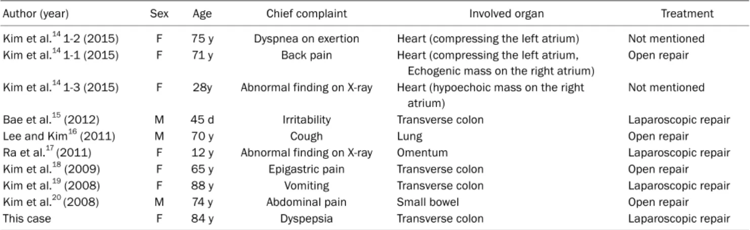
Table 1. Characteristics of Cases of Morgagni Hernia Cancer Reported in Korea
Author (year) Sex Age Chief complaint Involved organ Treatment
Kim et al.14 1-2 (2015) F 75 y Dyspnea on exertion Heart (compressing the left atrium) Not mentioned Kim et al.14 1-1 (2015) F 71 y Back pain Heart (compressing the left atrium,
Echogenic mass on the right atrium)
Open repair
Kim et al.14 1-3 (2015) F 28y Abnormal finding on X-ray Heart (hypoechoic mass on the right atrium)
Not mentioned
Bae et al.15 (2012) M 45 d Irritability Transverse colon Laparoscopic repair
Lee and Kim16 (2011) M 70 y Cough Lung Open repair
Ra et al.17 (2011) F 12 y Abnormal finding on X-ray Omentum Laparoscopic repair
Kim et al.18 (2009) F 65 y Epigastric pain Transverse colon Open repair
Kim et al.19 (2008) F 88 y Vomiting Transverse colon Laparoscopic repair
Kim et al.20 (2008) M 74 y Abdominal pain Small bowel Open repair
This case F 84 y Dyspepsia Transverse colon Laparoscopic repair
Morgagni hernias have been associated with the following syndromes and congenital defects: Down’s syndrome, Turner’s syndrome, Noonan syndrome, Prader–Willi syn- drome, tetralogy of Fallot, ventricular septal defects, sco- liosis, Morquio syndrome, connective tissue disorders, dex- trocardia, chest wall deformities, genitourinary abnormal- ities, and omphaloceles.5
The lack of a typical clinical presentation in cases of late-presenting Morgagni hernias leads to a delayed diag- nosis of the defect.7 Acute symptoms are rare and are usually due to large bowel obstruction. An X-ray is an effective diag- nostic tool. Radiographically, a Morgagni hernia appears as a fatty mass, and it can be difficult to differentiate from a prominent epicardial fat pad on the X-ray. It is important to distinguish Morgagni hernias from lipomas, teratomas, thy- momas, thymo-lipomas, liposarcomas, if traumatic dia- phragm rupture, pneumothorax and mediastinal tumors are the other types of hernias. A Morgagni hernia can sometimes mimic lung lesions, such as pulmonary tuberculosis or lung cancer. Therefore, other tools, such as ultrasonography and CT, should also be considered. Ultrasonography has been shown to be useful in assessing diaphragmatic hernias.
However, CT is the most sensitive tool, revealing anatomical details and complications, such as strangulation. A delay in the use of advanced diagnostic tools, such as CT or magnetic res- onance imaging, can lead to misdiagnoses and mistreatment.
Since Morgagni hernias are very rare, it is highly likely for clinicians to overlook them. However, a Morgagni hernia should be suspected in patients who present with un- explained symptoms of the digestive and respiratory systems.
If in doubt regarding the diagnosis of a Morgagni hernia, plain X-ray and CT or other examinations (echocardiography or duo-
denoscopy) should be considered, depending on the in- volved organ.8
A plain X-ray is a simple, cost-effective diagnostic tool.
However, as a Morgagni hernia can mimic other simple her- nias, CT is the most reliable imaging modality, despite radia- tion exposure and the use of contrast agents. Magnetic reso- nance imaging is not usually performed due to its high cost.
However, it can be used if contrast agent is contraindicated.
Other modalities, such as abdominal Ultrasonography (US), echocardiography, duodenoscopy, and UGI, can also be per- formed, depending on the involved organ, to improve the ac- curacy of the diagnosis and to evaluate the function of the organs.
Before the 1980s, every symptomatic CDH was treated as an emergency.9
Today, however, the consensus is that even asymptomatic cases of Morgagni hernias should not be considered as emergencies. However, it has been determined that they should be repaired surgically to avoid any complications.10 However, each case should be considered individually, with special attention to the age of patients. Surgery is associated with mortality risk, and general anesthesia can impose a se- vere strain on the lungs or heart of patients, especially in cas- es of older patients.11
Surgery is the treatment of choice for Morgagni hernias.
The first laparoscopic repair was reported by Kuster et al. in 1992.12 Subsequently, laparoscopic management became the general trend. Two main surgical approaches have been described for the treatment of these hernias: trans-abdomi- nal (open or laparoscopic) and trans-thoracic (open or thor- acosopic). The trans-abdominal approach is preferred when the diagnosis is certain, and the trans-thoracic approach is
warranted when it is uncertain. However, the trans-abdomi- nal approach has been linked to pneumonia and sepsis. The recurrence rate of Morgagni hernias is not clear, with about 2-15% of recurrent cases after surgery due to remnant sac or absorbable sutures.13
A search of PubMed using the terms “Morgagni hernia”
and “Korea” uncovered seven papers (Table 1).14-20 The pres- ent Morgagni hernia case was typical in terms of a right-sided hernia, transverse colon involvement, and treatment by lapa- roscopic repair via mesh. The most prominent differences compared with previously published reports in the literature were the duration and chronic nature of the symptoms (> 6 months), as well as the age of patients (84 years). In most cases involving Morgagni hernias, patients present to the hospital for acute episodes, such as severe abdominal pain, dyspnea, and DOE, or they are asymptomatic and are acci- dentally discovered on plain X-ray during routine health screening. In the present case, our patient experienced chronic worsening of symptoms for a duration of greater than 6 months. Although the patient visited her local clinic, the hernia was overlooked as a result of a failure to perform a plain X-ray. The present case provides a good example of how suspicion is the most important criterion in the diagnosis of a Morgagni hernia.
With regard to the anatomic distribution of Morgagni her- nias worldwide, Horton et al. reported that they were right sid- ed in about 91% of cases and that predisposing conditions were present in 41% of cases.5 Furthermore, they also found that women made up about 62% cases and that the average age of patients was 53 years. We found no significant differ- ences between the cases of Morgagni hernias in Korea as compared with those in other countries. The primary differ- ence between the cases of Morgagni hernias is the various organs involved.
The patient in the present case had no symptoms for al- most 70 years, except the occasional abdominal discomfort.
Her general health was not good due to age-related complications.
In deciding whether to operate, we used both duodenoscopy and UGI to evaluate the impact of the Morgagni hernia on the patient’s gastrointestinal function, although duodenoscopy is not commonly used as a diagnostic tool. Based on the re- sults of these examinations and a preoperative evaluation (pulmonary function test and echocardiography), we recom- mended surgery to the patient.
The lack of a unique clinical presentation of Morgagni her- nias can delay the diagnosis, especially in local clinics.
Considering the prevalence rate, Morgagni hernias should be considered in the differential diagnosis of patients present- ing with gastrointestinal disturbances and respiratory distress.
A plain X-ray, chest and abdominal CT, as well as ultrasonography can be used as diagnostic tools. The treatment of choice is laparoscopic surgery, but each case should be assessed on an individual basis.
REFERENCES
1. Harrington SW. Clinical manifestations and surgical treatment of congenital types of diaphragmatic hernia. Rev Gastroenterol 1951;18:243-256.
2. Al-Salem AH, Zamakhshary M, Al Mohaidly M, Al-Qahtani A, Abdulla MR, Naga MI. Congenital Morgagni's hernia: a national multicenter study. J Pediatr Surg 2014;49:503-507.
3. Marinceu D, Loubani M, O'Grady H. Late presentation of a large Morgagni hernia in an adult. BMJ Case Rep 2014;2014.
4. Donnelly LF, Sakurai M, Klosterman LA, Delong DM, Strife JL.
Correlation between findings on chest radiography and survival in neonates with congenital diaphragmatic hernia. AJR Am J Roentgenol 1999;173:1589-1593.
5. Horton JD, Hofmann LJ, Hetz SP. Presentation and management of Morgagni hernias in adults: a review of 298 cases. Surg Endosc 2008;22:1413-1420.
6. Eren S, Ciris F. Diaphragmatic hernia: diagnostic approaches with review of the literature. Eur J Radiol 2005;54:448-459.
7. Abraham V, Myla Y, Verghese S, Chandran BS. Morgagni-larrey hernia- a review of 20 cases. Indian J Surg 2012;74:391-395.
8. Tone K, Kiryu I, Yoshida M, Tsuboi K, Takagi M, Kuwano K.
Morgagni hernia with respiratory failure aggravated by non- invasive positive pressure ventilation: a case report and overview of the literature. Respir Investig 2014;52:203-208.
9. Frenckner B, Ehrén H, Granholm T, Lindén V, Palmér K. Improved results in patients who have congenital diaphragmatic hernia us- ing preoperative stabilization, extracorporeal membrane oxy- genation, and delayed surgery. J Pediatr Surg 1997;32:1185-1189.
10. Pousios D, Panagiotopoulos N, Piyis A, Gourgiotis S. Transthoracic repair of asymptomatic Morgagni hernia in an adult. Indian J Surg 2012;74:431-433.
11. Kozanhan B, Başaran B, Aygın F, Akkoyun İ, Özmen S. Anaesthetic management of laparoscopic Morgagni hernia repair in a patient with coexisting down syndrome, patent foramen ovale and pec- tus carinatum. Turk J Anaesthesiol Reanim 2016;44:44-46.
12. Kuster GG, Kline LE, Garzo G. Diaphragmatic hernia through the foramen of Morgagni: laparoscopic repair case report. J Laparoendosc Surg 1992;2:93-100.
13. Lamas-Pinheiro R, Pereira J, Carvalho F, et al. Minimally invasive repair of Morgagni hernia - A multicenter case series. Rev Port Pneumol (2006) 2016;22:273-278.
14. Kim SH, Kim MG, Kim SJ, et al. Unusual diaphragmatic hernias
mimicking cardiac masses. J Cardiovasc Ultrasound 2015;23:
107-112.
15. Bae MJ, I H, Kim DH, Jeong YJ, Kim YD, Cho JS. One-stage laparo- scopic repair of Morgagni and inguinal hernias in a two-month old male. Korean J Thorac Cardiovasc Surg 2012;45:415-417.
16. Lee SK, Kim DH. Congenital intercostal lung herniation com- bined with an unusual Morgagni's hernia. Korean J Thorac Cardiovasc Surg 2011;44:455-457.
17. Ra YJ, Huh U, Lee SG, Je HG. The laparoscopic repair of a Morgagni hernia in a child. Korean J Thorac Cardiovasc Surg
2011;44:80-82.
18. Kim HR, Hong TH, Lee YS, et al. Elective laparoscopic repair after colonoscopic decompression for incarcerated Morgagni hernia.
Gut Liver 2009;3:318-320.
19. Kim ES, Kang JY, Pyo CH, Jeon EY. A Morgagni diaphragmatic her- nia found after removal of mediastinal tumor. Ann Thorac Cardiovasc Surg 2008;14:175-177.
20. Kim SW, Jung SH, Kang SH. A case of left-sided Morgagni hernia complicating incarcerated small bowel hernia. Korean J Gastroenterol 2008;51:52-55.