대 한 밤사 선 의 학회 지 1996 ; 34( 2) : 165-169
경추전방고정술후의 자기공명영상소견 1
진 욱·최우석·오주혈·김의졸·윤 엽
목 적:경추전방고정술(이후 ACF ; Anterior cervical fusion) 후 보이는 변화의 자기공명영상(이후 MRI로 칭함)소견과 이의 유용성에 대하여 알아보고자 하였다.
대상 및 방법:추간판 탈출증 혹은 외상에 의한 경추 손상으로 ACF를 시행받은 후 임상적으로 추간 판염이나 척추염을 배제할 수 있었던 13명의 환자를 대상으로 수술후 1개월에서 1Q7H 월 사이에 관찰 되는 MRI 소견을 분석하였다.
결 과 :8/13여 1(62%) 에서 인두후부의 연부조직의 조영증강이 ACF 시행후(특히 초기 수개월 이내) 에 보이지만 주변 경추나 추간판과는 잘 구분되었다.2/4예 (50%) 에서는 이식된 골펀이 비교적 균일한 조영증강을 보였다. 또 9/13예 (69%) 에서 인공을편이나 이식된 골편은 수술후 시간에 관계없이 주변 구조물과 낮은 신호강도의 계면을 형성하여 잘 구분되었다. 수술후 남아있던 경추체와 추간판은 약
8/
13예 (62%) 에서 균일한 조영증강을 보였고, 특히 1예에서는 경추체 조영증강 정도가 2개월째보다 4개 월째 더감소되었다.
결 론:경추 추간판염이나 척추염이 없는 ACFOI 후의 경추 MRI소견은 주위와 잘 구분되는 인두후부 연부조직, 이식된 골편, 남아있는 경추체와 추간판의 비교적 균일한 조영증강 등이었다. MRI는 ACFOI
후의 소견과 수술후 염증과의 감별, 그리고 특히 이식된 골편이나 BOP의 추후관찰에 매우 유용한 검 사로사료된다.
서
론
최근 들어서 증가된 교통사고에 의한 경추손상이나 경추 간판 탈출증 (cervica1 disc herniation) 등을 치료하기 위 해 골편이식 (bone graft), 인공골편 (Bio1ogica1 Orthopedic Prosthesis : 이후 BOP) 혹은 금속 고정물 (metallic p1a te) 을 이용한 경추전방고정술 (Anterior Cervica1 Fusion : 이후 ACF) 의 시행이 많아지고 있다. 이에 저자들은 경추 전방 고정술 후에 엄상적으로 추간판염, 척추염 혹은 주변 연부조직의 염증소견없이 나타나는술후자기공명영상소 견과 MRI의 유용성 에 대 하여 알아보고자 본 연구를 시 행 하였다.
대상및방법
본원에서 1992년 5월부터 1995년 7월까지 추간판 탈출증 혹은 외상에 의한 경추손상으로 경추전방고정술을 시행받
1경희대학교 의과대학 진단방사선과학교실
이 논문은 1995년도 경희대학병원 연구비 지원에 의한 것임 이 논문은 1995년 8월 29일 접수하여 1996년 1월 3일에 채택되었음
고 자기공명영상을 시행한 환자 13명을 대상으로 하였다.
이들은 모두 자기공명영상 시행시까지 임상적 증상이나 검사결과(백혈구수나 ESR등)를 바탕으로 추간판염이나 척추염등의 부작용을 배제할 수 있었던 환자였으며, 이들 의 술후 1개월에서 10개월 사이 자기공명영상소견을분석 하였다. 이들 환자의 남녀비는 6:7 이었고, 연령분포는 23 세에서 65세로 평균연령이 45세였다. 이들중 4예는 골편이 식, 6예는 인공골편, 그리고 3예는 금속 고정물을 이용하여 수술을시행하였다.
사용한 기기는 1.5 tes1a unit(Toshiba MRFX
n
,Nasu, Japan) 로서, 척추 전용 표면코일 (surface coil)을 사용하였다. 13예 모두에서 스핀에코 (spin-echo) 방법으 로 T1 강조영상 (TR / TE
=
450 - 500/15 - 20) 빛 T2 강조 영상 (TR / TE=
1800 -2000 / 30 / 80) 의 시상 및 축상면을 얻었으며, Gd -DTPA(Magnevist, Schering, German) 로 체중 1Kg당 0.1 mmo1 (0.2 m1) 을 약 2-3분에 걸쳐 천천히 정맥주사하여 조영증강 영상을 얻었다. 이렇게 얻은 자기 공명영상 소견중 주변 구조물과 이식된 골편, 인공골편 흑 은 금속 고정물과의 구분, 남아있던 경추체나 추간판의 신 호강도 변화, 주변 연부조직 (특히 인두후부 연부조직 )의 신호강도 변화, 그리고 이식된 골편의 신호강도 등을 분석-165 -
대 한 방사 선 의 학회 지 1996: 34(2) : 165-169 하였다.
결 과
13예중 8예 (62%) 에서 ACF를 시행하는데 사용한 물질 혹은 골편에 관계없이 인두후부의 연부조직의 조영증강이 ACF 시행후(특히 초기 수개월 이내) 관찰되지만 주변 경 추나 추간판과는 잘 구분되 었고 (Fig. 1c, 2c, 3c), 술후 시 간이 경과할 수록 이러한 연부조직의 조영증강 정도는 감 소되었다 (Fig. 2c, 2d). 술후 남아있던 경추체와 추간판은 13예중 8예 (62%) 에서 조영증장을 보였고 (Fig. 1, 2), 특히 1예에서는 경추체 조영증강 정도가 2개월째보다 4개월째 더 감소되 었다 (Fig. 2).
골편을 이식했던 4예에서는 2예 (50%) 에서 이식된 골편 의 비교적 균일한 조영증강이 관찰되 었다 (Fig. 1). 또 삽입 된 골편이나금속고정불과주변 구조물과의 관계를보면,
13예중 9예 (69%) 에서 연공골편이나 이식된 골편이 술후 시간에 관계없이 주변구조물과 낮은 신호강도의 계면을 형성하여 잘 구분되었지만 (Fig. 1, 2) 일부 금속고정물의 경 우는 Metallic artifact 에 의 해 주변 구조물의 관찰이 어 렵기도하였다 (Fig. 3). 그에 비하여 인공골편은모두매우 낮은 신호강도를 보여 단순 X선 촬영에서와는 다르게 주 변 구조물과의 위치관계를 잘 알 수 있었고, 특히 전위를 보였던 1예에서 연부조직내 그 위치를 잘 볼 수 있었다.
고 찰
전방척추고정술 (Anterior cervical fusion)은 1950년대 Robinson등에 의해 소개된 이후(1) 추간판 탈출증, 척추 증 (spondylosis) , 그리고 외상등의 경추간판질환의 치료 로서 신경외과의와 정형외과의 모두에게 폭 넓게 받아들 여지고 있는 수술법이다 (2). 탈출된 추간판을 전방을 통한 접근으로 제거하는 것은 안전하고 척수와 경추신경근 (cer vical nerve root) 모두를 감압시켜줄 수 있다. 적용되는 원리로 Robinson등은 세가지의 이유를 들었는데 견고한 전방고정을 통해 신경근을 압박하는 뼈돌기 (os teophyte) 의 흡수가 일어나고, 전방접근은 척수의 손상을 방지할 수 있으며, 척수와 신경근을 압박할 수 있는 황색인대(liga
ment flavum) 와 후종인대 (posterior longitudinal liga- ment) 의 좌굴요절 (buckling) 이 골편을 삽업함으로써 추 간판의 공간 (disc space) 과 신경공 (neuroforamen) 의 높 이의 회복이 이루어져 감소될 수 있다는 설명이다(1). 이 러한 이유들로 전방접근법을 이용한 여러 다양한 수술법 이 통증, 신경근병증 (radiculopathy) , 그리고 척수병증
(myelopathy) 을 완화시키기위해 연구되고있다 (2).
전방척추고정술에 가장 많이 사용되는 것은 자가이식골 편 (autograft of bone fragment) (Fig. 1)으로 대개는 환자 자신의 장골(ilium) 의 일부를 사용하지만 (3) , 자가이식골
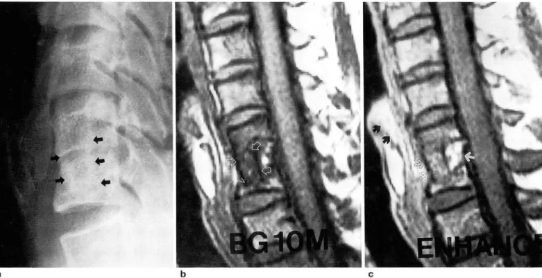
a b c
Fig. 1. A 44-year-old man with anterior cervical lusion(bone graft) after lall down a. Plain radiograph shows ill-delined margin 01 bone graft(arrows).
b. T1-weighted sagittal image 01 cervical spine shows relatively well delined bone graft(arrows) within vertebral bodies with low signal intensity intersurface.
c. On postcontrast T1-weighted sagittal image, dense enhancement 01 retropharyngeal soft tissue(black arrows) and posterior portion 01 the vertebral body(white arrow) are seen. Grafted bony Iragments show mild enhancement(black open arrows)
-166-
편을 사용시 장골 공여부(iliac donor site) 의 합병증 즉,
통증, 혈종, 혹은 감염등이 많이 보고되고 있어 (4, 5) 이에 대한 여러 대체물로 사체나 다른 이의 골편, 금속 고정물 (metallic plate)(Fig. 3) 이나 인공 골편 (Biological Or- thopedic Prosthesis: BOP)(Fig. 2) 등이 쓰이고 있다 (6,
7,8). 또한 경추체로부터 얻은 골편을 사용한 보고도 있다 (9).
척추수술후 MRI를 시행하는 경우 나타나는 소견으로 는 요추간판 탈출증의 수술후 변화가 추간판염의 경우와 비교되어 발표된 바 있다(1 0). 여기서 수술후 변화는 수술
진 욱외 ·경추전방고정술후의 자기공명영상
한 추간판이 Tl과 T2강조영상에서 모두 고신호 강도를 보 일 수 있고, Gd-DTPA 조영증강시 술후 초기(약 6개월) 까지는 이 부위에 국소적인 조영증강이 보일 수 있다 하였 다. 이러한 내용들은 본 연구에서도 적용이 가능하였고, 추 간판염시 보일 수 있는 추간판/추단판 (disc / end plate) 의 불균질성 조영증강과 추단판의 파괴와는 달리 비교적 균일한 조영증강을 보이는 남아있던 추체 /추간판과 주변 의 경추체, 시간경과에 따른 남아있던 경추체의 조영증강 감소로 추간판염과 술후 변화와는 감별이 가능하였다. 또 한 척추주변 근육의 조영증강도 알려져 있는데 수술후 3주
a b
c d
7
l
M
ω
Fig. 2. A 52-year-이 d man with anterior cervical lusion(BOP) after traffic accident a. Plain radiograph reveals bony delect in anteroinlerior portion 01 cervical vertebral body(arrows). BOP cannot be evaluated.
b. T1 -weighted sagittal image 01 cervical spine(2 months postop.) shows well demarcated BOP(arrows) with very low signal intensity in anterior portion 01 ver- tebral bodies and discs.
c. On postcontrast T1-weighted sagittal image(2 months postop.), highly enhanced retrophrayngeal soft tissue(black arrows)
" and remained posterior portion 01 the ver-
tebral bodies/discs(white arrows) are seen
d. After more than 2 months, postcontrast T1-weighted sagittal image(postoperative 4th month) reveals decreased enhance- ment in the retropharyngeal soft tissue and the vertebral bodies/discs(arrows)
대 한 방사 선 의 학회、7\: 1 1996; 34(2) : 165-169
a b c
Fig. 3. A 23-year-old lemale with anterior cervical lusion(metallic plate) after traffic accident a. On plain radiograph, metallic plate is well visible in anterior portion 01 cervical spine
b. On T1-weighted sagittal image 01 cervical spine, some metallic artilacts with low signal intensities are observed but spinal cord is well evaluated
c. On postcontrast T1-weighted sagittal image, retropharyngeal soft tissue is enhanced(arrows).
Fig. 4. A 52-year-old man with anterior cervical lusion(BOP) due to HIVD C4-5
T1-weighted sagittal image shows anterior displacement 01 B.
。 P. with low signal intensity(arrows)
- 168
째에 가장 큰 조영증강을 보이고, 6개월 이후까지도 조영 증강을 보일 수 있다한다(1 1).
MRI는 또한 이식된 골펀의 상태 특히 위치의 유지를 잘 알 수 있었다. 예를들면, 단순촬영에서는 그 경계의 확 실한 관찰이 불가능하던 자가이식골편이나 보이지 않던 BOP가 저신호강도의 경 계를 가지며 잘 파악되 었다.
BOP
의 전위는 직선 저신호강도의 위치변화와 주변 연부조직 의 조영증강으로 추적관찰이 용이하였다. 1 예에서는
BOP
수술후 추체에 고정되어 있지않고 전방전위된 것을 잘 관 찰할 수 있었다 (Fig.
4).
또, 자가골편이식의 경우 (3예), 그 골편의
Gd -DTPA
조영증강시 신호강도가 T1 강조영상에서 6개월째의 경우 조영층강되지 않았으나, 7개월과 107H월째의 것은 고신호 강도로 조영증강되었다 (Fig. 1). 이러한 소견이 시간의 경 과에 따라 이식된 자가골편의 자기인식이 받아들여졌음을 의미하는 MRI소견인지는더 많은증례와이에 따른추적 연구가 뒷받침되야 할 것같다.
결론적으로 경추전방고정술후 나타나는 MRI 소견으로 는 주위와 잘 구분되는 인두후부 연부조직, 이식된 골편,
그리고 남아있는 경추체와 추간판의 비교적 균일한 조영 증강 등이 었다.MRI는 ACF 이후의 소견과 수술후 염증과 의 감별, 그리고 특히 이식된 골편이나 단순촬영에서 관찰 이 불가능하였던 BOP의 추후관찰에 매우 유용한 검사로 사료된다.
진 욱 외 . 경추전방고정술후의 자기공명영상
*~ C그 고 헌
luation of frozen allografts versus autografts in anterior cervi- cal fusions. Clin Orthop 1976; 119 : 231-236
7. Cloward RB: Gas-sterilized cadaver bone grafts for spinal fusion operations. Spine 1980;5:4-10
1. Robinson RA, Walker AE, Ferlic OC, Wieching OK: The 8. Cloward RB: The treatment of ruptured lumbar intervertebral results of anterior interbony fusion of the cervical spine. J discs by vertebral body fusion: 111 method of use of banked Bone Joint Surg 1962; 44A ‘ 1569-1587 bone. Ann Surg 1952;136:987-991
2. Thomas AZ, Thomas BO: The use of freeze-dried allograft 9. Toyohiko 1, Kyosuke K, Nobuaki K, Shoji M :The surgical bone for anterior cervical fusions. Spine 1991 ;16(7) :726-9 technique of anterior fusion using bone grafts obtained from 3. Cloward RB: The anterior approach for removal of ruptured cervical vertebral bodies. J Neurosurg 1994; 80: 16-19
cervical disks. J Neurosurg 1958; 15: 602-617 10 임승재, 류경남, 최우석, 윤엽, 김기택 추간판 탈출증의 수술후 변화
4. Oepalma A, Rothman R, Lewinneck G, et al: Anterior 정상 및 추간판염의 자기공명영상 소견 대한방사선의학회지
interbody fusion for severe cervical disc degeneration. Surg 1994; 31 (2) : 223-228
Gynecol Obstet 1972; 134: 755-758 11. Boden SO, Oavis 00, Dina TS, et al. Contrast-enhanced MR 5. Whitecloud TS :Complications of anterior cervical fusion. Instr imaging performed after successf비 lumbar disc surgery: pro-
Course Lect 1976; 27: 223-227 spective study. Radiology 1992; 182 : 59-64 6. Brown MD, Malinin TI, Brown MD: A roentgenographic eva-
Journal of the Korean Radiological Society 1996; 34(2) : 165-169
Postoperative MR Findings of the Anterior Cervical Fusion
1Uk Jin, M.D., WOO Suk Choi, M.D., Joo Hyeong Oh, M.D.
Eui Jong Kim, M.D., Yup Yoon, M.D.
1 Department of Diagnostic Radiology, Kyung Hee University Hospital
Purpose: This study was conducted to describe the postopertive MRI findings with contrast enhancement fol- lowing anterior cervical fusion(ACF)
Materials and Methods: Thirteen patients after anterior cervical fusions for disc herniation or traumatic in- jury were studied with MRI 1 month to 10 months after operation. MRI findings were analysed with attention to the remained vertebral body, disc material, retropharyngeal soft tissue, grafted bone fragment, and biological orthopedic prosthesis(BOP).
Results: In 8/13 cases(62%), enhancement of the retropharyngeal soft tissue was seen with good demar- cation between soft tissue and surrounding vertebra or disc
In 9/13 cases(69%), grafted bone or BOP was well defined by intersurface with low signal intensity. In 4 cases of ACF with bone graft, 2 cases(50%) showed heterogeneous or homogeneous enhancement of grafted bone fragments. Remained vertebral bodies and discs showed enhancements in 8/13 cases(62 %).
Conclusion: Postoperative MR findings after ACF included good margination of enhanced retropharyngeal soft tissue, grafted bone, and remained vertebral bodies/discs. The MRI could be a useful study to differentiate findings after ACF from posoperative inflammations such as spondylitis or discitis, and be a good imaging mo- dality for follow up of grafted bony fragment or BOP.
Index Words : Spine, surgery Spine, MR
Address reprint requests to : Uk Jin, M.D., Department of Diagnostic Radiology, Kyung Hee University Hospital,
# 1 Hoeki강ong, Dongdaemun-ku, Seoul, 130-702 Korea. Tel. 82-2-958-8622 Fax.82-2-968-0787
m m
춘계학술대회
일 시 : 1996년 4월 26일 (금 )-27일 (토) 장 소:서울힐튼호텔
초록 마감.’96년 2월 21 일까지
춘계전공의연수교육
일 시 :4월 28일(일)
;야
〈그
^
--,
소 : 서울중앙병원 6층강당 저 1: 논문작성법
전공의평가고사
일 시 :5월 18일(토)
장 소:서울,부산,대구,전주
저19차 한일방사선학회 및 제 18차 전문의연수교육
일 시 :6월 21 일(금 )-22일(토)
장 소:용평리조트(강원도평창군소재) 초 록 마 감(한일학회) : ’96년 3월 31 일까지 사전등록 마감 1996년 4월 30일까지 연수교육 주제 . 전산화단층촬영술 (CT)
제 52차 학술대회 및 총회
일 시 : 1996년 10월 17( 목)-10월 19일(토) 장 소:호텔 롯데월드
추계전공의연수교육
일 시 : 1996년 10월 20일(일) 장 소:서울
주 저I
: Abdominal Radiology 제 5차 AFIP 강좌
일 시·미정