지방종은 가장 흔한 연부 조직 종양의 하나이나 골성 지방종은 비 교적 드문 종양으로 골수내, 피질골, 골막 주변에서 발생한다. 특 히 방골성 지방종은 골막 주위의 지방 조직에서 발생하며 전체 지 방종의 약 0.3% 정도를 차지하는 매우 드문 질환으로 대퇴골, 요 골, 경골, 비골 순으로 호발하는 것으로 알려져 있다.1,2) 국내에는 경골, 대퇴골, 수부 및 요골에 생긴 8예만이 보고되고 있으며, 대 개의 경우 증상이 없는 무통성 종괴를 주소로 내원하나 요골 근위 부에 발생한 방골성 지방종의 경우에는 후골간 신경을 압박하여 신경 마비 증상을 일으킬 수 있다.3-5) 저자들은 대퇴골 간부에서 발생한 과골증을 동반한 방골성 지방종 1예를 경험하였기에, 이 를 문헌 고찰과 함께 보고하고자 한다.
증례 보고
51세 여자 환자로 우측 중간 대퇴부 전내측에서 촉지되는 무통성 의 딱딱한 종괴를 주소로 내원하였다. 외상이나 감염 등 과거력상 특이 소견은 없었다. 우측 슬관절과 고관절의 운동 범위의 제한은 없었으나 종괴의 촉지시 미미한 압통이 관찰되었으며, 기타 신경 학적 이상 소견은 없었으며, 혈액학적 검사상 정상 소견이었다.
단순 방사선학적 검사에서는 우측 대퇴골 간부 원위 1/3 부위 에서의 불규칙하게 돌출된 골 융기가 관찰되었고, 그 주위에 주변
대퇴골의 과골증을 동반한 방골성 지방종: 증례 보고
Parosteal Lipoma of the Femur with Hyperostosis: A Case Report
김도연 • 전호승 • 전승주 • 노행기 • 강서구 • 송지웅 • 박병문*
성애병원 정형외과, *광명성애병원 정형외과
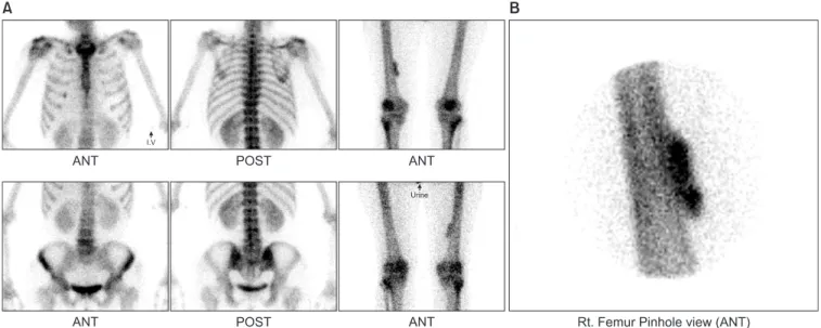
의 연부 조직과 잘 구분되는 선명한 타원형의 방사선 투과 음영이 관찰되었다(Fig. 1). 전신 골 주사 검사상 우측 대퇴골 간부 원위 1/3 부위의 전내측면에서 귓바퀴 모양으로 동위원소 추적자의 흡 수가 증가하는 소견을 보이고 있었으며, Pin-hole 조준기를 통한 영상에서 역시 높은 흡수 증가 소견을 보이고 있었다(Fig. 2). 전산 화 단층 촬영 검사상 우측 원위 대퇴골 내측에서 내측 피질골 내 측에서 형성된 석회화 소견과 함께 이를 둘러싸고 있는 최대 직경 이 8 cm 이상의 타원형의 경계가 명확하고 저신호 강도의 종괴가 관찰되었으며, 석회화 병변은 인접 피질골에 부착되어 있으나 골 수강과 연결되어 있지는 않았다(Fig. 3). 자기 공명 영상 검사에서
접수일 2012년 10월 15일 심사수정일 2012년 11월 20일 게재확정일 2012년 11월 22일
교신저자 노행기
서울시 영등포구 신길1동 451-5, 성애병원 정형외과 TEL 02-840-7233, FAX 02-840-7755
E-mail [email protected]
방골성 지방종은 골막 주위의 지방 조직에서 발생하는 매우 드문 형태의 지방종으로 대퇴골, 요골, 경골 및 비골 등에서 호발하는 것으로 알 려져 있다. 치료는 골 부착부를 포함한 지방 종괴를 절제하고 과골증을 동반시 골막의 제거를 원칙으로 한다. 저자들은 대퇴골 간부 내측에서 발병한 드문 방골성 지방종 1예에 대해서 문헌고찰과 함께 이를 보고하고자 한다.
색인단어: 대퇴골, 방골성 지방종, 과골증
Copyrights © 2012 by The Korean Bone and Joint Tumor Society
“This is an Open Access article distributed under the terms of the Creative Commons Attribution Non-Commercial License (http://creativecommons.org/licenses/by-nc/3.0/) which permits unrestricted non-commercial use, distribution, and reproduction in any medium, provided the original work is properly cited.”
대한골관절종양학회지:제18권 제2호 2012
Figure 1. Plain radiography: anterior view of the distal third of the right femur showing exuberant periosteal reaction in the medial cortical area associated with an increase of adjacent radiolucent soft parts (density of fatty tissue).
105
대퇴골의 과골증을 동반한 방골성 지방종
도 전산화 단층 촬영에서와 유사한 형태의 경계가 명확한 40×20
×80 mm 크기의 종괴가 관찰되었으며, 석회화 부위가 피질골에 맞닿아 있으나 골수강과 연결은 없었으며 병변 부위는 T1 강조 영 상에서 고신호 강도, T2 강조 영상에서 중간 신호 강도, 지방 억제 영상에서는 증강되지 않는 지방 조직과 유사한 음영으로 관찰되 었으며, 중앙 대퇴근과 내측 대퇴근을 압박하는 소견이 관찰되었 으나, 주위 연부조직으로의 침윤은 보이지 않았다(Fig. 4).
외과적 절제술은 과골증 부위 주변의 정상 골막을 포함하여 병
소 부위의 변연 절제술을 시행하였다(Fig. 5). 병리 조직학적 검사 소견상 국소 섬유화 및 과골화 병변을 가진 방골성 지방종으로 확 진하였다(Fig. 6). 수술 후 1년 6개월 추시 관찰 소견상 환자는 정 상 생활을 영위하고 있으며, 방사선 검사상 재발 소견은 보이지 않았다.
고 찰
방골성 지방종에 대한 최초의 기술은 1836년 Seerig에 의해 “Peri- osteal lipoma”였지만, 이후 Power에 의해서 “Parosteal lipoma”로 명명되어 지금까지 사용되고 있다.6) 골성 지방종은 골성 신생물 중 매우 드문 형태로, 일차성 골 종양의 0.1% 미만의 빈도로 발생 한다.6) 그러나 많은 지방종이 진단되지 않고 지내는 경우가 많기 때문에 그 유병율은 더 높을 것으로 추측된다.3) 감별해야 할 질환 으로는 결절종, 골육종, 혈관종, 섬유종 및 섬유 유종 등이 있으며, 이중 방골성 골육종(Parosteal osteosarcoma)과의 감별 진단이 매우 중요하다.1,7)
대개의 경우 무증상이나 때로는 압통을 동반하기도 하며, 종괴 의 압박으로 인해 인접 관절의 운동 혹은 감각신경의 장애를 초래 할 수도 있으나, 혈관 장애는 거의 발생하지 않는다. 본 증례는 압 통 및 운동 신경 압박 증상은 없었으며, 종괴가 약 7개월에 거쳐 서서히 커졌으나 악성 종양에서의 동통, 체중 감소등의 증상은 나 타나지 않았다.
방골성 지방종의 특징적인 방사선 소견으로 장관골에 인접하 여 주변 조직과 잘 구분되며, 보통 타원형 또는 원형의 방사선 투 과성의 지방 종물에 둘러싸인 과골 형성이 특징적이며 인접한 골 의 파괴나 팽창성 병변은 동반하지 않는다. 본 증례에서도 이와 같은 소견이 관찰되었다. 과골 또는 골막 반응등이 나타나는 현상 Figure 2. (A) An ear-shape increased tracer uptake area arising from the periosteum of the right lower medial femoral shaft. (B) The pinhole scan reveals the uptake to consist of speckled high uptake areas.
Figure 3. Computed tomography: Window of soft parts where an intramuscular expansive process can be seen around the femur, with the radiodensity of fat. And in the mass, partial overgrowing of cortical bone and seafood like bone forming calcifications are seen. Note the hyperostosis in the cortical medial area of the bone without medullary invasion and infiltrative appearance.
확한 종괴가 골 피질에 인접하여 위치하나 골수와의 연결성이 없 는 특징 등으로 악성 골종양과의 감별이 가능하기 때문에 아주 중 요한 진단 기법이라고 하겠다.7-9)
방골성 지방종에 대한 치료는 지방 종괴의 절제와 골막을 포함 의 원인은 아직 알려지지 않고 있다.8) 전산화 단층 촬영은 골 피
질과 인접 근육과의 관계를 관찰하는데 유용한 검사이며, 특히 자 기 공명 영상 검사는 골 피질과 인접 근육과의 관계 및 골 성장물 (bony excrescences)과 지방과의 관찰하는데 중요하며, 경계가 명
Figure 4. Magnetic resonance image: Under the intermediate vastus medialis muscle, the tumor mass is seen. The mass is attached to the femur cortex, and partial thickening of cortical bone is seen. And geographic irregular, hyposignal processes in T1 weight image is seen. Bone marrow involvement is not seen. And sharply defined capsule surrounding deep muscle is seen, too.
Figure 5. (A) Intraoperative photograph: yellowish, round shaped tumor attached to femur is seen. (B) The submitted specimen consists of a well demarcated fatty mass. (C) On section, it reveals yellow soft greasy fatty cut surface with some scattered bone spicules. And some crackled bone fragments, up to 1 cm, are also submitted.
107
대퇴골의 과골증을 동반한 방골성 지방종
한 골 성장물의 제거를 원칙으로 하며, 특히 종양의 크기가 크고 주위 신경을 압박해서 신경학적 증상이 유발된 경우 즉각적인 수 술적 치료가 필요하다. 종양 절제 후에는 재발이 거의 없고 악성 변화가 없는 것으로 되어 있다.1,10)
저자들은 대퇴골 간부에서 발생하여 서서히 진행된 과골증을 동반한 대퇴부 방골성 지방종 1예에 대하여 경험하였으며, 매우 드문 사례로 사료되어 문헌 고찰과 함께 보고하고자 하는 바이다.
참고문헌
1. Fleming RJ, Alpert M, Garcia A. A parosteal lipoma. AJR.
1962;87:1075-84.
2. Kim JY, Jung SL, Park YH, Park SH, Kang YK. Parosteal li- poma with hyperostosis. Eur Radiol. 1999;9:1810-2.
3. Canale ST, Beaty JH. Campbell’s operative orthopaedics. 11th ed. Philadelpia: Mosby; 2008. 871.
4. Moon N, Marmor L. Parostel lipoma of the proximal part of the radius. a clinical entity with frequent radial-nerve injury. J Bone Joint Surg Am. 1964;46:608-14.
5. Kong GM, Kim SH, Oh HK. Parosteal lipoma of the proximal radius causing posterior interosseous nerve palsy: a case re- port. J Korea Bone & Joint Tumor Soc. 2009;15:165-70.
6. Power DA. Parosteal lipoma, or congenital fatty tumor, con- nected with periosteum of femur. Trans Pathol Soc, London.
1888;39:270-2.
7. Murphey MD, Johnson DL, Bhatia PS, Neff JR, Rosenthal HG, Walker CW. Parosteal lipoma: MR imaging characteristics.
AJR Am J Roentgenol. 1994;162:105-10.
8. Rhee SK, Kang YK, Park WJ, Chung YG, Lee HJ. Parosteal lipoma with hyperostosis - two cases report. J Kor Bone Joint Tumor Soc. 2001;10:105-9.
9. Rodriguez-Peralto JL, Lopez-Barea F, Gonzalez-Lopez J, La- mas-Lorenzo M. Case report 821: Parosteal ossifying lipoma of femur. Skeletal Radiol. 1994;23:67-9.
10. Bispo Junior RZ, Guedes AV. Parosteal lipoma of the femur with hyperostosis: case report and literature review. Clinics (Sao Paulo). 2007;62:647-52.
Figure 6. Histologic findings of the parosteal lipoma. The lamellated mature bone surrounded by lipoma interposed with fibrous septa is shown. Adiposities were seen without cellular atypia (hematoxylin and eosin, ×200).
Parosteal Lipoma of the Femur with Hyperostosis: A Case Report
Do-Yeon Kim, Ho-Seung Jeon, Seung-Ju Jeon, Haeng-Kee Noh, Seo-Goo Kang, Ji-Ung Song, and Byeong-Moon Park*
Department of Orthopaedic Surgery, Sung-Ae Hospital, Seoul,
*Department of Orthopaedic Surgery, Kwang-Myung Sung-Ae Hospital, Kwangmyung, Korea
Parosteal lipoma is a rare kind of lipoma that occurring adipous tissue around the periosteum. It has been reported most commonly in the femur, the radius, the tibia and the fibular. Treatment consists of resecting the lipomatous tumor with further exeresis of the bone and periosteal excrescence in cases with hyperostosis. The authors report a rare case of parosteal lipoma occurring at the medial portion of the femur shaft with a review of the relevant literatures.
Key words: femur, parosteal lipoma, hyperostosis
Received October 15, 2012 Revised November 20, 2012 Accepted November 22, 2012 Correspondence to: Haeng-Kee Noh
Department of Orthopaedic Surgery, Sung-Ae Hospital, 451-5, Shingil-1-dong, Youngdeungpo-gu, Seoul 150-960, Korea
TEL: +82-2-840-7233 FAX: +82-2-840-7755 E-mail: [email protected]
Copyrights © 2012 by The Korean Bone and Joint Tumor Society
“This is an Open Access article distributed under the terms of the Creative Commons Attribution Non-Commercial License (http://creativecommons.org/licenses/by-nc/3.0/) which permits unrestricted non-commercial use, distribution, and reproduction in any medium, provided the original work is properly cited.”
The Journal of the Korean Bone and Joint Tumor Society Vol. 18, No. 2 (December 2012)