• 검색 결과가 없습니다.

Lipoma with Extraoral Swelling in the Labial Vestibule: Report of a Case

N/A
N/A
Protected

Academic year: 2021

Share "Lipoma with Extraoral Swelling in the Labial Vestibule: Report of a Case"

Copied!
4
0
0

로드 중.... (전체 텍스트 보기)

전체 글

(1)

267

J Korean Assoc Maxillofac Plast Reconstr Surg 2012;34(4):267-270 pISSN:1225-4207 eISSN: 2233-7296

Case Report

RECEIVED May 25, 2012, REVISED June 18, 2012, ACCEPTED July 13, 2012 Correspondence to Ju-Yeon Cho

Department of Dentistry, Dongsan Medical Center, Keimyung University School of Medicine 56, Dalseong-ro, Jung-gu, Daegu 700-712, Korea

Tel: 82-53-250-7805, Fax: 82-53-250-7802, E-mail: [email protected]

CC

This is an open access article distributed under the terms of the Creative Commons Attribution Non-Commercial License (http://creativecommons.org/licenses/

by-nc/3.0) which permits unrestricted non-commercial use, distribution, and reproduction in any medium, provided the original work is properly cited.

Lipoma with Extraoral Swelling in the Labial Vestibule:

Report of a Case

Ju-Yeon Cho, Ki-Young Nam

Department of Dentistry, Dongsan Medical Center, Keimyung University School of Medicine

Abstract

Lipoma is the most common benign neoplasm of the body with rare occurrence in the oral cavity. It represents 1∼4% of benign neoplasms of the mouth, which affect the buccal mucosa, floor of the mouth, tongue and lips. We report a case of lipoma in the labial vestibule with extraoral swelling, which could easily be misdiagnosed as an odontogenic abscess.

Excisional biopsy in this case revealed well-circumscribed masses, surrounded by a thin fibrous capsule and composed of sheets of mature adipocytes, arranged in a "chicken wire" configuration. After a computed tomography scan, excisoinal biopsy was done, and there were no recurrence after 5-month follow-up period.

Key words: Lipoma, Extraoral, Labial, Vestibule

Introduction

Lipomas are the most common soft tissue mesenchymal neoplasms with 15% to 20% of the cases involving the head and neck region and they are relatively rare in oral cavity, representing about 1% to 4% affecting the oral cav- ity[1].

Intraoral lipomas show higher prevalence in males over age 40[2,3]. In most cases the only symptom is a painless, palpable mass. The cheek is the most favored site, followed by the toungue, floor of mouth and buccal sulcus, lip, palate[3].

Soft and yellowish, well-encapsulated mass is the charac- teristics of intraoral lipoma, but painless swelling can be the only symptom if it lies deep inside the tissue. Therefore, differential diagnosis is important when asymptomatic

swelling occurs in oral cavity[4].

Surgical excision is the modality of treatment and malig- nant transformation or recurrence is rare[4,5].

Case Report

A 51-year-old, slim man with anti-hypertensive medi- cation was referred for evaluation of a swelling on his inner mucosa of lower lip.

The patient informed that the swelling was recognized a few days ago, and he was free of symptom and any trauma or operative history on the site.

Clinical examination revealed a soft swelling of similar

color to the surrounding mucosa measuring 3 cm on the

mucolabial fold of right mandibular incisor to premolar

area (Fig. 1) with extraoral swelling. But dental panoramic

(2)

268 Ju-Yeon Cho: Lipoma with Extraoral Swelling in the Labial Vestibule

J Korean Assoc Maxillofac Plast Reconstr Surg Fig. 1. Swelling of labial vestibule.

Fig. 2. Dental panoramic view.

Fig. 3. Transverse view of computed tomography scan.

Fig. 4. Excisional biopsy of lipoma.

Fig. 5. Lipoma with fibrous capsule.

view didn't show any specific problem of the affected site, so odontogenic infection was ruled out (Fig. 2).

Therefore, aspiration was done under the impression

of mucocele, but nothing was detected.

The computed tomography (CT) with enhancement re- vealed decreased radiodensity of fatty mass within the per- ioral muscle between the right mandibular body and lip, suggesting intramuscular lipoma (Fig. 3). Magnetic reso- nance imaging was not checked because the final diagnosis of CT confirmed intramuscular lipoma.

Excisional biopsy was performed under local anesthesia on 1 July 2011 (Fig. 4). Yellowish, globular mass covered by a fibrous capsule measuring 38×20 mm was easily dis- sected en block (Fig. 5).

It was far from buccal fat pad, and closely attached to

the periosteum of alveolar bone around mandibular lateral

incisors to premolar.

(3)

Ju-yeon Cho: Lipoma with Extraoral Swelling in the Labial Vestibule 269

Vol. 34 No. 4, July 2012

Fig. 7. Follow-up after 5 months.

Fig. 6. Microscopic view, mature adipose cells (H&E staining,

×200).

Histological examination revealed thin septa of con- nective tissue separating lobules of adipose tissue formed by mature, uniform adipose cells (Fig. 6).

The postoperative course was favorable, and there has been no recurrence until 5-month follow-up (Fig. 7).

Discussion

Benign lipomas are the most common mesenchymal tu- mors of soft tissue, but are relatively uncommon in the oral and maxillofacial region[6]. They occur in adult pa- tients most often between ages of 40 and 60 years[1,5]

and the most common locaton for lipomas in the max- illofacial region has been reported as the buccal muco- sa[1,7,8] or the parotid region[6].

Specific anatomic site reported for intraoral lipoma in- cluded the 30 cases in parotid region, 29 in buccal mucosa, 21 in lip, 17 in submandibular region, 15 in tongue, 6 in palate, 5 in floor of mouth, and only 2 cases in vestibule of 125 cases[6]. Only 4 patients noted prior trauma of the region and one patient with lipoma in the parotid gland had a Warthin's tumor.

The site predilection is probably related to the availability of adipose tissue, which is high in the buccal mucosa be- cause of the proximity of buccal fat pad and very low in palate[7].

Lipomas are divided into several histologic subtypes such as classic lipoma, fibrolipoma, intramuscular lipoma, minor salivary gland lipoma and spindle cell lipoma, which

have different proliferative activity[1,6,7].

The diffuse swelling-like appearance with extraoral swel- ling, unusual location in addition to sudden onset of the mass according to patient's report gave the impression of odontogenic infection or mucocele instead of lipoma[9].

Dental x-ray ruled out an odontogenic infection of men- tal space, because there was no caries, visible periodontitis nor periapical lesion. Although labial vestibule is the fre- quent site of mucocele, there was nothing aspirated, which ruled out a mucocele also.

Other soft tissue neoplasms were suspected after clinical examination, therefore CT scan was taken to assess the properties. CT scan revealed a decreased radiodensity of fat attenuation which proved a intramuscular lipoma[10]

and it was confirmed after histopathological examination.

The exact cause of lipoma is unknown, but chronic irrita- tion, trauma, hormones, and heredity have been implicated by several investigators[3,11-14], and there appears to be an increased incidence in obese individuals[15]. On the other hand, some authors suppose trauma does not cause the tumor, but contributes to the patient's recognition of pre-existing lipoma, or repeated mild trauma may trigger the proliferation of fatty tissue. But it is noteworthy that a lipoma has never appeared after closure of oro-antral fistula or in midfacial osteotomies where there's a lot of possible trauma with the buccal fat pad. However, lipomas have different metabolism from normal adipose tissue, the higher lipoprotein lipase activity of lipoma compared to normal fat tissue is likely contribute to the growth of tu- mor[16].

Oral lipomas usually do not show recurrence after con-

servative surgical excision, irrespective of the histopatho-

logic variant except intramuscular lipomas, where in-

(4)

270 Ju-Yeon Cho: Lipoma with Extraoral Swelling in the Labial Vestibule

J Korean Assoc Maxillofac Plast Reconstr Surg

complete surgical excision may result in a high recurrence rate[7]. Secondary changes and atrophy should not be con- fused with the malignant histologic features of a lip- osarcoma[6,7].

In this case, intraoral soft tissue mass in the labial vesti- bule, was misdiagnosed as abscess or mucocele at first[9], but proved a classic lipoma after biopsy although the rea- son was unclear.

Intraoral lipoma of labial vestibule is rare in its location and extraoral swelling of mentum area in our case made clinicians easily misdiagnose at first sight. Therefore, we present this confusing case with references.

References

1. Fregnani ER, Pires FR, Falzoni R, Lopes MA, Vargas PA.

Lipomas of the oral cavity: clinical findings, histological classification and proliferative activity of 46 cases. Int J Oral Maxillofac Surg 2003;32:49-53.

2. Perri de Carvalho AC, Martinelli C, Sanches MG. Congenital lipoma in the oral cavity. A case report. Quintessence Int 1987;18:799-802.

3. de Visscher JG. Lipomas and fibrolipomas of the oral cavity.

J Maxillofac Surg 1982;10:177-81.

4. Flaitz CM. Lipoma of the buccal vestibule. Am J Dent 2000;13:52.

5. Chen SY, Fantasia JE, Miller AS. Myxoid lipoma of oral soft tissue. A clinical and ultrastructural study. Oral Surg Oral Med Oral Pathol 1984;57:300-7.

6. Furlong MA, Fanburg-smith JC, Childers EL. Lipoma of the oral and maxillofacial region: Site and subclassification of 125 cases. Oral Surg Oral Med Oral Pathol Oral Radiol Endod 2004;98:441-50.

7. Manor E, Sion-vardy N, Joshua BZ, Bodner L. Oral lipoma:

analysis of 58 new cases and review of the literature. Ann Diagn Pathol 2011;15:257-61.

8. Tripp EA, Hartwell GR. Intraoral lipoma in the buccal vestibule. Gen Dent 1985;33:514-5.

9. Quinn JH. Lipoma of the oral labial vestibule. Oral Surg Oral Med Oral Pathol 1949;2:723-5.

10. Suh DW, Kim JY, Cho SW, et al. A lipoma of the mouth floor : A case report. J Korean Assoc Oral Maxillofac Surg 2009;35:41-4.

11. Hatziotis JC. Lipoma of the oral cavity. Oral Surg Oral Med Oral Pathol 1971;31:511-24.

12. Rapidis AD. Lipoma of the oral cavity. Int J Oral Surg 1982;11:30-5.

13. Macgregor AJ, Dyson DP. Oral lipoma. A review of the lit- erature and report of twelve new cases. Oral Surg Oral Med Oral Pathol 1966;21:770-7.

14. Turner H. Lipoma of the lip; report of a case. Oral Surg Oral Med Oral Pathol 1956;9:376-9.

15. Brooks JK, Scheper MA, Schwartz KG, Nikitakis NG. Oral lipoma: report of three cases. Gen Dent 2008;56:172-6.

16. Solvonuk PF, Taylor GP, Hancock R, Wood WS, Frohlich J.

Correlation of morphologic and biochemical observations in

human lipomas. Lab Invest 1984;51:469-74.

수치

Fig. 5. Lipoma with fibrous capsule.
Fig. 6. Microscopic view, mature adipose cells (H&E staining,

참조

관련 문서

(A) Interestingly, this has often been the case with English animal expressions.. As you may have observed, they make irregular twists and turns in their

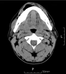
Osteomyelitis in the right distal femur with surrounding

This book contains hundreds of complete, working examples illustrating many common Java programming tasks using the core, enterprise, and foun- dation classes APIs.. Java Examples

The index is calculated with the latest 5-year auction data of 400 selected Classic, Modern, and Contemporary Chinese painting artists from major auction houses..

Time series of vertical cross section of potential vorticity and wind vector calculated by Case 1 along the A-A' line indicated at Fig.. Same

Mean and standard deviation of Labial margin, Lingual margin, and Incisal margin gaps at the three groups (unit : ㎛ )..

The labial surfaces of 24 extracted bovine incisors were used. For the ground enamel, flat enamel surfaces at labial aspect were ground. However, for

2) In between-group comparison before and after elastic band exercise, a significant difference was found in the percentage of body fat, fat-free