Volume 9, Number 2,October, 2006
인간 제대혈 유래 간엽줄기세포의 연골, 골 및 지방세포로의 분화 후 HLA-DR 발현양상의 차이
성균관대학교 의과대학 삼성서울병원 정형외과학교실 백금옥∙성지혜∙문정석∙하철원
= Abstract =
Difference in HLA-DR Expression of Human Umbilical Cord Blood Derived Mesenchymal Stem Cells after Tri-lineage Differentiation
Geum-Ok Baek, M.S., Ji-Hye Sung, M.S., Jeong-Seok Moon, M.D., Chul-Won Ha, M.D.
Department of Orthopedic Surgery, Samsung Medical Center, SungkyunKwan University, School of Medicine, Seoul; Medipost Biomedical Research Institute, Yongin, Korea*
Purpose: The aim of this study was to examine the expression of HLA-DR surface antigen in undifferentiat- ed human umbilical cord blood (hUCB) derived mesenchymal stem cells (MSCs) and after osteogenic, chon- drogenic, and adipogenic differentiation.
Materials and Methods: hUCB-derived MSCs were differentiated into osteogenic, chondrogenic, and adi- pogenic lineages. Differentiation was assessed by immunohistochemical staining and RT-PCR. The expression of HLA-DR was assessed with antihuman HLA-DR antibody in undifferentiated hUCB-derived MSCs and after tri-lineage differentiation.
Results: HLA-DR expression was negative in undifferentiated hUCB-derived MSCs and after osteogenic and adipogenic differentiation. However, HLA-DR surface antigen was expressed after chondrogenic differen- tiation.
Conclusion: The immunologic properties of hUCB-derived MSCs differ from known reports on bone mar- row derived MSCs from the results of this study. Careful immunological survey seems to be needed in case of considering the transplantation of hUCB-derived MSCs differentiated into chondrocytes or cartilaginous tissue.
Key Words: Human umbilical cord blood, Mesenchymal stem cells, Chondrogenic differentiation, Immuno- logic properties, HLA-DR
※ 통신저자: 하 철 원
서울특별시 강남구 일원동 50
성균관의대 삼성서울병원 정형외과학교실
TEL: 02) 3410-0275 FAX: 02) 3410-0084 E-mail: [email protected]
서 론
간엽줄기세포는 다양한 중간엽 조직에서 분리되 고 간엽계에서 유래하는 여러 조직으로 분화할 수 있는 능력을 가진 세포이다1,6,8,11,20)
. 골수 유래 간 엽줄기세포를 이용한 다양한 임상적 시도가 이루 어지고 있으며21,22) 최근에는 제대혈에서 비조혈성 간엽줄기세포가 분리 및 배양되고 이들의 세포 표 면항원의 표현양상이 골수 유래 간엽줄기세포와 비슷하며 여러 간엽 조직으로 분화된다고 보고된 바 있다9,10,17,24,25)
.
간엽줄기세포는 면역 반응을 유발하지 않거나 억제하는 특성이 있는 것으로 알려져 있으며 이러 한 특성은 간엽줄기세포의 다양한 세포 치료 시도 를 가능하게 한다1,14). 이러한 간엽줄기세포의 면 역 특성은 HLA class Ⅱ (DR) 표면항원의 발현 이 음성인 점이 중요한 요인으로 알려져 있는데
5,18,20)
, 골수 유래 간엽줄기세포는 분화 전과 후에 HLA class I은 발현하지만 HLA class Ⅱ 표면 항원은 발현하지 않는다고 알려져 있다1,5,16,18,20)
. 그러나, 제대혈 유래 간엽줄기세포는 분화 전에 HLA class Ⅱ 즉, HLA-DR 표면항원이 발현하 지 않는다고 알려져 있지만17,24,25), 분화된 후의 HLA-DR 발현은 아직 보고된 바가 없다.
본 연구에서는 인간 제대혈로부터 분리한 간엽 줄기세포를 골세포, 연골세포, 지방세포로 분화 유도한 후 HLA-DR 표면항원의 발현여부를 확
인하고자 하였다.
연구 대상 및 방법 1. 3가지 세포 계열로의 분화
인간 제대혈(제태연령:39.2~39.4주)로부터 Ficoll-Hypaque 용액(1.077 g/cm3, Sigma, St. Louis, MO)을 이용한 원심분리(400 g, 35 분) 방법으로 간엽줄기세포를 분리하였고24,25), 본 연구에 사용된 간엽줄기세포군에 대하여 유세포분 석기를 이용해 면역표현형을 분석한 결과 골수 유 래 간엽줄기세포 관련 항원(SH2, SH3, SH4) 양성, 조혈모세포항원(CD34) 음성, 조혈관련항원 (CD45, CD14) 음성, 조직적합항원(HLA-DR) 음성, 내피세포항원(CD31) 음성, 파골세포항원 (CD51/61) 음성임을 확인하였다24,25). 본 연구에서 는 총 15 유니트의 제대혈 중 6 유니트에서 얻은 제대혈 유래 간엽줄기세포군들이 사용되었다.
골, 연골 및 지방세포로의 분화는 기존에 보고된 방법을 따라 시행하였다24,25). 골세포로의 분화는 골 세포 분화배지(Dulbecco’s modified Eagle’s medium (DMEM), 0.1 μM dexamethasone, 10mM β-glycerol phosphate, 0.25 mM ascor- bic acid)에 4주간 분화 배양한 후 분화 후 1주, 2 주, 4주에 각각 Alizarin red S 염색과 Alkaline phosphatase (ALP) 염색을 시행하고, 골세포분
Table 1. Primer sequences and PCR conditions of target genes
Target Genes Primer Sequences Annealing Tm (�C) Size of PCR product
GAPDH 5’-GGGCTGCTTTTAACTCTGGT-3’
60 702 bp
5’-TGGCAGGTTTTTCTAGACGG-3’
Alkaline phosphatase 5’-CTACCAGCTCATGCATAACA-3’
58 251 bp
5’-GACCCAATGTAGTCCACA-3’
Type I Collagen 5’-TGGAGAGTACTGGATTGACC-3’
58 296 bp
5’-AGTGGTAGGTGATGTTCTGG-3’
Osteocalcin 5’-CAGCGAGGTAGTGAAGAGAC-3’
58 155 bp
5’-GATAGGCCTCCTGAAAGC-3’
Osteonectin 5’-ACATGGGTGGACACGG-3’
58 405 bp
5’-CCAACAGCCTAATGTGAA-3’
Osteopontin 5’-AGGAGTTGAATGGTGCATAC-3’
58 223 bp
5’-CTGACTTTGGAAAGTTCCTG-3’
화 관련유전자인 alkaline phosphatase, type I collagen, osteocalcin, osteonectin, osteopon- tin에 관해 역전사 중합효소연쇄반응을 시행하여 골세포로의 분화를 확인하였다. 역전사 중합효소연 쇄반응을 시행한 대상 유전자 및 각각의 anneal- ing 온도와 반응산물의 크기는 Table 1과 같다.
연골세포로의 분화는 연골세포 분화배지(α-mini- mum essential medium (α-MEM), 10 ng/ml TGF- β3, 0.1 μM dexamethasone, 50 μg/ml ascorbic acid, 40 μg/ml proline, 100 μg/ml sodium pyruvate, 50 mg/ml ITS+Premix)로 6주간 펠렛 배양하여 분화 후 2주, 4주, 6주에
A B
D E
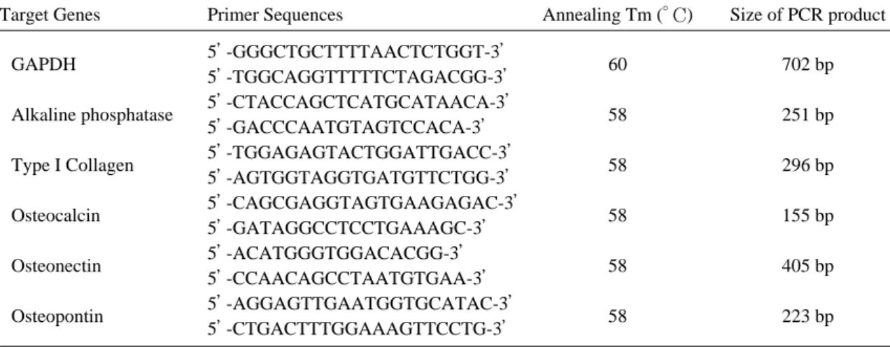
Fig. 1. hUCB-derived MSCs were differentiated into osteogenic, chondrogenic, and adipogenic lineages. Representa- tive results are shown. Osteogenic differentiation was confirmed by the formation of mineralized matrix (A), the expression of alkaline phosphatase (B) and the mRNA expression of osteogenic markers, alkaline phos- phatase (AP), type I collagen (ColI), osteocalcin (OC), osteonectin (ON) and osteopontin (OP) (C). Chondro- genic differentiation was evidenced by safranin O staining of cartilage matrix (D) and immunohistochemical staining for type Ⅱ collagen (E). Adipogenic differentiation was evidenced by the presence of intracellular lipid vacuoles using oil red O staining (F). Representative results are shown. A, B:×100, scale bar=500 ㎛;
D-F:×200, scale bar=200 μm.
C
F
각각 Safranin O 염색과 type Ⅱ collagen에 대한 면역염색을 시행하여 연골세포로의 분화를 확인하였다. Type Ⅱ collagen 면역 염색은 mouse antihuman type Ⅱ collagen (Onco- gene, Cambridge, MA)을 1:50으로 희석하여 4�C에서 12시간 반응시켰으며, DAKO Envi- sionTM+System, HRP (DAB) Kit (Dako Cytomation, Carpinteria, CA)를 이용하여 면 역 염색을 수행하였다.
지방세포로의 분화는 지방세포 분화배지 (DMEM, 1 μM dexamethasone, 0.5 mM IBMX, 0.2 mM indomethacin, 0.01 mg/ml h-insulin)에 4주간 분화 배양 후 Oil red O 염 색을 시행하여 지방세포로의 분화를 확인하였다.
2. HLA-DR 면역조직화학 염색
HLA-DR 표면항원의 발현 여부를 관찰하기 위
A B
D
E
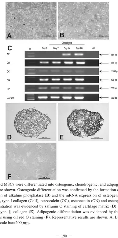
Fig. 2. HLA-DR expression was assessed before and after tri-lineage differentiation of hUCB-derived MSC popula- tion. HLA-DR immunostaining was performed on undifferentiated cells (A) and after osteogenic (B), adi- pogenic (C), and chondrogenic (D) differentiation. HLA-DR expression was positive in the chondrogenic dif- ferentiated cells. The human HLA-DR positive cells are shown in cell surface staining. Porcine bone was used as negative control (E), and human bone and human blood were used as positive control (F, G). Negative con- trol was only stained with hematoxylin as a nuclear counterstain. Representative results are shown. Original magnification, × 200.
C
F
G
해 분화하기 전과 골세포, 지방세포로 분화 유도한 후 4주, 연골세포로 분화 유도한 후 6주에 HLA- DR에 대한 면역염색을 시행하였다. 배양접시에서 배양한 세포는 4% paraformaldehyde로 고정시 켜 준비하고 펠렛은 4% paraformaldehyde로 고정시킨 후 7 μm 두께로 박리하여 슬라이드 절 편을 준비하였다. Mouse antihuman HLA class Ⅱ (DR)(Chemicon, Hampshire, Unit- ed Kingdom)을 1:100으로 희석하여 4℃에서 12 시간 반응시켰으며, DAKO EnvisionTM+System, HRP (DAB) Kit (DakoCytomation)를 이용하 여 면역 염색을 수행하였고, 대조염색으로 hema- toxylin 염색을 시행하였다. HLA-DR 면역염색 에 대한 음성대조시편은 돼지 골 조직을, 양성대조 시편은 사람 골 조직과 사람 혈액도말표본을 사용 하여 동일한 과정을 거쳐 면역 염색을 수행하였다.
결 과
1. 제대혈 유래 간엽줄기세포의 3가지 세포 계 열로의 분화 확인
제대혈로부터 분리한 간엽줄기세포를 골형성 배 지로 4주간 분화 유도한 결과, ALP의 발현 및 명확한 석회화(mineralized) 물질의 형성이 Alizarin red S 염색으로 확인되었고 골세포분화 관련유전자의 발현이 관찰됨으로써 골세포로의 분 화가 확인되었다(Fig. 1A-C). 연골형성 배지로 6주간 분화 유도한 후 연골 기질 염색인 Safranin O 염색을 수행한 결과 Safranin O 염색의 결과가 양성으로 나타나 연골기질의 형성 을 확인할 수 있었고 연골소강(lacunae)의 형성 을 관찰할 수 있었다. 관절 연골 특이 교원질인 type Ⅱ collagen의 면역염색 결과도 양성을 보 여 연골세포로의 분화가 확인되었다(Fig. 1D, E). 지방세포형성 배지로 4주간 분화 유도한 후 Oil red O 염색을 수행한 결과 세포내 지방체 염 색의 양성 소견이 보여 지방세포로의 분화가 확인 되었다(Fig. 1F). 실험에 사용된 6개의 모든 간 엽줄기세포군에서 골, 연골 및 지방세포로의 분화 를 확인할 수 있었다.
2. 제대혈 유래 간엽줄기세포의 HLA-DR 표면 항원 발현여부
제대혈 유래 간엽줄기세포의 분화전 HLA-DR 표면항원의 발현은 음성으로 확인되었다. 골세포 및 지방세포로 분화한 경우 HLA-DR 표면항원 의 발현이 계속 음성이었으나(Fig. 2A, B, C), 연골세포로 분화한 경우 펠렛의 단면에서 세포의 표면이 발색시약에 의해 발색되어 HLA-DR의 발현이 양성임을 확인하였다(Fig. 2D). 실험에 사용된 6개의 모든 간엽줄기세포군에서 동일한 염 색 양상을 확인할 수 있었다.
고 찰
간엽줄기세포는 다분화능(multipotentiality) 과 역분화과정을 통해 본래의 세포특성과 다른 종 류의 세포로 분화할 수 있는 가소성(plasticity) 을 가지고 있다고 알려져 있으며12,15,20,26)
, 손상받 은 장기로 가동화(mobilization)할 수 있어 손상 부위를 복구시킬 수 있다는 점 등이 알려지면서 간엽줄기세포를 통한 다양한 세포 치료 가능성이 제시되고 있다1,4,23). 여러 동물 실험에서 간엽줄기 세포가 손상된 조직을 복구하며 골, 연골, 근육 또는 심근 등의 재생에 이용될 수 있음이 보고된 바 있다2,3,7,13).
간엽줄기세포의 면역억제의 특성은 이식편대 숙 주반응(graft-versus-host disease)의 발생을 현저히 떨어뜨려 조직적합성 항원이 일치하지 않 는 간엽줄기세포의 다양한 세포 치료 시도를 가능 하게 한다1,14). 간엽줄기세포의 면역 특성은 HLA class Ⅱ (DR) 표면항원의 발현이 음성인 점과 B7-1, B7-2, CD40, CD40L과 같은 co-stimu- latory molecule의 발현이 낮은 점이 중요한 요 인으로 알려져 있다5,18,20).
서론에서도 언급한 바와 같이 제대혈 유래 간엽 줄기세포는 HLA class I과 HLA class Ⅱ의 발 현이 모두 음성이라고 보고되어있다17,24,25). 그러나 제대혈 유래 간엽줄기세포군을 골, 연골 및 지방 세포로 분화 후 이러한 표면항원의 발현 여부에 관한 연구는 저자들이 검색한 바로는 현재까지 보 고된 바 었다.
본 연구에서는 인간 제대혈로부터 유래한 간엽 줄기세포를 골세포, 연골세포, 지방세포로 분화시 킨 후 HLA-DR 표면항원의 발현 양상을 확인한 결과 골수 유래 간엽줄기세포와 다른 결과를 확인 할 수 있었다. 제대혈 유래 간엽줄기세포를 분화 시키기 전 실시한 HLA-DR 면역염색의 결과는 음성이었고, 이는 골수 유래 간엽줄기세포 연구에 서 보고된 바와 동일하였다17,24). 골세포와 지방세 포로 분화한 제대혈 유래 간엽줄기세포는 HLA- DR의 발현이 음성이었으나, 연골세포로 분화한 제대혈 유래 간엽줄기세포는 양성으로 확인되었 다. 이는 기존의 골수 유래 간엽줄기세포의 분화 전과 분화 후의 HLA-DR 표면 항원의 발현에 관 해 보고된 바와 다른 결과인 바, 인간 제대혈 유 래 간엽줄기세포는 골수 유래 간엽줄기세포와 면 역학적 특성이 다를 수도 있음을 시사한다.
최근 간엽줄기세포를 이용한 조직 재생 및 질환 치료의 활발한 연구가 진행되고 있으나, 간엽줄기 세포가 세포치료제로 이용되기 위해서는 분화세포 의 안전성 및 면역반응에 관한 정밀한 추적이 수 반되어야 한다. 본 연구의 결과로, 인간 제대혈 유래 간엽줄기세포를 연골세포나 조직으로 분화시 켜 이식하는 경우 그동안 골수 유래 줄기세포의 연구 결과에서 알려진 바와 다른 면역 특성을 나 타낼 수 있다고 생각된다. 제대혈 유래 간엽줄기 세포를 이용한 임상적 응용에 세심한 주의가 필요 하고, 제대혈 유래 간엽줄기세포의 면역학적 특성 에 대한 추가 연구가 필요할 것으로 사료된다.
결 론
인간 제대혈 유래 간엽줄기세포는 연골 세포로 분화한 후에 HLA-DR 표면항원의 발현이 양성 으로 확인되었으므로 골수 유래 간엽줄기세포에서 알려진 바와 다른 면역 특성을 나타낼 수 있다고 생각된다. 인간 제대혈 유래 간엽줄기세포를 연골 세포나 조직으로 분화시켜 이식하는 경우 그 면역 학적 특성에 대한 충분한 검토가 필요할 것으로 사료된다.
REFERENCES
01) Barry FP and Murphy JM: Mesenchymal stem cells: clinical applications and biological charac- terization. Int J Biochem Cell Biol, 36: 568-584, 2004.
02) Bruder SP, Kraus KH, Goldberg VM and Kadiyala S: The effect of implants loaded with autologous mesenchymal stem cells on the heal- ing of canine segmental bone defects. J Bone Joint Surg, 80-A: 985-996, 1998.
03) Bruder SP, Kurth AA, Shea M, Hayes WC, Jaiswal N and Kadiyala S: Bone regeneration by implantation of purified, culture-expanded human mesenchymal stem cells. J Orthop Res, 16: 155-162, 1998.
04) Caplan AI and Bruder SP: Mesenchymal stem cells: building blocks for molecular medicine in the 21st century. Trends Mol Med, 7: 259-264, 2001.
05) Deans RJ and Moseley AM: Mesenchymal stem cells: biology and potential clinical uses.
Exp Hematol, 28: 875-884, 2000.
06) Deasy BM, Jankowski RJ and Huard J: Mus- cle-derived stem cells: characterization and potential for cell-mediated therapy. Blood Cells Mol Dis, 27: 924-933, 2001.
07) De Bari C, Dell’’Accio F, Vandenabeele F, Vermeesch J, Raymackers JM and Luyten FP: Skeletal muscle repair by adult human mes- enchymal stem cells from synovial membrane. J Cell Biol, 160: 909-918, 2003.
08) D’’Ippolito G, Schiller PC, Ricordi C, Roos BA and Howard GA: Age-related osteogenic potential of mesenchymal stromal stem cells from human vertebral bone marrow. J Bone Miner Res, 14: 1115-1122, 1999.
09) Erices A, Conget P and Minguell JJ: Mes- enchymal progenitor cells in human umbilical cord blood. Br J Hematol, 109: 235-242, 2000.
10) Goodwin HS, Bicknese AR, Chien SN, Boguchi BD, Quinn CO and Wall DA: Multi-
lineage differentiation activity by cells isolated from umbilical cord blood: Expression of bone, fat, and neural markers. Biol Blood Marrow Trans, 7: 581-588, 2001.
11) Gronthos S, Franklin DM, Leddy HA, Robey PG, Storms RW and Gimble JM: Surface pro- tein characterization of human adipose tissue- derived stromal cells. J Cell Physiol, 189: 54-63, 2001.
12) Grove JE, Bruscia E and Krause DS: Plasticity of bone marrow-derived stem cells. Stem Cells, 22: 487-500, 2004.
13) Horwitz EM, Prockop DJ, Fitzpatrick LA, et al: Transplantability and therapeutic effects of bone marrow-derived mesenchymal cells in chil- dren with osteogenesis imperfecta. Nat Med, 5:
309-313, 1999.
14) Koc ON, Day J, Nieder M, Gerson SL, Lazarus HM and Krivit W: Allogeneic mes- enchymal stem cell infusion for treatment of metachromatic leukodystrophy (MLD) and Hurler syndrome (MPS-IH). Bone Marrow Transplant, 30: 215-222, 2002.
15) Krause DS, Theise ND, Collector MI, et al:
Multi-organ, multi-lineage engraftment by a sin- gle bone marrow-derived stem cell. Cell, 105:
369-377, 2001.
16) Le Blanc K, Tammik C, Rosendahl K, Zetter- berg E and Ringde´n O: HLA expression and immunologic properties of differentiated and undifferentiated mesenchymal stem cells. Exp Hematol, 31: 890-896, 2003.
17) Lee OK, Kuo TK, Chen WM, Lee KD, Hsieh SL and Chen TH: Isolation of multi potent mes- enchymal stem cells from umbilical cord blood.
Blood, 103: 1669-1675, 2004.
18) Majumdar MK, Keane-Moore M, Buyaner D, et al: Characterization and functionality of cell surface molecules on human mesenchymal stem cells. J Biomed Sci, 10: 228-241, 2003.
19) Minguell JJ, Erices A and Conget P: Mes- enchymal stem cell. Exp Biol Med, 226: 507-520, 2001.
20) Pittenger MF, Mackay AM, Beck SC, et al:
Multilineage potential of adult human mesenchy- mal stem cells. Science, 284: 143-147, 1999.
21) Ponticiello MS, Schinagl RM, Kadiyala S and Barry FP: Gelatin-based resorbable sponge as a carrier matrix for human mesenchymal stem cells in cartilage regeneration therapy. J Biomed Mater Res, 52: 246-255, 2000.
22) Quarto R, Mastrogiacomo M, Cancedda R, et al: Repair of large bone defects with the use of autologous bone marrow stromal cells. N Engl J Med, 344: 385-386, 2001.
23) Weissman IL: Translating stem and progenitor cell biology to the clinic: barriers and opportuni- ties. Science, 287: 1442-1446, 2000.
24) Yang SE, Ha CW, Jung MH, et al: Mesenchy- mal stem/progenitor cells developed in cultures from UC blood. Cytotherapy, 6: 476-486, 2004.
25) Yang SE, Yang YS and Ha CW: Isolation, characterization and tri-lineage differentiation of mesenchymal stem cells from human umbilical cord blood. J Korean Orthop Assoc, 39: 537-545, 2004.
26) Zipori D: Mesenchymal stem cells: harnessing cell plasticity to tissue and organ repair. Blood Cells Mol Dis, 33: 211-215, 2004.