https://doi.org/10.4174/astr.2018.94.3.129 Annals of Surgical Treatment and Research
Early disseminated recurrence after liver resection in solitary hepatocellular carcinoma
Jong Man Kim1, Nam-Joon Yi2, Choon Hyuck David Kwon1, Kwang-Woong Lee2, Kyung-Suk Suh2, Jae-Won Joh1
1Department of Surgery, Samsung Medical Center, Sungkyunkwan University School of Medicine, Seoul, Korea
2Division of HBP Surgery, Department of Surgery, Seoul National University College of Medicine, Seoul, Korea
INTRODUCTION
Liver resection is a curative treatment method for solitary liver cancer [1]. However, only 9%–27% of patients with hepato- cellular carcinoma (HCC) are eligible for surgical resection [2]. Although overall survival after resection of HCC has improved in recent years because improved surgical skills and postoperative management have led to a marked reduction in postoperative mortality rates. However, the recurrence of HCC after liver resection is a major surgical limitation because the recurrence rate remains high.
The pattern of recurrence after curative surgery for HCC is
vari able. Many hepatectomy patients developed intrahepatic re currence [3,4]. Intrahepatic recurrence of HCC can be divided into cases with solitary or a few nodules and those with multi- nodular recurrence.
Early recurrence, defined as recurrence within 1 year of liver resection, is one of the most important factors affecting the prognosis and survival of patients with HCC [5-7]. Tumor size, intrahepatic metastasis, microvascular invasion, serum α-FP, serum proteins induced by vitamin K antagonist-II, C-reactive protein, and serum alkaline phosphatase have been reported as risk factors associated with early recurrence [4,8-10]. Early recurrence of HCC in patients who undergo resection is still a Purpose: Although few hepatectomy patients develop unexpected early diffuse and multinodular recurrence in the rem- nant liver, the prognosis in such cases is often dismal. The aim of this study was to evaluate the risk factors of early disse- minated multinodular hepatocellular carcinoma (HCC) recurrence within 3 months after liver resection for solitary HCC.
Methods: Eighty-four patients who were diagnosed with recurrent HCC within 3 months after hepatectomy for solitary HCC were retrospectively reviewed. Disseminated HCC recurrence was defined as more than 10 tumors in both lobes and total tumor size >10 cm.
Results: Preoperative α-FP level, incidence of poor tumor grade, and presence of portal vein tumor thrombosis were higher in the patients with disseminated HCC recurrence than in those without disseminated HCC recurrence (P < 0.05).
Multivariate analysis showed that α-FP >1,000 ng/dL was a predisposing factor of disseminated HCC recurrence within 3 months after liver resection. The overall survival rate for patients without disseminated HCC recurrence was higher than that for patients with disseminated HCC recurrence (P < 0.001).
Conclusion: Early disseminated multinodular HCC recurrence in hepatectomy patients was associated with preoperative α-FP >1,000 ng/dL. Such patients should be frequently evaluated for the early detection of recurrent HCC for early intervention.
[Ann Surg Treat Res 2018;94(3):129-134]
Key Words: Hepatocellular carcinoma, Hepatectomy, Recurrence, Tumor biomarker, Survival
Reviewed January February March April May June July August September October November December
Received May 12, 2017, Revised July 7, 2017, Accepted July 25, 2017 Corresponding Author: Choon Hyuck David Kwon
Department of Surgery, Samsung Medical Center, Sungkyunkwan University School of Medicine, 81 Irwon-ro, Gangnam-gu, Seoul 06351, Korea Tel: +82-2-3410-1281, Fax: +82-2-3410-0040
E-mail: [email protected]
ORCID code: https://orcid.org/0000-0002-1082-3321
Copyright ⓒ 2018, the Korean Surgical Society
cc Annals of Surgical Treatment and Research is an Open Access Journal. All articles are distributed under the terms of the Creative Commons Attribution Non- Commercial License (http://creativecommons.org/licenses/by-nc/4.0/) which permits unrestricted non-commercial use, distribution, and reproduction in any medium, provided the original work is properly cited.
controversial issue [11].
Few patients who undergo liver resection develop unexpected early diffuse and multinodular recurrence in the remnant liver at first visit within 3 months after hepatic resection. The prognosis is often dismal if it occurs. There might be no benefit of hepatic resection if the patient dies due to early disseminated multinodular HCC recurrence shortly after the surgery. There- fore, the risk factors of early disseminated multinodular HCC recurrence should be evaluated.
In this retrospective study, we evaluated and identified risk fac tors of early disseminated multinodular HCC recurrence within 3 months after liver resection in patients with recurrent HCC by analysis of preoperative and pathological factors.
METHODS
Patients
This study included patients who underwent surgical resec- tion of solitary HCC in Samsung Medical Center or Seoul National University Hospital based on preoperative radiological images between January 2005 and December 2011. HCC was confirmed from pathologic results after hepatectomy. We included patients with recurrent HCC patients after liver resection in 2 centers (n = 661). This study was approved by the Samsung Medical Center Institutional Review Board (IRB) (approval number: SMC 2013-05-013) and Seoul National University Hospital IRB (approval number: H-1303-061-474).
Informed consent was exempted from the each IRB.
Inclusion criteria included primary curative resection, Child- Pugh class A, and HCC recurrence within 3 months after sur gery. Exclusion criteria were as follows: mixed HCC and cholan giocarcinoma on pathology; age <18 years; non-Korean ethnicity; hepatic resection in recurrent HCC patients; Child- Pugh class B or C; multiple preoperative tumors based on preoperative radiologic images; ruptured HCC; R1 resection;
extrahepatic metastasis or invasion; concurrent malignancy in other organs; history of malignancy other than HCC; pre vious locoregional therapies such as hepatectomy, radiation, trans- arterial chemoembolization (TACE), radiofrequency ablation (RFA), or percutaneous ethanol injection (PEI); intraoperative RFA; fibrolamellar HCC; or loss to follow-up after hepatectomy.
Demographic, preoperative laboratory findings, and patho logic data collected from electronic medical records were retro spec- tively reviewed. None of the patients in either group received postoperative adjuvant therapy before recurrence.
Surgery and pathology
Surgical and pathological procedures performed after liver resection have been described previously [3,4]. Liver function was evaluated using the Child-Pugh classification system. Adequate mobilization was achieved according to the part of the liver to
be resected. Selective clamping of the portal vein and hepatic artery was performed when feasible. One center routinely used Pringle maneuver during liver resection, but the other center used Pringle maneuver whenever necessary. If operators thought much bleeding during hepatectomy, they used Pringle maneuver in those cases. Parenchymal transection was performed using a Cavitron Ultrasonic Surgical Aspirator or Kelly-clysis under low central venous pressure. Major hepatectomy was defined as the resection of ≥3 segments and minor hepatectomy was defined as the resection of <3 segments. Postoperative histological assess ment and reporting included maximal tumor size, tumor grade, hemorrhage, necrosis, portal vein tumor thrombosis (PVTT), microvascular invasion, and intrahepatic metastasis. The histologic grade of HCC was assigned according to the Edmonson- Steiner system as well as differentiated (grade I), moderately differentiated (grade II), or poorly differentiated (grades III, IV) [12].
Surveillance after surgical resection
The procedures used for surveillance after liver resection have been described previously [8]. All patients were followed postoperatively one month. After then, all patients were followed every 2 or 3 months. Follow-up parameters were physical examination, serum α-FP, liver function tests, and chest x-rays. Abdominal CT was performed every 3 months or when recurrence was suspected. MRI and/or PET scans were performed if CT did not show definitive evidence of recurrence and tumor markers elevated. Disseminated HCC recurrence was defined as tumor number > 10 in both lobes and total tumor size >10 cm. Patients with intrahepatic recurrences were treated with RFA, TACE, liver resection, liver transplantation, or radiation according to functional liver reserve and the pattern of recurrence. Follow-up time was the length of time from surgery to the final follow-up or death.
Statistical analysis
All statistical analyses were performed using SPSS ver.
21.0 (SPSS, Inc., Chicago, IL, USA). Continuous variables are de scribed as median with range. Categorical variables are ex- pressed as number and percentage of subjects. Fisher exact test was conducted to evaluate differences in the frequencies of categorical variables between the groups. Mann-Whitney U analysis was conducted to evaluate the differences in continu- ous variables between the 2 groups. Binary logistic regression analysis was used to predict multinodular disseminated HCC recurrence within the first 3 months after liver resection. All tests were 2-sided, and statistical significance was defined as P <
0.05.
RESULTS
Baseline characteristics
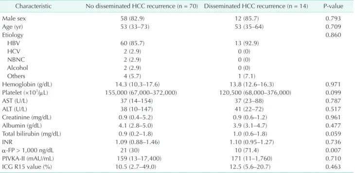
Eighty-four patients developed HCC recurrence within 3 months after liver resection. The baseline characteristics are summarized in Table 1. Disseminated HCC recurrence occurred in 14 patients (16.7%). There were no statistically significant differences in the sex, age, etiology for HCC, hemoglobin, platelet count, liver function tests, internal normalized ratio, indocyanine green value, or preoperative tumor size between the 2 groups. The preoperative median α-FP level was 6,295 ng/
dL (range, 6.9–356,800 ng/dL) in the patients with disseminated HCC recurrence and 164 ng/dL (range, 1.9–157,310 ng/dL) in the patients without disseminated HCC recurrence (P = 0.013).
Perioperative and pathologic characteristics
All patients had solitary nodule of HCC. The median tumor size was 5.5 cm (range, 2.2–17.0 cm) in the patients with disseminated HCC recurrence and 4.2 cm (range, 0.8–15.0 cm) in the patients without disseminated HCC recurrence (P = 0.140). The proportion of tumor grades 3 and 4 in the patients with disseminated HCC recurrence was 92.9% (n = 13), compared with 57.1% (n = 40) in the patients without disseminated HCC recurrence (P = 0.014). The incidence of PVTT was higher in the patients with disseminated HCC recurrence than in those without disseminated HCC recurrence (42.9% vs. 7.1%, P = 0.002). There were no statistically significant differences in extent of operation, tumor size, hemorrhage,
necrosis, microvascular invasion, intrahepatic metastasis, or hospitalization between the 2 groups (Table 2).
HCC recurrence and survival
The median follow-up duration was 9 months (range, 4–24 months) in the patients with disseminated HCC recurrence and 26 months (range, 4–101 months) in the patients without disseminated HCC recurrence (P < 0.001). At time of diagnosis of recurrent HCC, the median α-FP level was 9.385 ng/dL (range,
Table 1. Baseline characteristics
Characteristic No disseminated HCC recurrence (n = 70) Disseminated HCC recurrence (n = 14) Pvalue
Male sex 58 (82.9) 12 (85.7) 0.793
Age (yr) 53 (33–73) 53 (35–64) 0.709
Etiology 0.860
HBV 60 (85.7) 13 (92.9)
HCV 2 (2.9) 0 (0)
NBNC 2 (2.9) 0 (0)
Alcohol 2 (2.9) 0 (0)
Others 4 (5.7) 1 (7.1)
Hemoglobin (g/dL) 14.3 (10.3–17.6) 13.8 (12.6–16.3) 0.971
Platelet (×103/µL) 155,000 (67,000–372,000) 120,500 (68,000–376,000) 0.099
AST (U/L) 37 (14–154) 37 (23–88) 0.787
ALT (U/L) 38 (10–147) 41 (22–72) 0.517
Creatinine (mg/dL) 0.9 (0.4–5.2) 0.9 (0.6–1.2) 0.961
Albumin (g/dL) 4.1 (2.8–5.0) 3.9 (3.1–4.7) 0.477
Total bilirubin (mg/dL) 0.9 (0.2–1.8) 1.0 (0.6–1.8) 0.059
INR 1.09 (0.88–1.46) 1.10 (0.95–1.27) 0.736
αFP > 1,000 ng/dL 21 (30) 10 (71.4) 0.007
PIVKAII (mAU/mL) 159 (13–17,400) 171 (11–1,760) 0.710
ICG R15 value (%) 10.5 (2.7–49.0) 12.5 (5.6–20.7) 0.463
Values are presented as number (%) or median (range).
NBNC, non B non C; INR, internal normalized ratio; PIVKAII, proteins induced by vitamin K antagonistII; ICG, indocyanine green.
Table 2. Perioperative and pathologic characteristics Variable No disseminated
HCC recurrence (n = 70)
Disseminated HCC recurrence
(n = 14) Pvalue Extent of operation
(major) 18 (25.7) 6 (42.9) 0.209
Tumor size (cm) 4.2 (0.8–15.0) 5.5 (2.2–17.0) 0.140 Grade (3 and 4) 40 (57.1) 13 (92.9) 0.014 Pathologic
hemorrhage 28 (40.0) 7 (50.0) 0.151
Pathologic necrosis 54 (77.1) 11 (78.6) 0.907 Microvascular
invasion 45 (64.3) 11 (78.6) 0.367
Macrovascular
invasion 5 (7.1) 6 (42.9) 0.002
Intrahepatic
metastasis 10 (17.9) 1 (10.0) 0.539
Hospitalization (day) 10 (7–79) 12 (8–38) 0.327 Values are presented as number (%) or median (range).
5.8–427,000 ng/dL) in the patients with disseminated HCC recurrence and 171 ng/dL (range, 1.7–200,000 ng/dL) in the patients without disseminated HCC recurrence (P = 0.002).
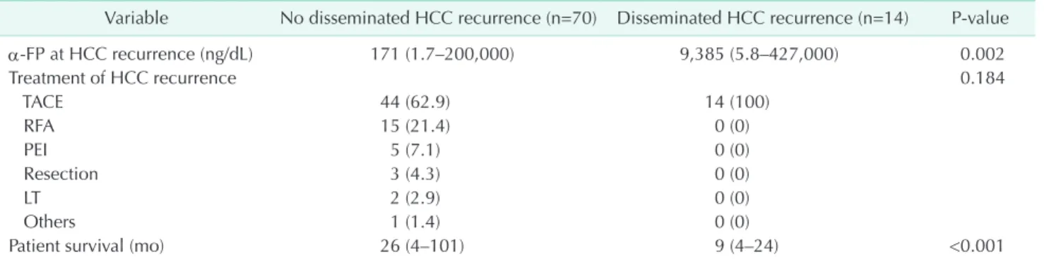
Multivariate analysis showed that α-FP >1,000 ng/dL (odds ratio, 5.333; 95% confidence interval, 1.095–25.985; P = 0.038) was a predisposing factor of disseminated HCC recurrence within 3 months after liver resection. Treatments for recurrent HCC patients are summarized in Table 3.
The overall survival rate at 1-year was 38.9% in the patients with disseminated HCC recurrence and 84.8% in the patients without disseminated HCC recurrence. The overall survival for patients without disseminated HCC recurrence was higher than that for patients with disseminated HCC recurrence (P < 0.001) (Fig. 1).
DISCUSSION
The present study showed that the incidence of rapid and diffuse recurrent tumors after hepatectomy is associated with poor prognosis. Within the observation period, the mortality rates of patients with disseminated HCC recurrences (within 3 months of liver resection) were higher than those of patients without disseminated HCC recurrences. Among the 70 patients with early nondisseminated intrahepatic recurrences within 3 months after hepatectomy, 47 died within the observation period. In contrast, 11 patients of the 14 patients with early disseminated intrahepatic recurrence were dead within 2 years after hepatic resection as a result of advanced HCC.
In the remnant liver after hepatectomy, tumor recurrence is recognized as intrahepatic metastasis caused by dissemination of cells in the portal vein or metachronous multicentric hepatocarcinogenesis [3-5]. Patients with solitary or a few recurring nodules can be treated using several different mo- dalities, including repeat liver resection, RFA, or liver trans- plantation, and have relatively high survival rates [13,14].
However, among these diverse patterns of recurrence, dis- seminated multinodular intrahepatic recurrence is thought
to be fatal not only because there is not amenable to effective treat ment strategy, but also because their recurrent tumors are usually aggressive [2,15]. These recurrence patterns inevitably mean a short remaining survival time [2,6,15].
If surgeons experience early disseminated multinodular HCC recurrence after liver resection in patients with solitary HCC, they can be disappointed and even embarrassed. However, the risk factors for early diffuse and multinodular recurrence within 3 months after liver resection are currently unclear.
Risk factors for early disseminated multinodular intrahepatic recurrence after surgical resection may be reliable prognostic factors of long-term survival. Therefore, understanding the patterns and risk factors of early disseminated multinodular intrahepatic recurrence of HCC after hepatectomy will help clinicians to identify high-risk patients during the preoperative evaluation. The present study showed that α-FP >1,000 ng/dL was a predisposing factor of disseminated multinodular HCC recurrence within 3 months after hepatic resection in patients Table 3. Hepatocellular carcinoma (HCC) recurrence
Variable No disseminated HCC recurrence (n=70) Disseminated HCC recurrence (n=14) Pvalue
αFP at HCC recurrence (ng/dL) 171 (1.7–200,000) 9,385 (5.8–427,000) 0.002
Treatment of HCC recurrence 0.184
TACE 44 (62.9) 14 (100)
RFA 15 (21.4) 0 (0)
PEI 5 (7.1) 0 (0)
Resection 3 (4.3) 0 (0)
LT 2 (2.9) 0 (0)
Others 1 (1.4) 0 (0)
Patient survival (mo) 26 (4–101) 9 (4–24) <0.001
TACE, transarterial chemoembolization; RFA, radiofrequency ablation; PEI, percutaneous ethanol injection; LT, liver transplantation.
Cumulativeoverallsurvivalrate(%)
1 10
Time from hepatectomy to death (yr) 0
1.0
0.8
0.6
0.4
0.2
9 8 7 6 5 4 3 2
Patients without disseminated HCC recurrence Patients with disseminated HCC recurrence
Patients without disseminated HCC recurrence-censored Patients with disseminated HCC recurrence-censored
P < 0.001
Fig. 1. Overall survival after liver resection. HCC, hepato cel
lular carcinoma.
with recurrent HCC.
When recurrent HCC appears immediately after liver resection for HCC, it is generally thought that metastases of occult cancer cells that were not detected either grossly or by imaging techniques were already presented at the time of surgery. If these occult micrometastases could be detected in the preoperative evaluation stage, surgical resection of the main tumor may be contraindicated. However, current clinical technology does not allow preoperative determination of micrometastatic lesions; therefore, the best available approach at the present time is the determination of risk factors of immediate postoperative fatal recurrence.
Previous data have shown that high preoperative α-FP level is a significant risk factor of early postoperative recurrence [5,6]. The present study also revealed similar result that serum α-FP level > 1,000 ng/dL is significant a risk factor of early disseminated multinodular HCC recurrence after hepatic resection in patients with solitary HCC. Tumor cells may be probably already present in the remnant liver, together with microscopic portal invasion, before liver resection in patients with high α-FP level [7]. Another study has reported that the liver regeneration process after liver resection can enhance the growth of occult metastases, which rapidly develop into overt metastasis [16]. Therefore, efforts to predict these micrometastases and systemic dissemination of cancer cells should not be overlooked. Present study revealed that poor differentiated grade and microvascular invasion were closely associated with early multinodular disseminated recurrence by univariate analysis. Another study reported that those factors were considered as predictive factors for recurrence beyond Milan criteria after hepatic resection [17]. However, the present study did not demonstrate that vascular invasion was an independent risk factor related to early disseminated multinodular HCC recurrence by multivariate analysis.
Hepatectomy patients with risk factors such as increased
preoperative α-FP levels, microvascular invasion, and poor differentiated grade had a dismal prognosis when early multinodular disseminated recurrence was developed after liver resection. Therefore, a possible strategy for improving patient survival may be early radiological evaluations within 2 weeks after surgery and early intervention.
This study has several limitations. First, the study is retrospective. Second, selection bias can occur due to inclusion of hepatectomy patients with preoperative radiologically solitary tumor and well-preserved liver function. Third, present study included only recurrent HCC patients after liver resection and excluded patients without HCC recurrence. Therefore, reports from other centers are also necessary to allow generalization of our findings.
In conclusion, early disseminated multinodular HCC recur- rence was associated with poor survival after liver resec tion.
Pa tients with α-FP >1,000 ng/dL in the preoperative period should be frequently evaluated for the early detection of recurrent HCC after hepatic resection. These findings indicate that the surgeon should explain the possibility of early disseminated multinodular HCC recurrence after surgery in pa tients with solitary HCC who have high serum α-FP level.
If there is a possibility of disseminated HCC recurrence after surgery, sequential locoregional therapies first and surgery may be helpful.
CONFLICTS OF INTEREST
No potential conflict of interest relevant to this article was reported.
ACKNOWLEDGEMENTS
This work was funded by the Korean Liver Cancer Associa- tion.
REFERENCES
1. Cherqui D, Laurent A, Mocellin N, Tayar C, Luciani A, Van Nhieu JT, et al. Liver resection for transplantable hepatocellular carcinoma: long-term survival and role of secondary liver transplantation. Ann Surg 2009;250:738-46.
2. Lee HY, Rhim H, Lee MW, Kim YS, Choi D, Park MJ, et al. Early diffuse recurrence of hepatocellular carcinoma after percu- taneous radiofrequency ablation: analysis
of risk factors. Eur Radiol 2013;23:190-7.
3. Kim JM, Kwon CH, Joh JW, Park JB, Lee JH, Kim SJ, et al. Outcomes after curative hepa tectomy in patients with non-B non-C hepatocellular carcinoma and hepa titis B virus hepatocellular carcinoma from non-cirrhotic liver. J Surg Oncol 2014;110:976-81.
4. Kim JM, Kwon CH, Joh JW, Park JB, Lee JH, Kim SJ, et al. Differences between hepa-
tocellular carcinoma and hepatitis B virus infection in patients with and without cirrhosis. Ann Surg Oncol 2014;21:458-65.
5. Imamura H, Matsuyama Y, Tanaka E, Ohkubo T, Hasegawa K, Miyagawa S, et al. Risk factors contributing to early and late phase intrahepatic recurrence of hepatocellular carcinoma after hepa tec- tomy. J Hepatol 2003;38:200-7.
6. Kim BW, Kim YB, Wang HJ, Kim MW.
Risk factors for immediate post-operative fatal recurrence after curative resection of hepatocellular carcinoma. World J Gastroenterol 2006;12:99-104.
7. Kamiyama T, Nakanishi K, Yokoo H, Kamachi H, Tahara M, Kakisaka T, et al.
Analysis of the risk factors for early death due to disease recurrence or progression within 1 year after hepatectomy in pa- tients with hepatocellular carcinoma.
World J Surg Oncol 2012;10:107.
8. Kim JM, Kwon CH, Joh JW, Park JB, Ko JS, Lee JH, et al. The effect of alkaline phos- phatase and intrahepatic metastases in large hepatocellular carcinoma. World J Surg Oncol 2013;11:40.
9. Kim JM, Kwon CH, Joh JW, Ko JS, Park JB, Lee JH, et al. C-reactive protein may be a prognostic factor in hepatocellular car ci- noma with malignant portal vein inva- sion. World J Surg Oncol 2013;11:92.
10. Kim JM, Hyuck C, Kwon D, Joh JW, Lee JH,
Paik SW, et al. Protein induced by vitamin K antagonist-II (PIVKA-II) is a reliable prog nostic factor in small hepatocellular car ci noma. World J Surg 2013;37:1371-8.
11. Yu SJ. A concise review of updated guide lines regarding the management of hepatocellular carcinoma around the world: 2010-2016. Clin Mol Hepatol 2016;
22:7-17.
12. Edmondson HA, Steiner PE. Primary car- cinoma of the liver: a study of 100 cases among 48,900 necropsies. Cancer 1954;7:
462-503.
13. Song KD, Lim HK, Rhim H, Lee MW, Kim YS, Lee WJ, et al. Repeated hepatic re sec- tion versus radiofrequency ablation for recurrent hepatocellular carcinoma after hepatic resection: a propensity score mat- ching study. Radiology 2015;275:599-608.
14. Lee S, Hyuck David Kwon C, Man Kim J, Joh JW, Woon Paik S, Kim BW, et al. Time of hepatocellular carcinoma recurrence
after liver resection and alpha-fetoprotein are important prognostic factors for sal- vage liver transplantation. Liver Transpl 2014;20:1057-63.
15. Park JH, Koh KC, Choi MS, Lee JH, Yoo BC, Paik SW, et al. Analysis of risk factors asso ciated with early multinodular re- currences after hepatic resection for hepa- to cellular carcinoma. Am J Surg 2006;192:
29-33.
16. Picardo A, Karpoff HM, Ng B, Lee J, Brennan MF, Fong Y. Partial hepatectomy accelerates local tumor growth: potential roles of local cytokine activation. Surgery 1998;124:57-64.
17. Fuks D, Dokmak S, Paradis V, Diouf M, Durand F, Belghiti J. Benefit of initial re sec tion of hepatocellular carcinoma fol lowed by transplantation in case of re- currence: an intention-to-treat analysis.
Hepatology 2012;55:132-40.