Brief Report
508 Ann Dermatol
Received January 12, 2016, Revised July 8, 2016, Accepted for publication August 13, 2016
Corresponding author: Yoshihiro Tokudome, Laboratory of Dermatological Physiology, Faculty of Pharmaceutical Sciences, Josai University, 1-1 Keyakidai, Sakado, Saitama 350-0295, Japan. Tel: 81-49-271-8140, Fax: 81-49-271-8140, E-mail: tokudome@ josai.ac.jp
This is an Open Access article distributed under the terms of the Creative Commons Attribution Non-Commercial License (http://creativecommons.org/
licenses/by-nc/4.0) which permits unrestricted non-commercial use, distribution, and reproduction in any medium, provided the original work is properly cited.
Copyright © The Korean Dermatological Association and The Korean Society for Investigative Dermatology porokeratosis of mibelli and disseminated superficial actinic
porokeratosis (DSAP). Ann Dermatol 2000;12:144-147.
3. McGuigan K, Shurman D, Campanelli C, Lee JB. Poro- keratosis ptychotropica: a clinically distinct variant of porokeratosis. J Am Acad Dermatol 2009;60:501-503.
4. Collgros H, Iglesias-Sancho M, Aldunce-Soto MJ. Recal-
citrant papules and plaques on perianal area and buttocks.
JAMA Dermatol 2014;150:1007-1008.
5. Pitney L, Weedon D, Pitney M. Porokeratosis ptychotropica:
a rare variant with discrete lesions. Australas J Dermatol 2015;56:e28-e29.
https://doi.org/10.5021/ad.2017.29.4.508
Glyceraldehyde-Derived Advanced Glycation End
Products Accumulate Faster Than N ε -(Carboxymethyl) Lysine
Mami Yokota, Marie Sekita, Yuri Okano
1, Hitoshi Masaki
1, Masayoshi Takeuchi
2, Yoshihiro Tokudome
Laboratory of Dermatological Physiology, Faculty of Pharmaceutical Sciences, Josai University, Saitama, 1School of Bioscience and Biotechnology, Tokyo University of Technology, Tokyo, 2Department of Advanced Medicine, Medical Research Institute, Kanazawa Medical University, Ishikawa, Japan
Dear Editor:
Advanced glycation end products (AGEs) are generated by the Maillard reaction between an aldehyde group and an amino group of a protein. The resulting protein degener- ation and inflammation are linked to both aging and hy- perglycemia1,2. Numerous carbonyl compounds are pres- ent in vivo, including reducing sugars such as glucose or fructose and intermediates of glucose metabolism. Glyce- raldehyde (GA), which is an intermediate product of both glycolysis and polyol metabolism, plays an important role in the pathogenesis of lifestyle-related diseases through the formation of glyceraldehyde-derived AGEs (Glycer-AGEs).
For example, Glycer-AGEs contribute to microvascular complications of diabetes, i.e., retinopathy and nephrop- athy and the malignancy of cancer via receptor for AGEs (RAGE) signal transduction followed by enhancement of intercellular ROS production3-5. It is thus expected that the
presence of Glycer-AGEs is highly related to intracellular metabolism in normal skin cells. However, the presence of Glycer-AGEs in epidermal cells, 95% of which are kera- tinocytes, has not been shown. We report an immunohis- tochemical detection of Glycer-AGEs in skin using a Glycer–AGE-specific antibody and the rates of AGE for- mation with GA and glyoxal (GO).
Skin samples were purchased from Biopredic International (Saint-Grégoire, France), fixed in 4% buffered paraformalde- hyde, and embedded in paraffin. After deparaffinization and antigen retrieval by heating in a microwave in 10 mM sodium citrate buffer (pH 6), sections were washed in 0.1%
phosphate-buffered saline (PBS) with Tween-20 for 30 min and prepared for immunohistochemistry6. Nonspecific staining was blocked by preincubation with 1% bovine se- rum albumin (BSA) in PBS for 1 h at room temperature.
Skin sections were incubated with the anti-Glycer–AGE
Brief Report
Vol. 29, No. 4, 2017 509 Fig. 1. Glyceraldehyde-derived advanced glycation end products (Glycer-AGEs) in sections of human skin from the abdominal area of individuals 28 and 63 years of age following immunohistochemical staining with anti-Glycer-AGEs antibody. Pretreatment with a blocking peptide resulted in significant loss of signal. Lower panels are shown with nuclear counterstaining. Bar=100 μm.
antibody (provided by Dr. Takeuchi, Kanazawa Medical University) at a 1:200 dilution in 1% BSA-PBS overnight at 4oC. For blocking peptide-treated samples, we used pri- mary antibody incubated with a blocking peptide for 1 h at RT. Bound antibodies were visualized with Alexa 488-conjugated secondary antibody at a 1:200 dilution in 1% BSA-PBS for 1 h at RT, and hoechst33258 was added for nuclear counterstaining. All images were obtained us- ing an IX71 microscope (Olympus, Tokyo, Japan).
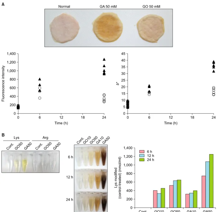
GA and GO were used as the glycation inducer to assay AGE formation in vitro. Skin was removed from 7-week-old female hairless (Hos:HR-1) mice (Hoshino Experiment Animal Center, Ibaraki, Japan), cleaned of subcutaneous fat, and mounted in modified vertical diffusion cells. The cells had an effective diffusion area of 1.77 cm2 and a re- ceptor compartment volume of 5.0 ml. For induction of glycation, the skin specimens were hydrated from the der- mal side for 6 to 24 h with PBS containing either 10 mM or 50 mM GO or GA. The receptor fluid was maintained at 32oC, and continuously agitated with a magnetic stirrer bar. After induction, the skin was placed on a white board, and the color was measured using a CR-400 chro- mameter (Konica-Minolta, Tokyo, Japan). Data were ex- pressed in the L*a*b* color space, and the b* was used as the yellow color value. The skin samples were then minced in 50% methanol and sonicated. Following cen- trifugation, AGEs were assayed in the supernatant by fluo- rescence with excitation at 365 nm and emission at 450 nm (SpectraMax M2e; Molecular Devices, Sunnyvale, CA, USA). To measure the modification rate of Lys, 10 mM or 50 mM of GO or GA was reacted with 10 mM of Lys in
PBS at 37oC for predetermined times. After hydrolysis in 6 N HCl for 8 h at 110oC, the concentration of modified Lys was measured by JLC-500 amino acid analyzer (JEOL Ltd., Tokyo, Japan).
The presence of Glycer-AGEs in the epidermis and dermis of human skin was demonstrated by immunohistochemistry, but they were not found in skin pretreated with blocking peptides. Glycer-AGEs were detected in stratum corneum and viable epidermis, and to a lesser extent, dermal cells.
No significant differences between young and old skin were observed (Fig. 1).
To describe the presence of Glycer-AGEs in epidermis, which has a turnover rate about 1 month, we compared the rates of AGE formation induced by GO and GA. The respective amounts of AGEs induced by GA, as indicated by fluorescence intensity and b* value, were 3.0 and 2.2 times higher than those of the AGEs induced by GO.
Interestingly, the intensity and b* values induced by GO peaked at 6 h, whereas they continued to increase when induced by GA (Fig. 2A). Finally, when the basic amino acids Lys, Arg reacted with glycation inducer, only Lys:GA showed a strong yellowish tint (Fig. 2B). Consistent with this, the modification rate of Lys from 6 to 24 h with 50 mM GA was greater than that of GO (26 vs. 6 nmol/h) (Fig. 2B).
Glycer-AGEs were present in both epidermis and dermis.
Because of the rapid turnover rate in the epidermis, der- mal AGEs present in long-lived substrates in the ECM such as collagen or elastic fiber have been the focus of research for a long time. Kawabata et al.7 first reported the pres- ence of AGEs in epidermis, the major one being Nε-(car-
Brief Report
510 Ann Dermatol
Fig. 2. (A) Macroscopic view of hairless mouse skin glycated using glyoxal (GO) or glyceraldehyde (GA). Rate of increase in advanced glycation end product (AGE) formation indicated by fluorescence intensity (excitation: 365 nm/emission: 450 nm) and b* value.
Glycation induced by 50 mM of GO: ○, or GA: ▲. (B) Macroscopic view of glycated amino acids before hydrolysis (6 h) and after hydrolysis (6, 12, 24 h). Rate of modification of Lys as determined by amino acid analysis.
boxymethyl)lysine (CML), derived from GO and they found that CML was localized primarily in epidermal kera- tin 10 and dermal ECM. However, we found that Gly- cer-AGEs were concentrated at both epidermal and der- mal cells. These results suggest that the accumulation of Glycer-AGEs in skin strongly reflects alterations in intra- cellular metabolism caused by lifestyle-related conditions like hyperglycemia. Though we couldn’t detect the differ- ence in Glycer-AGEs accumulation between young and
aged human skin, Glycer-AGEs may contribute to skin ag- ing via upregulation of RAGE expression in hyperglycemic condition8. Furthermore, the faster increase in fluo- rescence intensity and b* value in GA- versus GO-gly- cated skin suggests that Glycer-AGEs accumulate at early stages of hyperglycemia, playing a pro-inflammatory role via RAGE signaling and contribute to degeneration of proteins. In addition, we found a strong relationship be- tween a yellowish tint of the skin and faster Lys mod-
Brief Report
Vol. 29, No. 4, 2017 511
Received May 17, 2016, Revised August 18, 2016, Accepted for publication August 19, 2016
Corresponding author: Young Lee, Department of Dermatology, Chungnam National University School of Medicine, 282 Munhwa-ro, Jung-gu, Daejeon 35015, Korea. Tel: 82-42-280-7706, Fax: 82-42-280-7706, E-mail: [email protected]
This is an Open Access article distributed under the terms of the Creative Commons Attribution Non-Commercial License (http://creativecommons.org/
licenses/by-nc/4.0) which permits unrestricted non-commercial use, distribution, and reproduction in any medium, provided the original work is properly cited.
Copyright © The Korean Dermatological Association and The Korean Society for Investigative Dermatology
ification by GA. Further studies are required to elucidate the biological and physiological significance of Glycer-AGEs in skin, but our study suggests that they may influence normal human keratinocytes and fibroblasts by different mechanisms and accumulate to a different degree than GO-induced AGEs.
CONFLICTS OF INTEREST
The authors have nothing to disclose.
REFERENCES
1. Gkogkolou P, Böhm M. Advanced glycation end products:
Key players in skin aging? Dermatoendocrinol 2012;4:259-270.
2. Zhu P, Ren M, Yang C, Hu YX, Ran JM, Yan L. Involvement of RAGE, MAPK and NF-κB pathways in AGEs-induced MMP-9 activation in HaCaT keratinocytes. Exp Dermatol 2012;21:123-129.
3. Miura J, Yamagishi Si, Uchigata Y, Takeuchi M, Yamamoto H, Makita Z, et al. Serum levels of non-carboxymethyllysine advanced glycation endproducts are correlated to severity of
microvascular complications in patients with Type 1 diabetes.
J Diabetes Complicat 2003;17:16-21.
4. Takino J, Yamagishi S, Takeuchi M. Cancer malignancy is enhanced by glyceraldehyde-derived advanced glycation end-products. J Oncol 2010;2010:739852.
5. Abe R, Shimizu T, Sugawara H, Watanabe H, Nakamura H, Choei H, et al. Regulation of human melanoma growth and metastasis by AGE-AGE receptor interactions. J Invest Dermatol 2004;122:461-467.
6. Takeuchi M, Makita Z, Bucala R, Suzuki T, Koike T, Kameda Y. Immunological evidence that non-carboxymethyllysine advanced glycation end-products are produced from short chain sugars and dicarbonyl compounds in vivo. Mol Med 2000;6:114-125.
7. Kawabata K, Yoshikawa H, Saruwatari K, Akazawa Y, Inoue T, Kuze T, et al. The presence of N(ε)-(Carboxymethyl) lysine in the human epidermis. Biochim Biophys Acta 2011;1814:
1246-1252.
8. Park HY, Kim JH, Jung M, Chung CH, Hasham R, Park CS, et al. A long-standing hyperglycaemic condition impairs skin barrier by accelerating skin ageing process. Exp Dermatol 2011;20:969-974.
https://doi.org/10.5021/ad.2017.29.4.511
Scar Sarcoidosis Developed after Blepharoplasty in Acute Lymphoblastic Leukemia Patient
Sue-Jeong Kim, Ji-Young Kim, Myung Im, Young-Joon Seo, Jeung-Hoon Lee, Young Lee
Department of Dermatology, Chungnam National University School of Medicine, Daejeon, Korea
Dear Editor:
A 46-year-old woman presented with linear erythematous papules that developed from a 20-year-old scar on her left upper eyelid 2 weeks earlier. She had undergone an up-
per eyelid blepharoplasty 20 years earlier. She was treated with a topical steroid for 1 week, without improvement.
She felt some discomfort while opening her eyes. On physical examination, erythematous, firm, non-tender pap-