ISSN 2234-3806 • eISSN 2234-3814
http://dx.doi.org/10.3343/alm.2015.35.5.479
Multicenter Study of Antimicrobial Susceptibility of Anaerobic Bacteria in Korea in 2012
Yangsoon Lee, M.D.1, Yeon-Joon Park, M.D.2, Mi-Na Kim, M.D.3, Young Uh, M.D.4, Myung Sook Kim, M.T.5, and Kyungwon Lee, M.D.5
Department of Laboratory Medicine1, Hanyang University College of Medicine, Seoul; Department of Laboratory Medicine2, School of Medicine, The Catholic University of Korea, Seoul; Department of Laboratory Medicine3, University of Ulsan College of Medicine, Asan Medical Center, Seoul; Department of Laboratory Medicine4, Yonsei University Wonju College of Medicine, Wonju; Department of Laboratory Medicine5, Research Institute of Bacterial Resistance, Yonsei University College of Medicine, Seoul, Korea
Background: Periodic monitoring of regional or institutional resistance trends of clinically important anaerobic bacteria is recommended, because the resistance of anaerobic pathogens to antimicrobial drugs and inappropriate therapy are associated with poor clini- cal outcomes. There has been no multicenter study of clinical anaerobic isolates in Korea.
We aimed to determine the antimicrobial resistance patterns of clinically important anaer- obes at multiple centers in Korea.
Methods: A total of 268 non-duplicated clinical isolates of anaerobic bacteria were col- lected from four large medical centers in Korea in 2012. Antimicrobial susceptibility was tested by the agar dilution method according to the CLSI guidelines. The following antimi- crobials were tested: piperacillin, piperacillin-tazobactam, cefoxitin, cefotetan, imipenem, meropenem, clindamycin, moxifloxacin, chloramphenicol, metronidazole, and tigecycline.
Results: Organisms of the Bacteroides fragilis group were highly susceptible to piperacil- lin-tazobactam, imipenem, and meropenem, as their resistance rates to these three anti- microbials were lower than 6%. For B. fragilis group isolates and anaerobic gram-positive cocci, the resistance rates to moxifloxacin were 12-25% and 11-13%, respectively.
Among B. fragilis group organisms, the resistance rates to tigecycline were 16-17%. Two isolates of Finegoldia magna were non-susceptible to chloramphenicol (minimum inhibi- tory concentrations of 16-32 mg/L). Resistance patterns were different among the differ- ent hospitals.
Conclusions: Piperacillin-tazobactam, cefoxitin, and carbapemems are highly active β-lactam agents against most of the anaerobes. The resistance rates to moxifloxacin and tigecycline are slightly higher than those in the previous study.
Key Words: Anaerobe, Multicenter, Imipenem, Moxifloxacin, Tigecycline
Received: January 6, 2015 Revision received: January 28, 2015 Accepted: May 11, 2015
Corresponding author: Kyungwon Lee Department of Laboratory Medicine, Research Institute of Bacterial Resistance, Yonsei University College of Medicine, 50 Yonsei-ro, Seodaemun-gu, Seoul 120-752, Korea
Tel: +82-2-2228-2446 Fax: +82-2-313-0908 E-mail: [email protected]
© The Korean Society for Laboratory Medicine This is an Open Access article distributed under the terms of the Creative Commons Attribution Non-Commercial License (http://creativecom- mons.org/licenses/by-nc/3.0) which permits unrestricted non-commercial use, distribution, and reproduction in any medium, provided the original work is properly cited.
INTRODUCTION
Antimicrobial susceptibility testing (AST) may not be necessary for most clinical anaerobic strains isolated from routine anaero- bic culture. The CLSI suggests testing of isolates from serious infections such as bacteremia, brain abscess, endocarditis, os-
teomyelitis, and joint infection [1]. Additionally, any bacteria iso- lated from normally sterile body sites or associated with a failure to respond to empirical treatment should be tested [1]. Antimi- crobials that are potentially effective against anaerobic bacteria include β-lactams, combinations of β-lactams and β-lactamase inhibitors, metronidazole, chloramphenicol, clindamycin, mac-
rolides, tetracyclines, and fluoroquinolones [2].
Some anaerobic bacteria have become resistant to antimicro- bial agents, and some can develop resistance while a patient is receiving therapy [3]. Moreover, there are reports that the resis- tance of anaerobic pathogens to antimicrobials and inappropri- ate therapy are associated with poor clinical outcomes [4, 5].
These findings emphasize the importance of performing sus- ceptibility testing of organisms recovered from certain selected cases to guide therapeutic choices. In addition, regional sus- ceptibility patterns play a pivotal role in the empirical treatment of infections caused by anaerobic bacteria.
In Korea, the AST for anaerobe has been regularly performed at Yonsei University Hospital [6, 7, 13], but there has been no multicenter study of clinical anaerobic isolates. We aimed to de- termine and compare the antimicrobial resistance patterns for clinically important anaerobes collected from four medical cen- ters in Korea.
METHODS
1. Bacterial isolatesA total of 396 anaerobic isolates were prospectively collected at four tertiary-care hospitals (the Catholic University of Korea, CU;
University of Ulsan College of Medicine, UU; Yonsei University College of Medicine, YU; Yonsei University Wonju College of Medicine, YW) from June to December 2012 and transported to YU for anaerobic identification and AST, as previously reported [7]. During this period, the isolates were consecutively collected at each hospital and recovered as one isolate per patient. An- aerobes were isolated from blood, body fluid, and abscess specimens. Each isolate was identified by conventional methods [8], the ATB 32A system (bioMérieux, Marcy l’Etoile, France), or the VITEK MS (bioMérieux) matrix-assisted laser desorption ion- ization–time-of-flight mass spectrometry system. Propionibacte- rium acnes was excluded from the data analysis and AST. A to- tal of 268 randomly selected isolates were used for AST: 83 Bacteroides fragilis, 64 other B. fragilis group species, 16 Pre- votella spp., 6 Fusobacterium spp., 12 Veillonella spp., 15 Fine- goldia magna, 19 other gram-positive cocci, 26 Clostridium spp., and 27 other gram-positive bacilli.
2. Antimicrobial susceptibility testing
AST was performed by using the CLSI agar dilution method [1].
The medium used was Brucella agar (Becton Dickinson, Cock- eysville, MD, USA) supplemented with 5 mg/L hemin, 1 mg/L vitamin K1, and 5% laked sheep blood. The antimicrobial pow-
ders used were piperacillin and tazobactam (Yuhan, Seoul, Ko- rea), cefoxitin (Merck Sharp & Dohme, West Point, PA, USA), cefotetan (Daiichi Pharmaceutical, Tokyo, Japan), clindamycin (Korea Upjohn, Seoul, Korea), imipenem and metronidazole (Choong Wae, Seoul, Korea), chloramphenicol (Chong Kun Dang, Seoul, Korea), meropenem (Sumitomo, Tokyo, Japan), moxifloxacin (Bayer Korea, Seoul, Korea), and tigecycline (Wy- eth Research, Pearl River, NY, USA). For the piperacillin-tazo- bactam combination, a constant concentration of 4 mg/L tazo- bactam was used. The tigecycline breakpoints of ≤ 4 and ≥ 16 mg/L, suggested by the US Food and Drug Administration, were used in this study [9].
An inoculum of 105 colony forming units (CFU) was applied with a Steers replicator (Craft Machine Inc., Woodline, PA, USA), and the plates were incubated in an anaerobic chamber (Forma Scientific, Marietta, OH, USA) for 48 hr at 37°C. The minimum inhibitory concentration (MIC) of the antimicrobial agent was defined as the concentration at which there was a marked reduction in growth, such as from confluent colonies to a haze, <10 tiny colonies, or several normal-sized colonies [1].
B. fragilis ATCC 25285 and Bacteroides thetaiotaomicron ATCC 29741 were used as the controls.
3. Carbapenemase screening test and detection of the cfiA gene
Imipenem and EDTA-sodium mercaptoacetic acid double-disk synergy (IEDDS) tests were carried out on Brucella agar to screen for carbapenemase-producing B. fragilis isolates [9].
The cfiA gene and its upstream insertion sequence (IS) were detected by PCR as previously described [10].
RESULTS
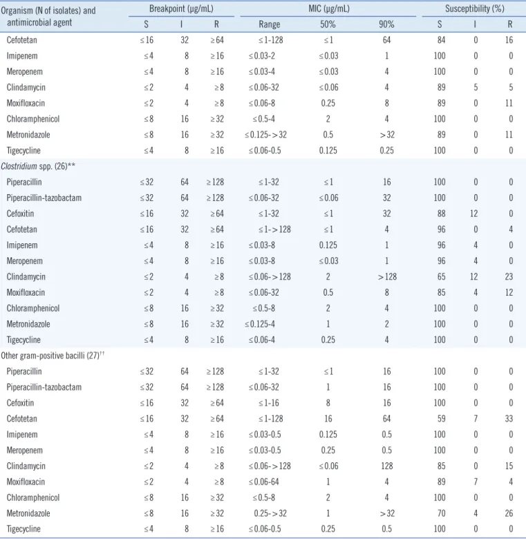
Table 1 shows the MICs of the antimicrobial agents and the re- sistance rates of the anaerobes tested. The resistance rates of B.
fragilis isolates and other B. fragilis group organisms to piper- acillin were 48-58%, whereas their resistance rates to piperacil- lin-tazobactam were 2-5%. Cefoxitin remained very active against B. fragilis, with only 4% of the isolates exhibiting resis- tance; however, 13% of other B. fragilis group isolates were re- sistant to this drug. Other B. fragilis group isolates were much more resistant to cefotetan, showing a 64% resistance rate. B.
fragilis group isolates showed resistance rates of only 0-6% to the carbapenems, which are the most active β-lactam drugs.
On the other hand, B. fragilis group isolates had high resistance rates of 52-80% to clindamycin. The resistance rates of the B.
Table 1. Activity of antimicrobials against 268 anaerobic bacteria isolated from four hospitals in Korea from June to December 2012 Organism (N of isolates) and
antimicrobial agent
Breakpoint (μg/mL) MIC (μg/mL) Susceptibility (%)
S I R Range 50% 90% S I R
Bacteroides fragilis (83)
Piperacillin ≤32 64 ≥128 2->256 64 >256 48 4 48
Piperacillin-tazobactam ≤32 64 ≥128 ≤0.06->128 0.5 8 96 1 2
Cefoxitin ≤16 32 ≥64 4-128 8 32 83 13 4
Cefotetan ≤16 32 ≥64 4->128 8 64 72 7 20
Imipenem ≤4 8 ≥16 0.06-16 0.125 1 96 0 4
Meropenem ≤4 8 ≥16 0.12-64 0.25 4 94 0 6
Clindamycin ≤2 4 ≥8 ≤0.06->128 >128 >128 47 1 52
Moxifloxacin ≤2 4 ≥8 0.25-32 0.5 8 86 2 12
Chloramphenicol ≤8 16 ≥32 2-8 4 4 100 0 0
Metronidazole ≤8 16 ≥32 0.125-4 1 2 100 0 0
Tigecycline* ≤4 8 ≥16 0.5-32 4 16 65 18 17
B. fragilis group, other (64)†
Piperacillin ≤32 64 ≥128 2->256 256 >256 38 5 58
Piperacillin-tazobactam ≤32 64 ≥128 ≤0.06->128 8 32 88 8 5
Cefoxitin ≤16 32 ≥64 ≤1-128 16 64 50 38 13
Cefotetan ≤16 32 ≥64 2->128 64 >128 19 17 64
Imipenem ≤4 8 ≥16 ≤0.03-32 0.5 2 97 0 3
Meropenem ≤4 8 ≥16 ≤0.03-8 0.25 2 98 2 0
Clindamycin ≤2 4 ≥8 ≤0.06->128 >128 >128 13 8 80
Moxifloxacin ≤2 4 ≥8 0.25-64 2 32 70 5 25
Chloramphenicol ≤8 16 ≥32 2-16 4 8 98 2 0
Metronidazole ≤8 16 ≥32 ≤0.125-8 2 4 100 0 0
Tigecycline ≤4 8 ≥16 ≤0.06-32 4 16 63 22 16
Prevotella spp. (16)‡
Piperacillin ≤32 64 ≥128 ≤1-64 16 32 94 6 0
Piperacillin-tazobactam ≤32 64 ≥128 ≤0.06 ≤0.06 ≤0.06 100 0 0
Cefoxitin ≤16 32 ≥64 ≤1-8 ≤1 4 100 0 0
Cefotetan ≤16 32 ≥64 ≤1-16 4 16 100 0 0
Imipenem ≤4 8 ≥16 ≤0.03-0.06 0.06 0.06 100 0 0
Meropenem ≤4 8 ≥16 ≤0.03-0.125 0.125 0.125 100 0 0
Clindamycin ≤2 4 ≥8 ≤0.06->128 ≤0.06 >128 63 0 38
Moxifloxacin ≤2 4 ≥8 ≤0.06-64 2 32 56 0 44
Chloramphenicol ≤8 16 ≥32 ≤0.5-8 2 8 100 0 0
Metronidazole ≤8 16 ≥32 0.25-16 1 16 81 19 0
Tigecycline ≤4 8 ≥16 0.125-4 0.25 2 100 0 0
Fusobacterium spp. (6)§
Piperacillin ≤32 64 ≥128 ≤1-2 NA NA NA NA NA
(Continued to the next page)
Organism (N of isolates) and antimicrobial agent
Breakpoint (μg/mL) MIC (μg/mL) Susceptibility (%)
S I R Range 50% 90% S I R
Piperacillin-tazobactam ≤32 64 ≥128 ≤0.06-1 NA NA NA NA NA
Cefoxitin ≤16 32 ≥64 ≤1-2 NA NA NA NA NA
Cefotetan ≤16 32 ≥64 ≤0.1 NA NA NA NA NA
Imipenem ≤4 8 ≥16 ≤0.03-0.5 NA NA NA NA NA
Meropenem ≤4 8 ≥16 ≤0.03 NA NA NA NA NA
Clindamycin ≤2 4 ≥8 ≤0.06-8 NA NA NA NA NA
Moxifloxacin ≤2 4 ≥8 0.125-4 NA NA NA NA NA
Chloramphenicol ≤8 16 ≥32 ≤0.5-2 NA NA NA NA NA
Metronidazole ≤8 16 ≥32 ≤0.125-0.25 NA NA NA NA NA
Tigecycline ≤4 8 ≥16 ≤0.06-0.25 NA NA NA NA NA
Veillonella spp. (12)||
Piperacillin ≤32 64 ≥128 ≤1-256 32 32 92 0 8
Piperacillin-tazobactam ≤32 64 ≥128 ≤0.06->128 8 16 92 0 8
Cefoxitin ≤16 32 ≥64 ≤1-8 ≤1 4 100 0 0
Cefotetan ≤16 32 ≥64 ≤1 ≤1 ≤1 100 0 0
Imipenem ≤4 8 ≥16 ≤0.03-0.5 0.25 0.5 100 0 0
Meropenem ≤4 8 ≥16 ≤0.03 ≤0.03 ≤0.03 100 0 0
Clindamycin ≤2 4 ≥8 ≤0.06-0.125 ≤0.06 0.125 100 0 0
Moxifloxacin ≤2 4 ≥8 ≤0.06-16 0.25 4 83 8 8
Chloramphenicol ≤8 16 ≥32 1-2 1 2 100 0 0
Metronidazole ≤8 16 ≥32 ≤0.125-16 2 4 92 8 0
Tigecycline ≤4 8 ≥16 0.25-2 1 1 100 0 0
Finegoldia magna (15)
Piperacillin ≤32 64 ≥128 ≤1 ≤1 ≤1 100 0 0
Piperacillin-tazobactam ≤32 64 ≥128 ≤0.06-0.5 ≤0.06 0.125 100 0 0
Cefoxitin ≤16 32 ≥64 ≤1-0.5 ≤1 ≤1 100 0 0
Cefotetan ≤16 32 ≥64 ≤1-4 ≤1 2 100 0 0
Imipenem ≤4 8 ≥16 ≤0.03-0.125 ≤0.03 0.06 100 0 0
Meropenem ≤4 8 ≥16 ≤0.03-0.125 0.06 0.125 100 0 0
Clindamycin ≤2 4 ≥8 ≤0.06->128 4 >128 47 13 40
Moxifloxacin ≤2 4 ≥8 ≤0.06-32 0.125 16 87 0 13
Chloramphenicol ≤8 16 ≥32 2-32 4 16 87 7 7
Metronidazole ≤8 16 ≥32 0.25-2 0.5 1 100 0 0
Tigecycline ≤4 8 ≥16 0.125-0.5 NA NA 100 0 0
Other gram-positive cocci (19)¶
Piperacillin ≤32 64 ≥128 ≤1-16 ≤1 8 100 0 0
Piperacillin-tazobactam ≤32 64 ≥128 ≤0.06-16 ≤0.06 8 100 0 0
Cefoxitin ≤16 32 ≥64 ≤1-16 ≤1 16 100 0 0
(Continued to the next page) Table 1. Continued
Organism (N of isolates) and antimicrobial agent
Breakpoint (μg/mL) MIC (μg/mL) Susceptibility (%)
S I R Range 50% 90% S I R
Cefotetan ≤16 32 ≥64 ≤1-128 ≤1 64 84 0 16
Imipenem ≤4 8 ≥16 ≤0.03-2 ≤0.03 1 100 0 0
Meropenem ≤4 8 ≥16 ≤0.03-4 ≤0.03 4 100 0 0
Clindamycin ≤2 4 ≥8 ≤0.06-32 ≤0.06 4 89 5 5
Moxifloxacin ≤2 4 ≥8 ≤0.06-8 0.25 8 89 0 11
Chloramphenicol ≤8 16 ≥32 ≤0.5-4 2 4 100 0 0
Metronidazole ≤8 16 ≥32 ≤0.125->32 0.5 >32 89 0 11
Tigecycline ≤4 8 ≥16 ≤0.06-0.5 0.125 0.25 100 0 0
Clostridium spp. (26)**
Piperacillin ≤32 64 ≥128 ≤1-32 ≤1 16 100 0 0
Piperacillin-tazobactam ≤32 64 ≥128 ≤0.06-32 ≤0.06 32 100 0 0
Cefoxitin ≤16 32 ≥64 ≤1-32 ≤1 32 88 12 0
Cefotetan ≤16 32 ≥64 ≤1->128 ≤1 4 96 0 4
Imipenem ≤4 8 ≥16 ≤0.03-8 0.125 1 96 4 0
Meropenem ≤4 8 ≥16 ≤0.03-8 ≤0.03 1 96 4 0
Clindamycin ≤2 4 ≥8 ≤0.06->128 2 >128 65 12 23
Moxifloxacin ≤2 4 ≥8 ≤0.06-32 0.5 8 85 4 12
Chloramphenicol ≤8 16 ≥32 ≤0.5-8 2 4 100 0 0
Metronidazole ≤8 16 ≥32 ≤0.125-4 1 2 100 0 0
Tigecycline ≤4 8 ≥16 ≤0.06-4 0.25 4 100 0 0
Other gram-positive bacilli (27)††
Piperacillin ≤32 64 ≥128 ≤1-32 ≤1 16 100 0 0
Piperacillin-tazobactam ≤32 64 ≥128 ≤0.06-32 1 16 100 0 0
Cefoxitin ≤16 32 ≥64 ≤1-16 8 16 100 0 0
Cefotetan ≤16 32 ≥64 ≤1-128 16 64 59 7 33
Imipenem ≤4 8 ≥16 ≤0.03-0.5 0.125 0.5 100 0 0
Meropenem ≤4 8 ≥16 ≤0.03-0.5 0.25 0.5 100 0 0
Clindamycin ≤2 4 ≥8 ≤0.06->128 ≤0.06 128 85 0 15
Moxifloxacin ≤2 4 ≥8 ≤0.06-64 1 4 89 7 4
Chloramphenicol ≤8 16 ≥32 ≤0.5-8 2 4 100 0 0
Metronidazole ≤8 16 ≥32 0.25->32 1 >32 70 4 26
Tigecycline ≤4 8 ≥16 ≤0.06-0.5 0.25 0.5 100 0 0
*US Food and Drug Administration breakpoints were used for tigecycline; †Bacteroides thetaiotaomicron (n=25), B. ovatus (n=8), B. vulgatus (n=8), Para- bacteroides distasonis (n=8), B. uniformis (n=4), B. salyersae (n=3), B. caccae (n=2), B. dorei (n=1), B. nordii (n=1), B. stercoris (n=1), Odoribacter splanchnicus (n=1), Bacteroides sp. (n=2); ‡Prevotella bivia (n=4), P. buccae (n=3), P. intermedia (n=3), P. denticola (n=1), P. disiens (n=1), P. mela- ninogenica (n=1), P. oralis (n=1); §Fusobacterium necrophorum (n=2), F. nucleatum (n=2), F. varium (n=1), Fusobacterium sp. (n=1); ||Veillonella par- vula (n=10), Veillonella sp. (n=2); ¶Parvimonas micra (n=7), Peptostreptococcus anaerobius (n=4), Peptoniphilus asaccharolyticus (n=3), Peptostrepto- coccus sp. (n=3), Streptococcus asaccharolyticus (n=2); **Clostridium perfringens (n=11), C. ramosum (n=2), C. tertium (n=2), C. baratii (n=1), C.
clostridioforme (n=1), C. paraputrificum (n=1), Clostridium sp. (n=8); ††Actinomyces meyeri (n=2), Actinomyces naeslundii (n=1), Actinomyces neuii (n=1), Actinomyces sp. (n=5), Bifidobacterium sp. (n=1); Collinsella aerofaciens (n=3), Eggerthella lenta (n=10), Eubacterium lentum (n=3), Eubacteri- um sp. (n=1).
Abbreviations: S, susceptible; I, intermediate; R, resistant; NA, not available/not applicable.
Table 1. Continued
fragilis group organisms to moxifloxacin and tigecycline were 12-25% and 16-17%, respectively. All B. fragilis group isolates were susceptible to chloramphenicol and metronidazole.
Prevotella isolates were susceptible to all antimicrobial agents tested, except for clindamycin (38% resistant) and moxifloxacin (44% resistant). The resistance rate to clindamycin was 40%
for F. magna and 5% for other gram-positive cocci. It should be noted that two isolates of F. magna showed non-susceptibility to chloramphenicol, with MICs of 16-32 mg/L. Clostridium isolates, including C. perfringens, were generally susceptible to the test drugs, except for clindamycin (23% resistant) and moxifloxacin (12% resistant). Other gram-positive bacilli such as Actinomy- ces, Bifidobacterium, Eggerthella, and Collinsella species were generally susceptible to the β-lactams, including piperacillin, but were resistant to cefoxitin (33%), metronidazole (26%), clindamycin (15%), and moxifloxacin (4%).
Table 2 shows the resistance rates of the B. fragilis group and other B. fragilis group isolates in each hospital and reveals some differences in resistance patterns among the hospitals. High re- sistance rates to cefotetan (33%) at YW and to moxifloxacin (22%) at CU were noted for B. fragilis isolates. Among non-B.
fragilis isolates, the highest resistance rates were observed to- ward piperacillin-tazobactam (13%) at YU and toward moxiflox- acin (57%) at CU.
Two imipenem-resistant B. fragilis isolates showed positive re- sults on the IEDDS test, whereas two imipenem-resistant B. the-
taiotaomicron isolates did not. The cfiA gene and its upstream IS elements were detected in two imipenem-resistant B. fragilis isolates.
DISCUSSION
This study is the first report of the antimicrobial susceptibility patterns of anaerobic clinical isolates collected from four institu- tions in Korea. Some results in this study (from YU) have been previously published [7], and these results were reanalyzed to- gether with the data from the other three hospitals. Among the anaerobes identified in clinical specimens, isolates from the B.
fragilis group are the most commonly encountered and are also more virulent and more resistant to antimicrobial agents than the other anaerobes [10]. Piperacillin was the most active of the ureidopenicillins against the B. fragilis group, with the organ- isms showing 38-48% resistance rates in this study. Piperacillin- tazobactam was active against nearly all strains of the B. fragilis group, with only 2-5% resistance rates in this study, in accor- dance with the less than 7% resistance in previous studies [12- 14].
The poor activity of clindamycin against the B. fragilis group is recognized worldwide and has been reported in several stud- ies [15-17]. High rates of resistance to clindamycin among B.
fragilis group isolates have also been reported in Korea [10, 14], and the recent anaerobic isolates tested in this study showed resistance rates of 52-80%. Moxifloxacin was recently intro- duced for the treatment of skin and soft tissue infections [18].
The resistance rates (12-25%) to moxifloxacin of B. fragilis group organisms in this study were slightly higher than the 11- 18% rates reported in 2010 in Korea [14] but lower than the 34-55% rates in US hospitals [1].
Jacobus et al. [19] reported that the geometric mean MICs of tigecycline for Parabacteroides distasonis were significantly higher than those for other Bacteroides species. Karlowsky et al.
[16] noted that 14% of B. fragilis isolates and 31% of B. the- taiotaomicron isolates were resistant to tigecycline, compared with 5% of B. fragilis isolates and 3-7% of other B. fragilis group isolates in the study by Snydman et al. [15]. Our data showed tigecycline resistance rates of 16-17% for the B. fragilis group isolates.
Overall, Prevotella and Fusobacterium isolates were more susceptible to the antimicrobials than B. fragilis group organ- isms. The resistance rates to moxifloxacin were as low as 24%
and 36% for Prevotella isolates in Belgium [18] and USA [20], respectively, whereas 44% of Prevotella isolates were resistant Table 2. Comparison of resistance rates of Bacteroides fragilis and
other Bacteroides spp. isolates by hospital
Antimicrobial agent
Resistance rates (%) of B. fragilis/resistance rates (%) of other B. fragilis group isolates CU (23/14)* YU (22/24) UU (17/16) YW (21/10)
Piperacillin 52/57 23/79 53/50 67/20
Piperacillin-tazobactam 0/0 0/13 6/0 0/0
Cefoxitin 0/0 5/17 12/25 0/0
Cefotetan 22/57 9/75 18/63 33/50
Imipenem 0/0 0/8 18/0 0/0
Meropenem 4/0 5/0 18/0 0/0
Clindamycin 48/98 41/92 59/75 62/50
Moxifloxacin 22/57 5/17 12/13 10/20
Chloramphenicol 0/0 0/0 0/0 0/0
Metronidazole 0/0 0/0 0/0 0/0
*Number of B. fragilis/other B. fragilis group isolates.
Abbreviations: CU, the Catholic University of Korea; YU, Yonsei University College of Medicine; UU, University of Ulsan College of Medicine; YW, Yon- sei University Wonju College of Medicine.
to moxifloxacin in this study. Papaparaskevas et al. [21] re- ported that moxifloxacin resistance was prevalent among Pre- votella and Bacteroides species in Greece. Moreover, species variation was noted, with the highest non-susceptible rates be- ing detected among Prevotella oralis (90%) and Prevotella bivia (80%). The discovery in this study of two F. magna isolates that were non-susceptible to chloramphenicol is interesting, since chloramphenicol-resistant anaerobic gram-positive cocci have not been reported.
In this study, there were some differences in the geographical patterns of resistance, and even differences in resistance pat- terns among the different hospitals in a single city, perhaps due in part to variability in the patterns of prescribing drugs. The CLSI recommends that hospitals conduct at least one annual AST surveillance to elucidate local patterns of resistance. Over- all, isolates from UU B. fragilis were more resistant to cefoxitin (12% vs. 5%), imipenem (18% vs. 0%), and meropenem (18%
vs. 5%) (drugs highly active against group organisms) than those from the other hospitals. The difference in resistance rates among hospitals may be important when selecting appropriate antimicrobial treatment options, although susceptibility testing is not generally performed for individual patient isolates.
Carbapenem resistance is usually mediated by metallo-β- lactamase, which is encoded by the cfiA gene in the presence of IS elements that activate the gene [22]. In the present study, two imipenem-resistant B. fragilis isolates carried the cfiA gene with upstream IS elements.
In conclusion, piperacillin-tazobactam, cefoxitin, imipenem, meropenem, metronidazole, and chloramphenicol remain ac- tive against most anaerobic isolates. The 2012 rates of resis- tance to moxifloxacin and tigecycline for B. fragilis group isolates were slightly higher than those reported in 2010 [14]. There were some differences in resistance patterns among the differ- ent hospitals. Continuous monitoring is necessary to detect changes in resistance patterns at regional centers and hospitals.
Authors’ Disclosures of Potential Conflicts of Interest
No potential conflicts of interest relevant to this article were re- ported.
Acknowledgments
This study was supported by a CMB-Yuhan research grant of Yonsei University College of Medicine for 2012 (6-2012-0048).
REFERENCES
1. Clinical and Laboratory Standards Institute. Methods for antimicrobial susceptibility testing of anaerobic bacteria; Approved standard, 8th ed.
M11-A8. Wayne, PA: Clinical and Laboratory Standards Institute, 2012.
2. Brook I, Wexler HM, Goldstein EJ. Antianaerobic antimicrobials: spec- trum and susceptibility testing. Clin Microbiol Rev 2013;26:526-46.
3. Pumbwe L, Chang A, Smith RL, Wexler HM. Clinical significance of overexpression of multiple RND-family efflux pumps in Bacteroides fra- gilis isolates. J Antimicrob Chemother 2006;58:543-8.
4. Goldstein EJ, Solomkin JS, Citron DM, Alder JD. Clinical efficacy and correlation of clinical outcomes with in vitro susceptibility for anaerobic bacteria in patients with complicated intra-abdominal infections treated with moxifloxacin. Clin Infect Dis 2011;53:1074-80.
5. Lassmann B, Gustafson DR, Wood CM, Rosenblatt JE. Reemergence of anaerobic bacteremia. Clin Infect Dis 2007;44:895-900.
6. Lee K, Shin HB, Chong Y. Antimicrobial resistance patterns of Bacteroi- des fragilis group organisms in Korea. Yonsei Med J 1998;39:578-86.
7. Yim J, Lee Y, Kim M, Seo YH, Kim WH, Yong D, et al. Antimicrobial sus- ceptibility of clinical isolates of Bacteroides fragilis group organisms re- covered from 2009 to 2012 in a Korean hospital. Ann Lab Med 2015;
35:94-8.
8. Jousimies-Somer HR, Summanen P, et al. eds. Wadsworth-KTL anaero- bic bacteriology manual, 6th ed. Belmont, CA: Star Publishing Co., 2002:55-70.
9. The US Food and Drug Administration. FDA Approved Drug Products Label for Tygacil, NDA no. 021821 (approved on May 23, 2013). http://
www.accessdata.fda.gov/drugsatfda_docs/nda/2005/21-821_Tygacil.
cfm.
10. Roh KH, Kim S, Kim CK, Yum JH, Kim MS, Yong D, et al. New cfiA vari- ant and novel insertion sequence elements in carbapenem-resistant Bacteroides fragilis isolates from Korea. Diagn Microbiol Infect Dis 2010;66:343-8.
11. Citron DM and Hecht DW. Susceptibility test methods: anaerobic bacte- ria. In: Versalovic J, Carroll KC, Funke G, et al., eds. Manual of clinical microbiology. 10th ed. Washington, DC: ASM Press, 2011:1204-14.
12. Koeth LM, Good CE, Appelbaum PC, Goldstein EJ, Rodloff AC, Claros M, et al. Surveillance of susceptibility patterns in 1297 European and US anaerobic and capnophilic isolates to co-amoxiclav and five other anti- microbial agents. J Antimicrob Chemother 2004;53:1039-44.
13. Snydman DR, Jacobus NV, McDermott LA, Ruthazer R, Golan Y, Gold- stein EJ, et al. National survey on the susceptibility of Bacteroides fragi- lis group: report and analysis of trends in the United States from 1997 to 2004. Antimicrob Agents Chemother 2007;51:1649-55.
14. Lee Y, Park Y, Kim MS, Yong D, Jeong SH, Lee K, et al. Antimicrobial susceptibility patterns for recent clinical isolates of anaerobic bacteria in South Korea. Antimicrob Agents Chemother 2010;54:3993-7.
15. Snydman DR, Jacobus NV, McDermott LA, Golan Y, Hecht DW, Gold- stein EJ, et al. Lessons learned from the anaerobe survey: historical perspective and review of the most recent data (2005-2007). Clin Infect Dis 2010;50(S1):S26-33.
16. Karlowsky JA, Walkty AJ, Adam HJ, Baxter MR, Hoban DJ, Zhanel GG.
Prevalence of antimicrobial resistance among clinical isolates of Bacte- roides fragilis group in Canada in 2010-2011: CANWARD surveillance study. Antimicrob Agents Chemother 2012;56:1247-52.
17. Katsandri A, Avlamis A, Pantazatou A, Petrikkos GL, Legakis NJ, Papa- paraskevas J, et al. In vitro activities of tigecycline against recently iso- lated Gram-negative anaerobic bacteria in Greece, including metronida- zole-resistant strains. Diagn Microbiol Infect Dis 2006;55:231-6.
18. Wybo I, Piérard D, Verschraegen I, Reynders M, Vandoorslaer K, Claeys G, et al. Third Belgian multicentre survey of antibiotic susceptibility of anaerobic bacteria. J Antimicrob Chemother 2007;59:132-9.
19. Jacobus NV, McDermott LA, Ruthazer R, Snydman DR. In vitro activi- ties of tigecycline against the Bacteroides fragilis group. Antimicrob Agents Chemother 2004;48:1034-6.
20. Citron DM, Goldstein EJ, Merriam CV, Lipsky BA, Abramson MA. Bacte- riology of moderate-to-severe diabetic foot infections and in vitro activity of antimicrobial agents. J Clin Microbiol 2007;45:2819-28.
21. Papaparaskevas J, Pantazatou A, Katsandri A, Houhoula DP, Legakis NJ, Tsakris A, et al. Moxifloxacin resistance is prevalent among Bacte- roides and Prevotella species in Greece. J Antimicrob Chemother 2008;
62:137-41.
22. Sóki J, Edwards R, Hedberg M, Fang H, Nagy E, Nord CE, et al. Exami- nation of cfiA-mediated carbapenem resistance in Bacteroides fragilis strains from a European antibiotic susceptibility survey. Int J Antimicrob Agents 2006;28:497-502.