498
통신저자:박 상 진
전남 화순군 화순읍 일심리 160번지 전남대학교 의과대학 정형외과학교실 화순전남대학교병원 관절센터
TEL: 061-379-7676ㆍFAX: 061-379-7681 E-mail: [email protected]
Address reprint requests to Sang Jin Park, M.D.
Department of Orthopedics, Chonnam National University School of Medicine, Center for Joint Disease, Chonnam National University Hwasun Hospital, 160, Ilsim-ri, Hwasun-eup, Hwasun-gun, Jeonnam 519-809, Korea Tel: +82.61-379-7676, Fax: +82.61-379-7681
E-mail: [email protected]
슬괵건과 Ligament Anchor 나사를 이용한 관절경적 전방 십자인대 재건술의 중기 추시 결과
송은규ㆍ선종근ㆍ박상진ㆍ박주권
전남대학교 의과대학 정형외과학교실, 화순전남대학교병원 관절센터
The Midterm Results of Arthroscopic Anterior Cruciate Ligament Reconstruction with Hamstring Tendon and Ligament Anchor Screw
Eun Kyoo Song, M.D., Jong Keun Seon, M.D., Sang Jin Park, M.D., and Ju Kwon Park, M.D.
Department of Orthopedics, Chonnam National University School of Medicine, Gwangju, Center for Joint Disease, Chonnam National University Hwasun Hospital, Jeonnam, Korea
Purpose: To evaluate the clinical and radiological results of an arthroscopic ACL reconstruction with a hamstring tendon and Ligament Anchor (LA) screw.
Materials and Methods: 48 cases among 47 patients with a minimum 5 year follow up after the ACL reconstruction were examined. The clinical results (Lysholm Knee score, Tegner activity scale, Lachmann test, Pivot-shift test, and complications) were evaluated, and the radiological results (osteoarthritis, bony tunnel enlargement, instrumented anterior laxity test with TelosⓇ) were as- sessed.
Results: The Lysholm Knee score showed significant improvement from 65.8 to 96.7 at the final follow up. The Tegner activity scale also showed significant improvement from 3.5 preoperatively to 6.0 at the final follow-up. The Lachmann test showed that 42 cases had converted to negative.
The Pivot-shift test was negative in 39 cases and mild positive in 9 cases. The side to side difference using the instrumented anterior laxity test was 12.5 mm preoperatively and 3.2 mm at the final follow-up. Degenerative osteoarthritis was observed in 9 (19%) knees. The femoral and tibial tunnel were widened by 21.6 and 20.9% in the AP view and 16.3 and 19.0% in the lateral view, respectively (p<0.05).
Conclusion: An ACL reconstruction with a hamstring tendon and LA screw can restore the knee stability with satisfactory clinical results and few complications.
Key Words: Anterior cruciate ligament, Arthrosopic ACL reconstruction, Hamstring tendon, LA screw
서 론
전방 십자 인대는 슬관절의 안정성에 중요한 역할을 하며, 손상시 적절히 치료하지 않으면 만성 불안정성이 유발될 수 있고, 반월상 연골과 관절연골의 손상을 초래 하여 관절염으로 진행될 수 있다12,14,19). 전방십자인대 재건술시 보편적으로 이용되는 자가 이식물로는 골-슬
개건-골과 슬괵건이 있다. 골-슬개건-골 이식물은 술 후 슬개-대퇴 관절의 동통과 대퇴 신전력의 약화, 슬개 건 파열 등의 문제가 발생할 수 있어4) 최근에는 슬괵건을 이용한 방법이 증가하는 추세이다.
Steiner 등27)은 슬괵건을 이용한 재건술에 있어서, 대 퇴부에 슬괵건의 견고한 고정이 매우 중요하다고 보고하
Table 1. Kellgren and Lawrence's Classification
Grade Definition
I Minute osteophyte II Definite osteophyte
III Moderate diminution of joint space
IV Joint space impaired with subchondral sclerosis
Fig. 1. Radiographic measurement of the bony tunnel. The actual tunnel size was calculated by correcting the measured tunnel size with a correction ratio that was obtained by dividing the real diameter of the LA screw by the measured diameter of the LA screw. The bony tunnel was measured at the point of the maximal tunnel width in well-defined sclerotic margins.
였는데 대퇴부 고정방법에 쓰이는 재료로는 생체 흡수성 간섭 나사(Bioscrew, Linvatec, FL), 교차 강선인 RIGID fix system (Mitek, Johnson and Johnson, MA, USA), EndoButtonⓇ (Acufex Microsurgical, INC.
MA, USA), Ligament Anchor screwⓇ (Sulgo, Pyung- tak, Korea) 등이 알려져 있다. 그리고 현재까지 다양한 이식물과 그 고정방법을 이용한 전방십자인대 재건술의 초기 안정성에 대한 연구는 많이 이루어지고 있지만, 중 장기 추시의 결과에 대한 연구는 거의 없는 실정이다.
본 연구의 목적은 슬괵건과 자체 고안한 LA나사를 이 용한 전방 십자인대 재건술 후 5년 이상 중기 추시상 그 임상적, 방사선학적 결과를 알아보고자 하였다.
대상 및 방법
1. 대상
1997년 7월부터 2001년 7월까지 슬괵건과 LA나사를 이용한 관절경적 전방십자인대 재건술을 시행 받고, 술 후 최소 5년 이상(평균 7.0년) 추시가 가능하였던 47명, 48예를 대상으로 하였으며, 남자가 37명, 여자가 10명이 었다. 수술시 평균나이는 32 (19-59)세였다. 동반손상 은 반월상 연골 손상이 31예로 가장 많았고, 내측 측부인 대 손상이 10예, 내측과 외측 측부인대가 같이 손상된 경 우 1예 있었으며, 후방 십자인대 손상이 2예 동반되었다.
2. 임상적 및 방사선적 평가
임상적 결과는 환자의 주관적인 통증 정도, 부종, 불안 정성, 이학적 검사 및 Lysholm Knee score, 운동 능력 (Tegner activity scale) 및 합병증 유무를 조사하였고, 방사선학적으로 관절염의 유무, 골 터널의 확장 및 TelosⓇ (Telos stress device; Austin & Associate, Inc., Pol- ston, US) 기구를 이용하여 술 전후에 전방 전위 정도를 측정하고 이를 정상측과 비교하였다11). 골관절염의 평가 는 Kellgren과 Lawrence 방법17)을 이용하였으며 명확
한 골극이 관찰되는 Grade II 이상을 골관절염으로 정의 하였다(Table 1). 술 후 골터널 확장 정도를 측정하기 위 하여 전후방 및 측면 방사선사진을 찍어 경화성 가장자리 의 가장 넓은 부위를 골터널과 수직인 부분에서 측정하 고, 실제 LA 나사의 두께를 이용하여 방사선의 확대를 보정하였다(실제 터널 크기=계측된 터널 크기×<실제 LA나사 두께/계측된 LA나사 두께>)(Fig. 1).
결 과
1. 임상결과
슬관절의 통증은 술 전 39예(81%)에서 최종 추시상 3 예(6%)로 감소하였고, 26예(54%)에서 있었던 부종은 3 예(6%)로 감소하였다. 6예(12.5%)에서 대퇴사두근 위축 이 남아있었다. Tegner activity scale은 술 전 평균 3.5 (3-5)점에서 최종 추시 상 6.0 (4-8)점으로 향상되었다.
축구가 가능한 7점 이상인 경우는 15예(31%)였고, 32예 (69%)에서 4-6점으로 여가 생활을 할 수 있는 정도의 스포츠가 가능하였다.
술 전 5예에서 관찰되었던 슬관절 신전 부족현상이 최 종 추시상 단 1예에서도 관찰되지 않았고, 최종 추시상 저명한 운동 범위의 제한을 호소한 경우는 없었다. La-
Table 2. Clinical Results
Preop. Last follow up Lysholm knee score (p<0.05) 66 (37-75) 97 (77-100) Tegner activity scale 3.5 (3-5) 6.0 (4-8)
7 (Scoccer) 15 (31%)
4-6 (Recreational sports) 32 (69%)
3-5 (Light work to cycling) 48 (100%) Lachman test (p<0.05)
0 (0%) 42 (88%)
+ 19 (40%) 4 (9%)
++ 17 (35%) 2 (3%)
+++ 12 (25%)
Pivot-Shift test (p<0.05)
10 (22%) 39 (81%)
+ 27 (56%) 9 (19%)
++ 5 (10%)
+++ 6 (12%)
Table 3. Radiology Results
Preop. Last follow up Osteoarthritic change (total) 1 (2%) 9 (19%)
II 1 (2%) 4 (8.5%)
III 4 (8.5%)
IV 1 (2%)
Side to side difference by 12.5 mm 3.2 mm TelosⓇ stress view
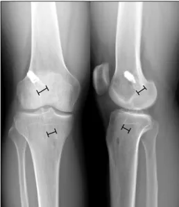
Fig. 2. (A) Anteroposterior and lateral radiographs before surgery showed no evidence of osteoarthritis. (B) Anteroposte- rior and Lateral radiographs of a 67 year old patient taken 7.8 years after the reconstruction showed evidence of grade III osteoarthritis on the all com- partments of the reconstructed knee.
chmann 검사는 술 전 경도 양성 19예(40%), 중등도 양 성 17예(35%), 고도 양성 12예(25%)에서 최종 추시상 음성 42예(88%), 경도 양성 4예(9%)로 호전되었다(p<
0.05). Pivot-shift 검사는 술 전 경도 양성 27예(56%), 중등도 양성 5예(10%), 고도 양성 6예(12%)에서 최종 추 시상 음성 39예(81%), 경도 양성 9예(19%)로 호전되었 다(p<0.05)(Table 2).
Lysholm 점수는 술전 평균 65.8 (37-95)점에서 술후
평균 96.7 (77-100)점으로 호전되었다(p<0.05).
2. 방사선적 결과 1) 골관절염의 정도
최종 추시 상 총 48예 중 9예(19%)에서 골관절염이 관 찰되었고, 그 중 Grade II가 4예(8%), Grade III가 4예 (8%), Grade IV 1예(2%) 관찰되었다. 부위별로는 6예에 서 슬관절 내측 구획에 관절염이 관찰되었고, 외측 구획 에 3예, 내측 및 외측 구획에 2예, 슬개 대퇴 관절에 1예, 전 구획에 1예 관찰되었다(Fig. 2)(Table 3).
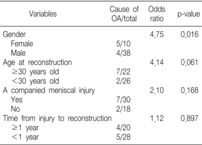
골관절염 발생에 영향을 미치는 인자 중 성별은 여자에 서 5예(50%), 남자에서 4예(11%)에서 골관절염이 발생 하여, 여자에서 남자에서보다 통계적으로 유의하게 높은 발생률을 보였다(p=0.016). 재건술 당시의 연령이 평균
Table 4. Factors Affecting the Development of Osteoarthritis (OA) after the Anterior Cruciate Ligament Reconstruction
Cause of Odds
Variables p-value
OA/total ratio
Gender 4.75 0.016
Female 5/10
Male 4/38
Age at reconstruction 4.14 0.061
≥30 years old 7/22
<30 years old 2/26
A companied meniscal injury 2.10 0.168
Yes 7/30
No 2/18
Time from injury to reconstruction 1.12 0.897
≥1 year 4/20
<1 year 5/28
Fig. 3. The radiographs taken 6.7 years after surgery. (A) An- teroposterior and lateral radio- graphs of the left knee show proper placement of the graft and LA screw. (B) Side to side difference in the instrumented anterior laxity test with TelosⓇ had improved to 2 mm at the follow up.
연령 30세 이상인 경우, 7예(32%)에서, 평균 연령 30세 이하인 경우 2예(8%)에서 골관절염이 발생하여 평균 연 령 30세 이상에서 높은 발생률을 보였지만 통계적으로 유의한 차이를 보이지 않았다(p=0.061). 반월상 연골 손 상이 동반된 경우 7예(23%)에서, 연골판 손상이 동반되 지 않은 경우 2예(11%)에서 골관절염이 발생하여 반월상 연골 손상 동반시 높은 발생률을 보였지만 통계적으로 의 미는 없었다(p=0.168). 수상 후 재건술까지의 기간은 1 년 이상인 군에서 4예(20%), 그렇지 않는 군에서 5예 (18%)의 골관절염이 발생하여 유의한 차이를 보이지 않
았다(p=0.897)(Table 4).
2) 골터널의 확장
대퇴터널은 전후면 방사선 사진상 최종 추시에 12.2 (10.2-14.7) mm로 술중의 터널에 비해 약 21.6%의 확 장을 보였으며(p<0.05), 측면 사진상 최종 추시에 11.6 (10.2-15.0) mm로 술중의 터널에 비해 약 16.3%의 확 장을 보였다(p<0.05). 경골측 터널은 전후면 방사선 사 진상 최종 추시상 9.2 (7.2-11.0) mm로 술중의 터널에 비해 약 20.9%의 확장을 보였으며(p<0.05), 측면 사진 상 최종 추시 상 8.9 (7.1-10.4) mm로 술중의 터널에 비해 약 19.0%의 확장을 보였다(p<0.05).
3) TelosⓇ 스트레스 부하 방사선 검사
TelosⓇ 스트레스 부하 방사선 검사 상 술 전 평균 12.5 (6-26) mm의 양측간 차이를 보이던 것이 최종 추시 상 평균 3.2 (0-6) mm로 호전되었다(p<0.05)(Fig. 3).
3. 합병증
최종 추시상 이식건 공여부위 절개부의 감각이상이 15 예(31%), 슬관절 운동시의 연발음이 7예(14%), 대퇴사 두근의 위축은 6예(12%)에서 발생하였다. 심부 감염이 나 관절 강직은 관찰되지 않았다.
고 찰
전방십자인대 손상 시 기능의 조기회복을 위해 관절경 하 전방십자인대 재건술이 주로 이용된다10). 과거 골-슬 개건-골을 이용한 전방십자인대 재건술이 널리 이용되 었고, 이에 대한 장기 추시 결과상 많은 보고에서 만족스 러운 결과를 보고하였다1,5,22).
현재 슬괵건을 포함한 다양한 이식물과 고정 방법 등이 소개되고 있고, 이를 이용한 전방십자인대 재건술의 초 기 안정성에 대한 연구는 많이 이루어지고 있다7). 그러나 슬괵건을 이용한 전방십자인대 재건술의 장기 추시 결과 는 그 보고가 많지 않은 실정이다.
송 등24)은 LA나사와 슬괵건을 이용한 전방십자인대 재건술의 2년 추시의 연구에서 우수한 초기 안정성을 보 고한 적이 있다. 본 연구는 최소 5년 이상(평균 7년)의 중기 추시 결과로 Lysholm 점수는 술 후 평균 97점으로 호전되었고, Lachmann 검사는 술 후 42예(88%)에서 음성, Pivot shift 검사는 39예(81%)에서 음성으로 전환 되어 만족스러운 임상적 결과를 보였다.
전방십자인대 재건술 이후 발생할 수 있는 합병증으로 감염, 심부정맥 혈전증, 운동 범위의 제한, 관절 섬유화, 결절, 슬개하 구축 증후군, 신경 혈관의 손상, 대퇴 사두 근의 위축 등27)이 알려져 있다. 본 연구에서는 이식건 공 여부위의 감각이상이 가장 많은 빈도를 보였고, 슬관절 운동시의 연발음, 대퇴 사두근의 위축 등이 발생하였으 나, 심부 감염이나 명백한 관절 운동 범위의 제한 등은 관찰되지 않았다. 대퇴 사두근의 위축은 전방십자인대 재건술 후 보이는 가장 많은 합병증 중의 하나로 슬개골 상연에서 약 10 cm 상방의 대퇴부의 둘레를 재서 건측과 1 cm 이상 차이를 보이는 경우를 대퇴 사두근 위축으로 정의하였는데, 6예(12.5%)에서 발생하였고 이는 2년 추 시 이후 변하지 않았다. 저자는 자가 슬개건을 이용한 전 방십자인대 재건술 후 평균 23개월 추시에서 약 57%의 대퇴사두근의 위축을 보고하였는데27) 슬괵건을 이용한 본 연구에서 보다 높은 발생률을 보였다. 재수술을 시행 한 경우는 1예에서 있었는데, 이는 축구 도중 재 수상 당 한 경우로 수상 전 Lysholm 점수는 100점, Tegner activity scale은 7점으로, 재수술의 원인은 외상에 의한 이식물의 파열에 의한 것이었다.
전방십자인대 재건술 후 골관절염 발생률에 있어서 Jomha 등16)은 자가 슬개건을 이용한 재건술 후 7년 추
시 후 72%의 발생률을 보고하였고, Shelbourne과 Gray21) 는 11%의 발생률을 보고하였다. 골관절염의 위험인자로 는 재건술 당시의 환자 연령, 성별, 동반된 반월상 연골 파열, 수상 후 재건술까지의 기간, 전방 불안정성 및 지 속적인 아탈구 등이 언급되나 아직 명확한 관계에 대해서 는 밝혀지지 않았다2,3,9,13,22)
. 송 등25)은 자가 슬개건을 이용한 재건술 후 10년 추시 후에 약 41%의 골관절염 발생률을 보고하였고, 그 영향 인자로 재건술 당시의 연 령, 수상 후 재건술까지의 기간, 반월상 연골 손상의 동 반 여부 등을 보고하였다.
슬괵건 이용한 본 연구에서는 9예에서 골관절염이 발 생하였으며 골관절염 발생에 영향을 미치는 인자로 성별 은 여자에서 50%에서 발생하여 남자에서(11%) 보다 통 계적으로 유의하게 높은 발생률을 보였다(p=0.016). 재 건술 당시의 연령이 평균 연령 30세 이상인 경우 32%에 서, 평균 연령 30세 이하인 경우 8%에서 골관절염이 발 생하였고 반월상 연골 손상이 동반된 경우 23%에서, 연 골판 손상이 동반되지 않은 경우 11%에서 골관절염이 발 생하여 수술 당시 연령이 많거나 반월상 연골 손상 동반 시 높은 발생률을 보였지만 통계적으로 의미는 없었다.
하지만 수상 후 이환 기간과의 관계에 대해서는 의의 있 는 결과를 보여주지 못했다.
슬괵건 이용한 본 연구와 자가 슬개건을 이용한 전방십 자 인대 재건술 후 골관절염의 발생 빈도를 단순 비교하 는 것은 어려우며 이는 수술시의 나이, 반월상 연골의 손 상유무, 이환 기간의 차이 등의 환자요인과 수술 방법의 차이, 그리고 추시 기간의 차이 등을 고려해 볼 수 있겠으 며 특히 10년 이상의 장기 추시 결과에 대한 비교 연구가 이루어져야 할 것으로 사료된다. 일반적으로 전방십자인 대 재건술 후 골터널의 확장은 대부분 술 후 첫 1 개월 사이에 일어나고, 약 2-3년 후에는 더 이상의 확장은 일 어나지 않는다고 알려져 있는데15), 본 연구에서도 같은 결과를 얻었다. 2년 추시 상 대퇴골 전후면, 측면, 경골의 전후면, 측면 방사선 촬영에 각각 21.7%, 16.5%, 20.7%, 18.9% 의 확장을 보였고 5년 이상의 중기 추시 에서는 21.6%, 16.3%, 20.9%, 19.0%로 더 이상의 확장 증가 소견은 관찰되지 않았다.
다른 고정 방법을 이용한 전방십자인대 재건술에서도 단순 방사선사진상 대퇴 및 경골 터널 크기의 증가는 많 이 보고되어 있다6,8,12). L'Insalata 등18)은 EndoButtonⓇ
을 이용한 전방십자인대 재건술 후 대퇴측 골터널의 증가 를 전후면 사진상 30.2±17.2%, 측면 사진상 28.1±
14.7%로, 경골측 골터널은 전후면 사진상에서 20.9±
13.4% 측면 사진상에서 25.5±16.7%로 본 연구에 비해 보다 많은 대퇴 및 경골 터널 확장을 보고하였다. 이는 EndoButtonⓇ을 이용하여 대퇴측에 고정하는 방법이 LA 나사를 이용한 방법에 비교하여 이식건의 대퇴골 고 정위치와 경골 고정위치 사이의 길이가 길기 때문에 슬관 절의 운동에 따른 이식건의 소위 wind-shield 효과나 bungee효과 때문으로 유추된다. 골터널의 확장 정도가 슬관절의 불안정과 연관이 없다고 보고되고 있으나12), 골터널의 확장에 이식건의 골터널 내에서의 운동이 관여 할 것으로 보이며 안정적인 대퇴측 고정이 장기 추시시 좀 더 유리한 영향을 미치리라 사료된다.
TelosⓇ 기기를 이용한 전방전위 검사상, 경골의 전방 전위가 건측과의 차이가 3 mm 이내를 우수로 판정한다 면, 본 연구에서는 약 75%에서 우수한 결과를 보였고, 주관적인 불안정성은 2예에서 호소하여 슬관절 안정성에 있어서도 우수한 결과를 보였다.
결 론
Ligament Anchor (LA) 나사와 슬괵건을 이용한 전방 십자인대 재건술은 5년 이상의 중기 추시 상 임상적, 방 사선학적으로 우수한 결과를 보였고 안정적인 슬관절의 기능을 회복할 수 있었으며, 합병증이 적은 효과적인 수 술방법으로 생각된다.
참고문헌
1. Aglietti P, Buzzi R, Giron F, Simeone AJ, Zaccherotti G: Arthroscopic-assisted anterior cruciate ligament reconstruc- tion with the central third patellar tendon. A 5-8-year follow-up. Knee Surg Sports Traumatol Arthrosc, 5: 138-144, 1997.
2. Aglietti P, Zaccherotti G, De Biase P, Taddei I: A com- parison between medial meniscus repair, partial meniscectomy, and normal meniscus in anterior cruciate ligament recon- structed knees. Clin Orthop Relat Res, 307: 165-173, 1994.
3. Almekinders LC, Pandarinath R, Rahusen FT: Knee sta- bility following anterior cruciate ligament rupture and surgery.
The contribution of irreducible tibial subluxation. J Bone Joint
Surg Am, 86: 983-987, 2004.
4. Aune AK, Ekeland A, Cawley PW: Interference screw fixation of hamstring vs patellar tendon grafts for anterior cruciate ligament reconstruction. Knee Surg Sports Traumatol Arthrosc, 6: 99-102, 1998.
5. Bach BR Jr, Tradonsky S, Bojchuk J, Levy ME, Bush- Joseph CA, Khan NH: Arthroscopically assisted anterior cruciate ligament reconstruction using patellar tendon autograft. Five- to nine-year follow-up evaluation. Am J Sports Med, 26: 20-29, 1998.
6. Balkfors B: The course of knee-ligament injuries. Acta Orthop Scand, 198(Suppl): 1-99, 1982.
7. Brown CH Jr, Steiner ME, Carson EW: The use of ham- string tendons for anterior cruciate ligament reconstruction.
Technique and result. Clin Sports Med, 12: 723-756, 1993.
8. Daniel DM, Stone ML, Dobson BE, Fithian DC, Ross- man DJ, Kaufman KR: Fate of the ACL-injured patient. A prospective outcome study. Am J Sports Med, 22: 632-644, 1994.
9. Ferretti A, Conteduca F, De Carli A, Fontana M, Mariani PP: Osteoarthritis of the knee after ACL reconstruction. Int Orthop, 15: 367-371, 1991.
10. Fetto JF, Marshall JL: The natural history and diagnosis of anterior cruciate ligament insufficiency. Clin Orthop Relat Res, 147: 29-38, 1980.
11. Franklin JL, Rosenberg TD, Paulos LE, France EP:
Radiographic assessment of instability of the knee due to rupture of the anterior cruciate ligament. A quadriceps- contraction technique. J Bone Joint Surg Am, 73: 365-372, 1991.
12. Furman W, Marshall JL, Girgis FG: The anterior cruciate ligament. A functional analysis based on postmorterm studies.
J Bone Joint Surg Am, 58: 179-185, 1976.
13. Gillquist J, Messner K: Anterior cruciate ligament re- construction and the long-term incidence of gonathrosis. Sports Med, 27: 143-156, 1999.
14. Giove TP, Miller SJ, Kent BE, Sanford TL, Garrick JG:
Non-operative treatment of the torn anterior cruciate ligament.
J Bone Joint Surg Am, 65: 184-192, 1983.
15. Giron F, Aglietti P, Cuomo P, Mondanelli N, Ciardullo
A: Anterior cruciate ligament reconstruction with double- looped semitendinosus and gracilis tendon graft directly fixed to cortical bone: 5-year results. Knee Surg Sports Traumatol Arthrosc, 13: 81-91, 2005.
16. Jomha NM, Borton DC, Clingeleffer AJ, Pinczewski LA:
Long-term osteoarthritic changes in anterior cruciate ligament reconstructed knees. Clin Orthop Relat Res, 358: 188-193, 1999.
17. Kellgren JH, Lawrence JS: Radiological assessment of osteo- arthrosis. Ann Rheum Dis, 16: 494-502, 1957.
18. L’Insalata JC, Klatt B, Fu FH, Harner CD: Tunnel expansion following anterior cruciate ligament reconstruction:
a comparison of hamstring and patellar tendon autografts.
Knee Surg Sports Traumatol Arthrosc, 5: 234-238, 1997.
19. McDaniel WJ Jr, Dameron TB Jr: Untreated ruptures of the anterior cruciate ligament. A follow-up study. J Bone Joint Surg Am, 62: 696-705, 1980.
20. Noyes FR, Barber-Westin SD: Anterior cruciate ligament reconstruction with autogenous patellar tendon graft in patients with articular cartilage damage. Am J Sports Med, 25:
626-634, 1997.
21. Shelbourne KD, Gray T: Anterior cruciate ligament recon- struction with autogenous patellar tendon graft followed by accelerated rehabilitation: A two- to nine-year followup. Am
J Sports Med, 25: 786-795, 1997.
22. Sommerlath K, Lysholm J, Gillquist J: The long-term course after treatment of acute anterior cruciate ligament ruptures. A 9 to 16 year followup. Am J Sports Med, 19:
156-162, 1991.
23. Song EK, Kim HS, Park CH: Parapatellar complications after ACL reconstruction using bone-patellar tendon-bone autograft. J Korean Orthop Assoc, 34: 917-921, 1999.
24. Song EK, Lee KB, Shin SG, Kim HJ: Clinical results of anterior cruciate ligament reconstruction using hamstring tendon. J Korean Orthop Sports Med, 1: 21-25, 2002.
25. Song EK, Seon JK, Yoon TR, Seo HY, Yim JH: Osteoar- thritis after ten year follow-up of ACL reconstruction using patellar tendon autograft. J Korean Orthop Assoc, 41: 122-128, 2006.
26. Song EK, Yoon TR, Jung JW, Jeong KC: Widening of bony tunnel after ACL reconstruction using hamstring tendon with ligament anchor (LA) screw. J Korean Arthro Soc, 5: 69-73, 2001.
27. Steiner ME, Hecker AT, Brown CH Jr, Hayes WC:
Anterior cruciate ligament graft fixation. Comparison of hamstring and patellar tendon grafts. Am J Sports Med, 22:
240-246, 1994.
= 국문초록=
목 적: 슬괵건과 LA (Ligament Anchor) 나사를 이용한 관절경적 전방십자인대 재건술 후 임상적 방사선적 결과를 알아보았다.
대상 및 방법: 최소 5년 이상 추시가 가능하였던 47명, 48예를 대상으로 하여 임상적(통증, 부종, 불안정성, 이학적 검사 및 Lysholm Knee Score, Tegner 운동 지수 및 합병증) 및 방사선학적(관절염, 골 터널의 확장 및 TelosⓇ 기구를 이용한 전방 전위) 평가를 하였다.
결 과 : Lysholm Knee score는 술 전 65.8점에서 96.7점으로, Tegner 운동 지수는 3.5점에서 6점으로 향상되었 다. Lachmann 검사는 42예(88%)에서, Pivot-shift 검사는 39예(81%)에서 음성으로 전환되었다(p<0.05).
TelosⓇ 전방 전위 검사는 술 전 12.5 mm에서 술 후 3.2 mm로 감소하였다(p<0.05). 술 후 퇴행성 변화는 9 (19%)예에서 보였으며, 대퇴와 경골 터널은 전후면 방사선 사진상 21.6, 20.9%, 측면 사진상 16.3, 19.0%
확장되었다.
결론 : 슬괵건과 LA 나사를 이용한 관절경적 전방십자인대 재건술은 5년 이상의 중기 추시상 안정적인 슬관절의 기능을 회복할 수 있었고, 합병증도 적은 효과적인 수술방법으로 생각된다.
색인 단어: 전방십자인대, 관절경적 재건술, 슬괵건, LA 나사