18
Clinical Significance of Prostate Cancer Diagnosed by 12 Core Biopsy
Chan Soo Park, Sang Eun Lee1, Hyeon Jeong2
From the Department of Urology, Seoul National University College of Medicine, Seoul, and the 1Seoul Bundang Hospital, Bundang, and the 2Seoul Municipal Boramae Hospital, Seoul, Korea
Purpose: Twelve-core biopsy improves the detection rate of prostate cancer;
however,the clinical significance of these additionally detected tumors has not been established. We retrospectively evaluated the clinical difference between prostate cancers detected by only the 12 core biopsy and by the 6 core biopsy from patients underwent radical prostatectomy.
Materials and Methods: Between February 2000 and April 2005, 124 men underwent radical prostatectomy after 12-core biopsy at Seoul National University College of Medicine, Bundang Seoul National University Hospital, Seoul National University Boramae Hospital. Among the 12-core biopsy group, we compared the subset with standard sextant cores (Group A) and the subset with lateral sextant cores (Group B). The low-risk group was defined as patients with a serum prostate-specific antigen (PSA) level less than 10.0ng/ml, a Gleason score of the final pathologic findings≤6, no pelvic lymph node metastasis and no lymphadenectomy performed, and a T stage≤T2a.
Results: There was no statistically significant difference between low-risk patients in group A and group B (p=0.0814). Groups A and B were then divided into three groups according to preoperative PSA levels of less than 10.0ng/ml, 10.0ng/ml to less than 20.0ng/ml, and greater than 20.0 ng/ml. The results of the subset analysis were the same for all three PSA categories. There was no statistically significant difference when we performed the analysis between group A and group B according to the Gleason score of the final pathologic findings and the TNM stage (p=0.0814), and there was also no statistically significant difference when we performed the analysis between group A and group B according to only the Gleason score of the final pathologic findings (p=0.5026).
Conclusions: The prostate cancers diagnosed by means of 12-core biopsy did not appear to be of a clinically lower risk than those detected with 6-core biopsy. We have firmly shown that 12-core biopsy is a more clinically effective procedure than 6-core biopsy. (Korean J Urol 2009;50:18-22) Key Words: Prostatic neoplasms, Biopsy, Significance
대한비뇨기과학회지 제 50 권 제 1 호 2009
서울대학교 의과대학
비뇨기과학교실, 1분당서울대학교
비뇨기과학교실, 2서울대학교
보라매병원 비뇨기과학교실
박찬수ㆍ이상은1ㆍ정 현2
접수일자:2008년 7월 30일 채택일자:2008년 10월 22일
교신저자: 정 현
서울대학교 보라매병원 비뇨기과
서울시 동작구 신대방동 425 156-752
TEL: 02-840-2486 FAX: 02-831-2826 E-mail: [email protected]
서 론
1989년 Hodge 등1이 6부위 전립선 생검을 발표한 이후, 6부위 전립선 생검은 전세계적으로 전립선암 검출을 위한 표준적인 방법으로 자리 잡았다. 그러나 최근의 많은 연구 들이 기존의 6부위 생검은 전립선 용적에 따른 고려가 없고
임상적으로 의미 있는 전립선암의 상당 부분을 진단해내지 못하여, 전립선암 검출에 있어 대략 30%의 위음성률을 갖 고 있다고 보고하였다.2,3 이러한 점을 극복하기 위하여 생 검 수를 늘려 전립선암 검출률을 높이려는 다양한 시도가 있었으며, 많은 연구들이 전립선 생검 수를 증가시킴으로 써 6부위 전립선 생검보다 유의하게 전립선암 검출을 향상 시켰다고 보고하였다.3-11
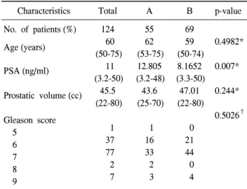
Table 1. Clinical characteristics of patients who underwent radical prostatectomy
Characteristics Total A B p-value
No. of patients (%) Age (years)
PSA (ng/ml)
Prostatic volume (cc)
Gleason score 5
6 7 8 9
124 60 (50-75)
11 (3.2-50)
45.5 (22-80)
1 37 77 2 7
55 62 (53-75) 12.805 (3.2-48)
43.6 (25-70)
1 16 33 2 3
69 59 (50-74) 8.1652 (3.3-50)
47.01 (22-80)
0 21 44 0 4
0.4982*
0.007*
0.244*
0.5026†
A: prostate cancer was detected only at the far lateral areas (lateral apex, mid-lobe and base-7, 8, 9, 10, 11, 12 sites), B: prostate cancer was detected at including medial sites, PSA: prostate-specific antigen, Gleason score: post-operative final pathologic findings. *:
statistical analysis by Student's t-test, †: statistical analysis by Fisher’s exact test
확장생검법이 6부위 전립선 생검법에 비해 높은 진단율 을 보인다는 것은 이미 여러 연구에서 알려져 있지만, 임상 적으로 의미 있는, 암을 진단하는가에 대해서는 아직 많은 논란의 여지가 있다. 진단율의 향상을 보이는 확장생검법 으로만 발견할 수 있는 전립선암 환자군이 6부위 전립선 생검법에 의해 발견할 수 있는 전립선암 환자군과 비교할 때 임상적 중요도가 떨어질 수 있다는 것이다. 하지만 Eskew 등12이 시행한 13부위 전립선 생검법에 의해 진단된 전립선 암 환자군의 전립선암은 6부위 생검법에 의해 진단된 전립 선암 환자군의 전립선암에 비해 임상적으로 차이를 보이지 않았다. 13부위 전립선 생검법은 전립선암 진단율의 향상 을 보이고 6부위 생검법과 비교하여 추가로 발견된 전립선 암이 임상적 차이가 없어 진단 및 치료에 중요한 수단이 된다는 것이다. 하지만 이 연구는 대상군이 적다는 한계점 을 가지고 있다.
우리는 이처럼 12부위 확장생검법에 의해 발견될 수 있 는 전립선암이 6부위 생검법에 의해 발견된 전립선암에 비 해 임상적 중요성의 차이가 있는지에 대해 수술 전의 혈청 PSA, 최종 병리 결과의 Gleason score, 양측 골반 림프절절 제술의 림프절 전이 유무, TNM stage T stage를 고려하여 후향적 연구를 통하여 알아보고자 한다.
대상 및 방법
2000년 2월부터 2005년 4월까지 서울대학교병원, 분당 서울대학교병원, 서울대학교 보라매병원 비뇨기과를 방문 하여 PSA 수치가 3.0ng/ml 이상인 경우이거나, 직장수지검 사에서 경결이 만져지거나 또는 경직장초음파검사에서 비 교적 잘 경계 지어지는 저반향 병변 (hypoechoic lesion)과 같 은 전립선암이 의심되는 환자에서 12부위 전립선 생검을 시행 받은 1,275명 중 392명이 전립선암을 진단받았다. 이 중 근치적 전립선적출술을 시행한 124명의 환자를 대상으 로 하였다. 이 환자들은 제한적 양측 골반 림프절절제술을 같이 시행하였다.
혈청 PSA는 직장수지검사와 경직장초음파검사를 시행 하기 전에 채혈하였으며 chemiluminescent microparticle im- muno assay를 이용한 Architecti2000sr (Abbot raboratories, Ame- rica), immunoradiometric assay를 이용한 cobra quantum (A anberra company, France)으로 측정하였다.
조직검사는 환자가 측와위를 취한 상태에서 경직장초음 파 유도하에 시행되었다. Acuson사의 Sequiona 512 기종의 EC-10C5 탐촉자 (transducer)를 이용한 경직장초음파로 전 립선의 용적을 측정한 후 18-gauge needle을 장착한 생검용 총 (spring biopsy gun)으로 초음파 유도하에 조직검사를 시
행하였다. 모든 환자에서 기존의 6부위 생검 부위 외에 양 측 외측 면의 첨부, 중간, 기저부에서 각 1번씩 총 12부위 생검을 실시하여, 각 부위에 명칭을 붙인 포르말린 통에 넣 어 병리과에 의뢰하였다.
양엽의 정중선에서 조직검사를 시행하는 통상적인 6부 위 생검과 동일한 부위인 전립선 내측을 1, 2, 3, 4, 5, 6번으 로 구분 지었으며 외측을 7, 8, 9, 10, 11, 12번으로 구분지어 외측 7, 8, 9, 10, 11, 12번에서만 전립선암이 진단된 A 군과 내측을 포함한 병변에서도 전립선암이 진단된 B군으로 대 상을 분류하였다. B군은 6부위 생검법에서 전립선암이 진 단된 경우와 동일한 것으로 간주하였다.
저위험군(low risk group)은 2007년 AUA guideline을 원용 하였으며 수술 전 시행한 PSA 10ng/ml 이하, 최종 병리 결 과 Gleason score 6 이하, 양측 골반 림프절절제술에서 음성 이며 TNM stage T2a 이하인 그룹으로 정의하였다.13 두 대상군 간에 저위험군이 유의하게 차이가 있는 지를 비교하였으며, p값이 0.05 미만인 경우 통계적으로 유의하 다고 판정하였고, 통계프로그램은 SPSS (ver 11.0)를 이용하 여 t-test, Fisher's exact test, chi-square test를 시행하였다.
결 과
환자의 평균 연령은 65세 (50-75)였으며, 전립선 평균 용
Table 2. PSA and pathological characters of 2 groups (A and B) Low risk
group
High and moderate
risk group p-value*
A B
11 20
44
49 0.251
Low risk group: PSA≤10, Gleason score≤6, TNM stage: ≤T2a, N0, M0, A: prostate cancer was detected only at the far lateral areas (lateral apex, mid-lobe and base-7, 8, 9, 10, 11, 12 sites), B:
prostate cancer was detected at all sites, including the medial sites, PSA: prostate-specific antigen. *: statistical analysis by chi-squre test
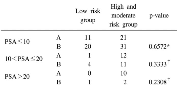
Table 3. PSA distribution of 2 groups (A and B) Low risk
group
High and moderate risk group
p-value
PSA≤10
10<PSA≤20
PSA>20
A B A B A B
11 20 1 4 0 1
21 31 12 11 10 2
0.6572*
0.3333†
0.2308† Low risk group: Gleason score≤6, TNM stage: ≤T2a, N0, M0, A: prostate cancer was detected only at the far lateral areas (lateral apex, mid-lobe and base-7, 8, 9, 10, 11, 12 sites), B: prostate cancer was detected at all sites, including the medial sites, PSA:
prostate-specific antigen. *: statistical analysis by chi-squre test,
†: statistical analysis by Fisher’s exact test 적은 45cc (22-80), 수술 전 시행한 PSA는 평균 11ng/ml (3.2-
50)이었다.
12부위 전립선 생검을 시행한 뒤 근치적 전립선적출술을 시행한 124명의 환자 중 A군은 55명이었으며, 내측을 포함 한 병변에서도 전립선암이 진단된 B군은 69명이었다. A군 과 B군의 PSA는 통계적으로 차이가 있었으며 A군이 더 높 게 측정되었으나 (p=0.007), 나이 (p=0.4982), 전립선 용적 (p=0.244)에서는 차이를 보이지 않았다. 또한 두 군 간에 최 종 병리 결과의 Gleason score도 통계적으로 차이가 없었다
(p=0.5026) (Table 1).
저위험군에 대해 비교하였을 때 A, B군 간에 통계적으로 차이가 없었으며 (p=0.251) (Table 2), A와 B군의 수술 전의 PSA를 고려하여 PSA 10ng/ml 이하, PSA 10ng/ml 초과 20ng/ml 이하, PSA 20ng/ml 초과인 군으로 대상군을 층화하 여 비교하였을 때도 모두 통계적으로 유의한 차이를 보이 지 않았다 (Table 3). 수술 전 PSA를 고려하지 않고 최종 병 리 결과 Gleason score, Pathologic TNM stage를 가지고 A, B군을 비교하였을 때도 통계적으로 유의한 차이를 보이지 않았다 (p=0.0814).
고 찰
Hodge 등1이 6부위 전립선 생검을 기술한 이후 6부위 전 립선 생검은 가장 보편적으로 쓰이는 생검 방법이다. 그러 나 최근 연구들은 기존의 6부위 전립선 생검은 종양의 크기 나 위치, 또는 전립선 용적에 대한 고려가 없으며 위음성률 이 높아 종양의 20-30%를 놓칠 수 있다고 보고하고 있다.8,14 이에 생검 숫자를 늘려 진단율을 높이려는 여러 시도가 있 어 왔으며 확장생검법이 기존의 6부위 생검법에 비해 합병 증은 비슷하면서 유의하게 진단율을 높인다고 주장하여 최 근에는 생검 수를 늘리는 경향이 있다.
Durkan 등15과 Ravery 등8은 12부위 생검법에서 각각 19%
와 6.6%의 진단율 상승을 보고하였다. Kim 등16이 시행한 전립선특이항원 및 전립선용적에 따른 12부위 확장생검법 의 유용성에서도 12부위 생검법은 기존 6부위 전립선 생검 법에 비해 전립선암 진단율을 전체 환자에서 4.9%, 전립선 암 환자 중에서 16.0% 증가시켰으며, PSA 수치와 전립선 용적을 동시에 고려해 볼 때, 상대적으로 작은 전립선 용적 을 가진 경우를 제외한 거의 대부분의 경우에서 의미 있는 진단율의 향상을 보였다. 또한 Lee 등11은 직장수지 검사 소 견이 정상이고 혈청 PSA 치가 4 이상 20 이하인 환자에서 생검수를 늘리면 암의 진단율을 높일 수 있다고 하였으며 Moon 등17은 10군데 생검을 시행하여 진단율이 23.4% 향상 되었다고 보고하였다.
확장생검법이 6부위 전립선 생검법에 비해 높은 진단율 을 보인다는 것은 이미 여러 연구에서 알려져 있지만, 임상 적으로 의미 있는 암을 진단하는가에 대해서는 아직 많은 논란의 여지가 있다.12 일부에서는 확장생검법으로 전립선 암의 진단율은 상승되었으나 임상적으로 중요성이 떨어지 는 암이 진단되어 치료되고 있다는 견해도 있는 것이다. 또 한 Freedland 등18은 저위험군의 전립선암 환자들은 즉각적 인 치료가 반드시 필요한 것은 아니며, 진단 후 180일 안에 치료를 할 경우 암의 진행에 영향을 주지 않는다는 결과도 발표하였다.
하지만 Eskew 등12은 13부위 전립선 생검으로 전립선암 을 진단하였을 때 6부위 전립선 생검에 비해 진단율의 향상 을 보일 뿐만 아니라 진단된 전립선암이 임상적으로 유의 한 차이를 보이고 있지 않음을 보고하였다. 13부위 전립선 생검으로 전립선암이 진단되어 근치적 전립선적출술을 시 행한 21명의 환자 그룹을 6부위 전립선 생검으로 전립선암
이 진단될 수 있었던 10명과 13부위 전립선 생검으로만 진 단될 수 있는 11명으로 대상 그룹을 나누어 비교하였다. 두 대상 그룹의 전립선암은 tumor volume, ploidy status, Gleason score, TNM pathological stage를 가지고 비교하였으며 두 그 룹간에 유의한 차이를 보이지 않았다. 그렇지만 이 연구는 대상군이 적다는 한계점을 가지고 있으며, 이를 보강하기 위해 저자들은 더 많은 환자를 대상군으로 연구를 진행하 였다.
Cintra와 Billis19는 Gleason scoring system이 tumor grade를 가장 효과적으로 반영하고 높은 intra-observer reproducibility 를 보인다고 보고하였다. 또한 Epstein 등20은 TNM stage가 예후 인자로 가장 유용함을 보고하였으며, 높은 TNM stage 는 국소 재발 및 전이를 높인다고 보고하였다. Gleason score 와 TNM stage가 임상적으로 의미 있는 지표가 될 수 있음을 보여준 것이다.
Kim 등21은 12부위 생검 및 외측 6부위 생검법은 표준 6 부위 생검법에 비하여 근치적 전립선적출술 조직과의 높은 Gleason score의 일치도를 보였으며 임상적으로 무의미한 암은 증가하지 않아 표준 6부위 생검을 대신한 생검법으로 이용될 수 있으리라 기대했다. 저자들이 시행한 연구에서 도 저위험군은 유의한 차이가 없음을 보여 주었다. 또한 PSA 10ng/ml 이하, PSA 10ng/ml 초과 20ng/ml 이하, PSA 20ng/ml 초과인 군으로 대상군을 층화하여 비교하였을 때 도 동일한 결과를 확인할 수 있었다. Gleason score, patholo- gic TNM stage를 가지고 A, B군을 비교하였을 때도 통계적 으로 유의한 차이를 보이지 않았다.
생검 수를 결정할 때 진단율과 함께 같이 고려해야 할 사 항으로 6부위 생검과 12부위 생검 환자가 느끼는 고통이나 불편함의 차이, 직장출혈, 패혈증 등의 합병증 발생빈도 등 이 있다. Eskew 등4이 시행한 13부위 전립선 생검법에서는 80%의 환자들이 시간이 지나면 사라지는 혈뇨를 호소하였 으나 이러한 높은 발생률은 중앙 내측에서 시행한 생검이 요도를 천자하면서 생긴 것이다. Kim 등16이 시행한 전립선 특이항원 및 전립선용적에 따른 12부위 확장생검법의 유용 성에서도 12부위 생검법은 시행 당일 또는 시행 다음날 환 자에게 혈뇨와 혈변의 유무, 발열이나 요폐 등 입원을 요하 는 비율을 확인한 결과, 두 방법 간에 유의한 차이가 없어 확장생검법이 기존의 6부위 생검법에 비해 합병증을 유의 하게 증가시키지 않는 것으로 보고하였다. 또한 Naughton 등22도 환자의 삶의 질에 대한 영향과 합병증 발생은 6부위 생검군과 12부위 생검군 간에 차이가 없음을 보고하였다.
저자들이 시행한 이번 연구의 제한점은 6부위 생검 환자 군과 12부위 생검 환자군의 직접적인 비교가 아닌 12부위 확장 생검을 시행 후 기존의 6부위 생검 위치에 해당되는
생검 결과와 12부위 확장생검에서만 검출될 수 있는 환자 의 결과를 비교하는 것이었다. 따라서 내측의 6부위가 기존 의 6부위 위치와 완전히 동일하다는 가정하에 분석을 시행 하였고, 각 병원의 경험이 풍부한 방사선과 전문의가 시행 하였으므로 이 가정에는 문제가 없다고 생각한다. 이에 대 한 정확한 비교를 위해서는 6부위 생검법과 12부위 확장생 검법을 직접적으로 비교하는 전향적인 연구가 추가적으로 필요하리라 생각한다. 또한 저자들의 경우 외측에서만 전 립선암이 진단된 경우가 거의 반 수에 달하며, 내측을 포함 한 B그룹이 A그룹보다 PSA가 유의하게 낮은 것은 후향적 연구로 인하여 전립선절제술 환자를 고를 때의 selection bias가 있음을 배제할 수 없기 때문이다.
기존의 연구를 통하여 12부위 생검법은 6부위 생검법과 비교할 때 유병률의 차이는 없으며 전립선암의 진단율을 향상시킬 수 있음을 알 수 있었고, 저자들의 연구를 통하여 6부위 생검법으로 진단 가능한 전립선암과 비교할 때 임상 적으로도 저위험군의 차이가 없음을 알 수 있었다.
결 론
기존의 많은 연구에서 12부위 확장생검법은 6부위 전립 선 생검보다 유의하게 전립선암 검출을 향상시켰다고 보고 되었다. 저자들은 이번 연구를 통하여 12부위 확장생검법 으로만 전립선암 진단이 가능한 환자들은 6부위 생검법으 로 진단이 가능한 전립선암과 비교하였을 때 임상적으로 의미가 적은 저위험군에서 두 군 간에 차이가 없음을 보여 주었다. 전립선암의 진단율을 향상시키며, 진단된 암에서도 임상적으로 의미 있는 전립선암을 검출하는 12부위 확장생 검법을 표준적 진단 방법으로 널리 활용하여 전립선암의 진단과 치료에 기여할 것으로 기대하는 바이다.
REFERENCES
1. Hodge KK, McNeal JE, Terris MK, Stamey TA. Random sys- tematic versus directed ultrasound guided transrectal core biopsies of the prostate. J Urol 1989;142:71-4
2. Naughton CK, Smith DS, Humphrey PA, Catalona WJ, Keetch DW. Clinical and pathologic tumor characteristics of prostate cancer as a function of the number of biopsy core: a retrospective study. Urology 1998;52:808-13
3. Levine MA, Ittman M, Melamed J, Lepor H. Two consecutive sets of transrectal ultrasound guided sextant biopsies of the prostate for the detection of prostate cancer. J Urol 1998;
159:471-5
4. Eskew LA, Bare RL, McCullough DL. Systematic 5 region prostate biopsy is superior to sextant method for diagnosing
carcinoma of the prostate. J Urol 1997;157:199-202 5. Stewart CS, Leibovich BC, Weaver AL, Lieber MN. Prostate
cancer diagnosis using a saturation needle biopsy technique after previous negative sextant biopsies. J Urol 2001;166:86-91 6. Babaian RJ, Toi A, Kamoi K, Troncoso P, Sweet J, Evans R,
et al. A comparative analysis of sextant and an extended 11-core multisite directed biopsy strategy. J Urol 2000;163:
152-7
7. Presti JC Jr, Chang JJ, Bhargava V, Shinohara K. The optimal systematic prostate biopsy scheme should include 8 rather than 6 biopsies: results of a prospective clinical trial. J Urol 2000;163:163-6
8. Ravery V, Goldblatt L, Royer B, Blanc E, Toublan M, Bocon- Gibod L. Extensive biopsy protocol improves the detection rate of prostate cancer. J Urol 2000;164:393-6
9. Norberg M, Egevad L, Holmberg L, Sparen P, Norlen BJ, Busch C. The sextant protocol for ultrasound-guided core biopsies of the prostate underestimates the presence of cancer.
Urology 1997;50:562-6
10. Yeo BG, Lee ES, Byun SS. Peripheral 10 sites prostate biopsy:
is it really effective? Korean J Urol 2003;44:851-4
11. Lee SB, Kim CS, Ahn H. Comparative analysis of sextant and extended prostate biopsy. Korean J Urol 2004;45:524-9 12. Eskew LA, Woodruff RD, Bare RL, McCullough DL. Prostate
cancer diagnosed by the 5 region biopsy method is significant disease. J Urol 1998;160:794-6
13. Thomspson I, Thrasher JB, Aus G, Burnett AL, Canby-Hagino ED, Cookson MS, et al. Guideline for the management of clinically localized prostate cancer: 2007 update. J Urol 2007;
177:2106-31
14. Stamey TA. Editorial comment: systematic 5 region prostate
biopsy for prostate cancer diagnosis. J Urol 1997;157:199 15. Durkan GC, Sheikh N, Johnson P, Hildreth AJ, Greene DR.
Improving prostate cancer detection with an extended-core transrectal ultrasonography-guided prostate biopsy protocol.
BJU Int 2002;89:33-9
16. Kim TB, Lee SE, Jeong H. The effectiveness of 12 core biopsy protocol according to prostate-specific antigen (PSA) level and prostate volume. Korean J Urol 2006;47:1166-71
17. Moon KH, Cheon SH, Kim CS. Systematic 10-site prostate biopsy is superior to sextant method for diagnosing carcinoma of the prostate. Korean J Urol 2000;41:1178-82
18. Freedland SJ, Kane CJ, Amling CL, Aronson WJ, Presti JC Jr, Terris MK. A delay of radical prostatectomy and risk of biochemical progression in men with low risk prostate cancer.
J Urol 2006;175:1298-303
19. Cintra ML, Billis A. Histologic grading of prostatic adeno- carcinoma: intraobserver reproducibility of the Mostofi, Gleason, and Bocking grading systems. Int Urol Nephrol 1991;23:449- 54
20. Epstein JI, Pizov G, Walsh PC. Correlation of pathologic findings with progression after radical retropubic prostatec- tomy. Cancer 1993;71:3582-93
21. Kim YJ, Chang IH, Gil MC, Hong SK, Byun SS, Lee SE.
Concordance of Gleason scores between prostate needle biopsy and radical prostatectomy specimens according to the number of biopsy cores. Korean J Urol 2006;47:482-8
22. Naughton CK, Ornstein DK, Smith DS, Catalona WJ. Pain and morbidity of transrectal ultrasound guided prostate biopsy: a prospective randomized trial of 6 versus 12 cores. J Urol 2000;163:168-71