INTRODUCTION
Fluorescence guidance has been actively investigated as a tool that facilitates the gross total resection of brain tumors and has emerged as an additional preoperative technique, es- pecially for surgeries involving high-grade gliomas [1-3].
Several reports that describe the application of fluorescence- guided resection of metastatic brain tumors, meningiomas [4], hemangioblastomas [5], pleomorphic xanthoastrocytomas, and ependymomas have been published, demonstrating the usefulness of the technique.
Extraventricular neurocytoma (EVN) is a rare type of brain tumor that displays a diverse range of clinical behaviors with- out established predictive markers. The limited number of EVNs and the heterogeneous radiological findings make the diagnosis of these tumors difficult prior to surgery, and glio- ma is the most common radiological tentative preoperative diagnosis. The degree of resection, however, is very important for patient prognosis in both classic and atypical EVNs [6,7].
We describe two cases of EVN that involved resections us- ing 5-aminolevulinic acid (5-ALA) for fluorescence guidance.
Interestingly, the fluorescent reaction of the tissue in one case differed from the reaction in the other case; however, the in- dividual fluorescent reactions were consistent with the patho-
5-Aminolevulinic Acid Fluorescence Discriminates the Histological Grade of Extraventricular Neurocytoma
Sang Woo Song1, Young-Hoon Kim2, Sung-Hye Park3, Chul-Kee Park2
1Department of Neurosurgery, Konkuk University Hospital, Seoul, Korea
Departments of 2Neurosurgery, 3Pathology, Seoul National University College of Medicine, Seoul, Korea
Received March 30, 2013 Revised April 19, 2013 Accepted April 20, 2013 Correspondence Chul-Kee Park
Department of Neurosurgery, Seoul National University College of Medicine, 101 Daehak-ro, Jongno-gu, Seoul 110-744, Korea Tel: +82-2-2072-0347
Fax: +82-2-741-8594 E-mail: nsckpark@paran.com
Extraventricular neurocytomas are rare brain tumors that have a diverse range of clinical characteris- tics. We describe two cases involving fluorescence-guided resection of extraventricular neurocytoma using 5-aminolevulinic acid (5-ALA) and evaluate the efficacy of the technique. We found that the tu- mor reactions to 5-ALA differed depending on the histologic grade. This finding shows that the 5-ALA fluorescence reaction may potentially be used as a biomarker of the clinical behavior of these tumors.
To our knowledge, this is the first report in which fluorescence-guided resection was utilized for the re- section of extraventricular neurocytomas.
Key Words Extraventricular neurocytoma; Fluorescence guided surgery; 5-aminolevulinic acid.
logic grading. To the best of our knowledge, this is the first re- port on the use of fluorescence-guided resection of EVN.
CASE REPORT
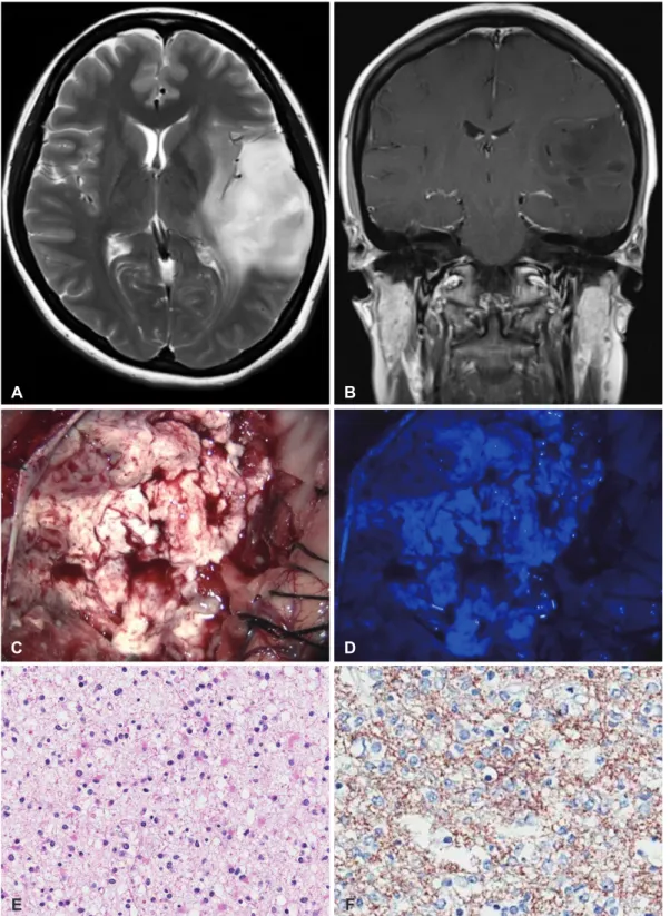
Case 1A 33-year-old woman presented with a 6-month history of severe headaches. Magnetic resonance (MR) imaging reve- aled a lesion with diffuse high T2 signal intensity, focal areas of cystic changes, and increased perfusion in the left tempo- ral lobe (Fig. 1A, B). Based on a presumed diagnosis of low- grade glioma with possible anaplastic foci, the patient under- went fluorescence-guided surgical resection. The patient was administered 20 mg/kg of 5-ALA 4 hours before the induc- tion of general anesthesia. A Leica M720 OH5 microscope (Leica, Wetzlar, Germany) equipped with an FL400 Fluores- cence Module was used for the investigation of 5-ALA fluo- rescence. Resection of the tumor was performed using intra- operative evoked potential monitoring, and all operative fields were assessed for fluorescence. There was no fluorescence at all in the operative field (Fig. 1C, D) and maximal safe resec- tion was achieved. The pathological diagnosis was classic ex- traventricular neurocytoma (World Health Organization gr- ade II). Hematoxylin-eosin staining showed that the tumor was
composed of uniform round cells with or without clear cyto- plasm. Some scattered cells showed astrocytic differentiation (Fig. 1E), and the mitotic rate was low (2/10 high power field).
Immunohistochemical staining was positive for glial fibrillary
acidic protein and synaptophysin, and the Ki-67 labeling in- dex was 2.84% (Fig. 1F). The patient developed postoperative transient sensory aphasia, from which she fully recovered wi- thin one month. No adjuvant treatment was administered, and
A
C
E
B
D
F
Fig. 1. A: A transaxial T2-weighted MR image showing a diffuse high signal mass in the left temporal lobe. B: The mass was not enhanced in the coronal T1-weighted MR image with contrast. C and D: No fluorescence reaction was observed in any tumor tis- sue during surgery. E: The tumor composed of uniform round cells with or without clear cytoplasm (hematoxylin-eosin staining). F:
Immunohistochemical staining for synaptophysin showing diffuse, strong reactivity in the tumor cells. MR: magnetic resonance.
no evidence of recurrence was observed at the regular imag- ing follow-up at 24 months after surgery.
Case 2
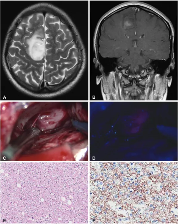
A 16-year-old girl presented with an episode of convulsion without any neurological deficits. MR images showed an in- traaxial mass with faint focal enhancement and high T2 signal
intensity in the right high frontal lobe (Fig. 2A, B). The patient underwent fluorescence-guided tumor resection after a pre- sumed diagnosis of low-grade glioma with anaplastic foci. The same procedure as that described for the above Case 1 was per- formed. Fluorescence was observed in some areas of the tu- mor tissue (Fig. 2C, D), and frozen biopsies were obtained from multiple sites. Three tissues that showed strong red flu-
A
C
E
B
D
F
Fig. 2. A and B: A preoperative MRI scan showing a right high frontal mass with high signal intensity on a T2-weighted image and focal enhancement on a T1-weighted image with contrast. C and D: Intraoperatively, the mass showed positive fluorescence reac- tion and was red after illumination with ultrawave light of 440-nm wavelength. E: The tumor was composed of monotonous round cells with clear cytoplasm (hematoxylin-eosin staining). F: Immunohistochemical staining for synaptophysin showing diffuse, strong reactivity in the tumor cells. MRI: magnetic resonance imaging.
orescence was confirmed as high-grade tumors, whereas all other tissues that showed no fluorescence were determined to be low-grade tumors. During the tumor resection, motor-ev- oked potentials were decreased. We therefore decided not to proceed with the resection. The lesion with the positive fluo- rescence was totally removed grossly, but some remnant tu- mor was observed on postoperative MR images. The patient developed transient foot drop postoperatively, which almost completely resolved one month later. The histological diag- nosis was compatible with atypical EVN, which was compos- ed of monotonous round cells with a clear halo (Fig. 2E, F). As- trocytic differentiation was also found, and endothelial hyper- plasia and focal necrosis were present. The mitotic rate was 4/10 HPF, and the Ki-67 labeling index was elevated at 7.3%.
The patient received adjuvant radiotherapy due to the residual tumor and atypical features. At the 28-month follow-up, the patient returned to her normal activities without any clinical or radiological evidence of tumor recurrence.
DISCUSSION
Since central neurocytomas were first described by Has- soun et al. [8] in 1982, there has been a series of reports about neurocytomas that arise from the brain parenchyma without any connection to the ventricular system [9]. These so-called ‘ex- traventricular neurocytomas’ were named as the counterpart to central neurocytomas, which are typically located in the lat- eral ventricle near the foramen of Monro [6]. In the recent 2007 World Health Organization classification, EVN was recogniz- ed as a new entity to distinguish it from central neurocytomas [10]. EVNs are rare, and their clinical characteristics still need to be evaluated. The radiological findings of EVNs mimic gl- iomas and are effectively treated with complete resection, re- gardless of histological grade [6,7]. When EVNs display atypi- cal histological features such as vascular proliferation, necro- sis, increased mitotic activity and an increased proliferation index, they are classified as atypical EVNs [6,7]. However, the impact of these histological features on predicting the clinical behavior of these tumors is unknown, and the total resection of the tumor is the best indicator of prognosis, regardless of histological grade [6,7]. Rades et al. [7] reviewed 85 cases of atypical neurocytomas, including 21 cases of atypical EVN, and concluded that patients who underwent complete resec- tion achieved better local control and better survival rates compared to patients who underwent incomplete resection.
They also showed that the patients who underwent incomplete resection appeared to benefit from radiotherapy. Brat et al.
[6] reported on 35 EVNs from three large institutes and sh- owed that none of the 14 completely resected tumors recurred within the follow-up period, whereas ten of the 19 incom-
pletely removed tumors recurred, regardless of adjuvant ra- diotherapy. Recurrence occurred in five of ten classic EVNs and five of nine atypical EVNs after incomplete removal. In the Brat report, only one of ten atypical EVNs was completely re- sected.
Gross total resection of benign brain tumors plays a signif- icant role in prognosis. In malignant brain tumors, gross total resection and overall prognosis seem to be highly correlated [2]. Fluorescence-guided resection of brain tumors has been extensively studied as a potential tool to help neurosurgeons achieve maximal resection of gliomas. Recently, fluorescence- guided surgery for brain tumors using 5-ALA has become more widespread in clinical practice. The proposed mecha- nisms of the increased 5-ALA uptake into neoplastic brain tissue include the following: induced low permeability of the blood-brain barrier, the presence of adaptable specific mem- brane transporters, the reduced activity of ferrochelatase, the lowering of the available iron due to chelators, the increased activity of the enzymes that are involved in protoporphyrin IX synthesis, elevated intracellular 5-ALA uptake, and decel- erated protoporphyrin IX outflow from the cell [1]. Various types of tumors were investigated for the use of 5-ALA for tu- mor resection, and the efficacy of the technique has been de- monstrated in the literature [4,5]. To our knowledge, however, the fluorescence reaction for EVN has yet to be reported.
In our 2 cases, the maximal safe resection under fluoresc- ence guidance and intraoperative evoked potential monitor- ing was performed in both patients who had EVN adjacent to eloquent areas, and there were no permanent postoperative neurological deficits. There were some areas that exhibited the fluorescence reaction for 5-ALA within the tumor tissue, and these areas correlated with histological EVN grade. We discovered that the 5-ALA fluorescence reaction may help increase the likelihood of complete resection of highly malig- nant cell burden and may facilitate the intraoperative diag- nosis of histological grade in EVNs. Furthermore, the histo- logical grading system of EVNs has not been systemically de- termined, the 5-ALA fluorescence reaction may have a po- tential role as a biomarker for the prognosis of EVN. Further study on the clinical experience with the 5-ALA fluorescence reaction in EVNs is needed to confirm its role in the clinical evaluation and treatment of these tumors.
In summary, the 5-ALA fluorescence reaction discrimi- nated the histological grade of EVN and was helpful in terms of achieving complete resection and determining the intraop- erative diagnosis in atypical cases. This finding shows that the 5-ALA fluorescence reaction may be a potential biomarker of the clinical behavior of EVNs.
Conflicts of Interest
The authors have no financial conflicts of interest.
REFERENCES
1. Pogue BW, Gibbs-Strauss S, Valdés PA, Samkoe K, Roberts DW, Paulsen KD. Review of Neurosurgical Fluorescence Imaging Methodologies.
IEEE J Sel Top Quantum Electron 2010;16:493-505.
2. Stummer W, Reulen HJ, Meinel T, et al. Extent of resection and survival in glioblastoma multiforme: identification of and adjustment for bias.
Neurosurgery 2008;62:564-76; discussion 564-76.
3. Stummer W, Pichlmeier U, Meinel T, et al. Fluorescence-guided surgery with 5-aminolevulinic acid for resection of malignant glioma: a ran- domised controlled multicentre phase III trial. Lancet Oncol 2006;7:
392-401.
4. Coluccia D, Fandino J, Fujioka M, Cordovi S, Muroi C, Landolt H. In- traoperative 5-aminolevulinic-acid-induced fluorescence in meningio- mas. Acta Neurochir (Wien) 2010;152:1711-9.
5. Utsuki S, Oka H, Sato K, Shimizu S, Suzuki S, Fujii K. Fluorescence di- agnosis of tumor cells in hemangioblastoma cysts with 5-aminolevu- linic acid. J Neurosurg 2010;112:130-2.
6. Brat DJ, Scheithauer BW, Eberhart CG, Burger PC. Extraventricular neurocytomas: pathologic features and clinical outcome. Am J Surg Pa- thol 2001;25:1252-60.
7. Rades D, Fehlauer F, Schild SE. Treatment of atypical neurocytomas.
Cancer 2004;100:814-7.
8. Hassoun J, Gambarelli D, Grisoli F, et al. Central neurocytoma. An electron-microscopic study of two cases. Acta Neuropathol 1982;56:
151-6.
9. Louis DN, Swearingen B, Linggood RM, et al. Central nervous system neurocytoma and neuroblastoma in adults--report of eight cases. J Neu- rooncol 1990;9:231-8.
10. Louis DN, Ohgaki H, Wiestler OD, et al. The 2007 WHO classification of tumours of the central nervous system. Acta Neuropathol 2007;114:
97-109.