http://dx.doi.org/10.4078/jrd.2012.19.2.73
73
<Received:November 2, 2011, Revised:November 21, 2011, Accepted:November 22, 2011>
Corresponding to:Young Mo Kang, Division of Rheumatology, Kyungpook National University School of Medicine, 50, Samdeok-dong, Jung-gu, Daegu 700-706, Korea. E-mail:[email protected]
pISSN: 2093-940X, eISSN: 2233-4718
Copyright ⓒ 2012 by The Korean College of Rheumatology
This is a Free Access article, which permits unrestricted non-commerical use, distribution, and reproduction in any medium, provided the original work is properly cited.
βig-h3 절편-RGD 재조합단백의 만성염증성 관절염 치료효과
장지애1,2ㆍ강진희1,2ㆍ사금희1,2ㆍ한승우1ㆍ서재석1ㆍ김경훈1ㆍ남언정1ㆍ김인산2,3ㆍ강영모1,2,3
경북대학교 의학전문대학원 류마티스내과학교실1, 생화학세포생물학교실2, 경북대학교 세포기질연구소3
Therapeutic Effect of a Recombinant βig-h3 Fragment-RGD Peptide for Chronic Inflammatory Arthritis
Ji Ae Jang1,2, Jin Hee Kang1,2, Keum Hee Sa1,2, Seung Woo Han1, Jae Seok Seo1, Kyung Hoon Kim1, Eon Jeong Nam1, In San Kim2,3, Young Mo Kang1,2,3
Division of Rheumatology, Departments of Internal Medicine1, Biochemistry and Cellular Biology2, Cell and Matrix Research Institute3, Kyungpook National University School of Medicine, Daegu, Korea
Objective. βig-h3 is a 68kDa extracellular matrix protein which is overexpressed in synovial tissues of rheumatoid arthritis (RA). Previous results proved that βig-h3 frag- ments are relevant to adhesion and migration of synovial fibroblast and angiogenesis through interaction with αvβ 3 integrin. We designed a recombinant βig-h3 protein consisting of a fas-1 domain and RGD motif and evaluated the therapeutic efficacy in RA.
Methods. Inhibitory effect of adhesion and migration of NIH3T3 cell line was evaluated in 96 well microtiter and transwell plates coated with βig-h3. Clinical arthritis in- dex was evaluated after treating CIA mice with MFK12.
Immunohistochemical staining in synovial tissues were performed. Expression of transcripts and proteins of in- flammatory mediators were analyzed by semi-quantitative RT-PCR and immunoblotting.
Results. Recombinant protein consisted of 4th fas-1 domain
truncated for H1 and H2 sequences and RGD peptide (MFK12), had M.W. of 10.4kDa. βig-h3 mediated adhe- sion and migration of NIH3T3 cell line were significantly inhibited in a dose-dependent manner. Arthritis severity and incidence were efficiently reduced when CIA mice were treated with MFK12 at 30 mg/kg/day compared with the control. Immunohistochemical staining of joint tissues in MFK12 treated mice exhibited reduced angiogenesis. In treated mice, expression of transcripts regarding in- flammatory mediators was markedly suppressed and im- munoblotting of ICAM-1 and RANKL from whole extract of hind paws also showed a significant reduction.
Conclusion. This study shows that MFK12 is effective in treating RA, although further study is warranted to im- prove the therapeutic efficacy.
Key Words. Rheumatoid arthritis, Inflammation, βig-h3, collagen-induced arthritis, Fas-1
서 론
류마티스관절염은 만성 염증성 자가면역질환으로서 조 직학적으로 초기부터 활막조직 표층에서 섬유아세포양 활
막세포(이하 활막세포)의 증식이 관찰되며, 질병이 진행됨 에 따라 염증세포의 침윤이 증가되어 활막조직의 과증식, 혈관신생, 세포외기질의 축적이 나타난다 (1,2). 과증식된
활막조직은 단백분해효소가 다량 발현되는 파누스를 형성 하며, 인접한 연골 및 뼈의 파괴를 일으킨다. 또한 염증세 포에서 분비된 TNF-α 및 IL-1β와 같은 염증매개물질이 활막세포와 대식세포의 활성화에 관여하며, 부착분자 및 기질금속 단백분해효소(Matrix metalloproteinase, MMP)의 생산 및 분비를 촉진시킴으로써 연골 및 뼈의 세포외기질 분해를 촉진시킨다 (3).
세포외기질단백은 류마티스 관절조직에 증식되어있는 활 막세포, 혈관내피세포, 염증세포의 부착 및 이주, 증식 및 분화를 조절함으로써 염증반응의 유지와 진행에 관여한다 (4,5). 세포외기질에는 콜라겐, glycoprotein, 그리고 proteo- glycan 등이 있으며, 그 중에서 transforming growth factor β (TGF-β)에 의해 유도되는 βig-h3는 상피세포, 내피세포, 케라틴 형성세포, 섬유아세포 및 단핵세포 등 다양한 세포 의 부착, 이주 및 분화 등 기능조절에 관여한다 (6).
βig-h3는 4개의 fasciclin 1 (fas-1) 상동도메인과 카르복실 기 말단의 아르기닌-글라이신-아스파라트산(RGD) 펩타이 드로 구성되어 있는 68 kDa의 기질 단백이다 (7). Fas-1 도 메인에는 티로신(Y)과 히스티딘(H)으로 구성된 YH18모티 프가 있으며 이는 다양한 인테그린과의 상호작용을 통해 각막 (6) 및 신세뇨관 상피세포 (8), 혈관의 내피세포 (9,10) 및 폐섬유아세포 등과 같은 다양한 세포에서 부착 및 이주 조절에 관여한다. 또한 RGD 펩타이드 역시 vitronection, fi- bronectin, osteopontin과 같은 세포외기질의 구성요소로서 인테그린과 결합하여 세포의 부착 및 이주, 증식, 분화에 도 관여하며, 다양한 세포의 염증매개물질의 발현을 조절 함으로써 염증반응 및 종양의 진행에 관여한다 (11-13).
이전 연구에서 βig-h3가 류마티스관절염 활막조직에 과 발현되어있음을 (14) 규명한 바 있으며, 활막세포의 β ig-h3-매개 부착저해 실험을 통해 두 번째와 네 번째 fas-1 도메인 각각의 YH18 모티프의 차단효과를 조사한 결과, 네 번째 fas-1 도메인의 YH18에서 더 효과적임을 규명한 바 있다 (15). 하지만 YH18 모티프는 최소활성 단편으로 100 μmol/L 이상 높은 농도에서 활성을 나타내며 효율이 매우 낮았다. 이러한 문제를 극복하기 위해 YH18 모티프 보다 좀 더 효과적으로 기질-인테그린 상호작용을 차단할 수 있는 새로운 구조의 펩타이드 개발이 필요하다.
본 연구에서는 βig-h3 네 번째 fas-1 도메인과 RGD 펩타 이드를 재조합하여 마우스 유래 섬유아세포인 NIH3T3 세 포주를 이용하여 부착 및 이주 조절기전을 조사하고, 만성 염증성 관절염 동물모델에서 그 치료효과 및 작용기전을 규명하고자 하였다.
대상 및 방법 재조합 단백 합성
야생형 마우스 βig-h3 유전자로부터 H1 및 H2 서열을 제 거(ΔH1ΔH2)한 4번째 fas-1 도메인(아미노산 548-620)과 카르복실기 말단의 RGD 펩타이드로 구성된 재조합 단백을
합성하였다 (6,10). βig-h3의 ΔH1ΔH2 네 번째 fas-1 도메 인의 프라이머(forward: 5’-TATCATATGCAAGCCATGCC- 3’, reverse: 5’-TCACCGAATTCCATGATGTC-3’)를 이용하여 pol- ymerase chain reaction (PCR)을 통해 증폭한 유전자를 pET- 29b (Novagen, Darmstadt, Germany) 발현벡터의 NdeI/ EcoRI 자리에 삽입하였다. RGD 코돈을 포함한 프라이머(forward:
5’-AATTCGGAGGACGAGGAGATGGAGGAC-3’, reverse: 5’-T CGAGTCCTCCATCTCCTCGTCCTCCG-3’)를 이용하여 RGD 유전자를 EcoRI/XhoI 자리에 삽입시켰다. 발현벡터는 BL21 (DE3) 대장균에 넣고 배양하였으며, Isopropyl-β-D-Thiogal- actopyranoside (IPTG) 유도생산하여 단백을 발현시켰다.
MFK12 단백을 순분리 과정에서 Triton X-114를 이용하여 지질다당체(LPS)를 제거한 후 LPS 제거 칼럼을 두 번 연속 통과시켜 0.1 EU/마우스 이하로 유지하였다.
세포 부착 및 이주 저해실험
마우스의 섬유아세포인 NIH3T3 세포 부착저해실험을 위 해 96-well microtitre plate (Costar, Cambridge, MA)에 5 μg/
mL의 재조합 마우스 βig-h3를 넣고 4oC에서 하룻밤 동안 코팅한 후, 실온에서 2% bovine serum albumin (BSA)이 함 유된 phosphate buffered saline (PBS)를 넣어 비특이적인 결 합을 차단시켰다. NIH3T3 세포(5×10⁵ cells/mL)에 재조합 단백인 MFK12를 37oC에서 1시간 동안 전처치한 후, 0.1 mL을 코팅된 well에 넣고 37oC에서 2시간 동안 배양하였 으며, 부착되지 않은 세포는 제거하였다. 각 well에 3.75 mM p-nitrophenyl-N-acetyl-D-glycosaminide (hexosaminidase substrate)와 0.25% Triton X-100이 포함된 50 mM citrate buffer (pH 5.0)를 넣고 37oC에서 2시간 동안 배양하였다.
효소활성 중지를 위해 5 mM EDTA (pH 10.4)가 포함된 50 mM glycine buffer로 처리한 후 Multiscan MCC/340 micro- plate reader (Molecular device, Sunnyvale, CA)를 이용하여 405 nm에서 흡광도를 측정하였다 (6).
NIH3T3 세포 이주 저해실험을 위해 βig-h3 (5 μg/mL)를 구멍크기가 8 μm인 transwell (Costar, Cambridge, MA)의 아랫면을 코팅한 후, 2% BSA로 비특이적인 세포의 결합 을 차단시켰다. NIH3T3 세포(1×10⁶ cells/mL)와 MFK12를 37oC에서 1시간 동안 전처치한 후, 0.1 mL씩 transwell 윗칸 에 넣고 37oC에서 6시간 동안 배양하였다. 세포를 4% par- aformaldehyde로 고정한 후, 0.2% crystal violet으로 염색하 여 임의의 9장을 현미경으로 관찰하여 세포 수를 측정하 였다 (14).
콜라겐 유도 관절염 마우스 모델 제작
콜라겐 유도 관절염(collagen-induced arthritis; CIA) 모델 은 6∼8주령의 DBA/1J (SLC Inc., Hamamatsn, Japan) 마우 스를 이용하였다. 우형 제 2형 콜라켄(bovine CII, Chond- rex, Redmond, Washington)을 complete Freund adjuvant (Chondrex, Redmond, Washington)와 혼합하여 마우스 꼬리
에 CII 100 μg를 피내 주사하였으며, 면역화한 21일 후에 CII와 incomplete Freund adjuvant (Chondrex, Redmond, Washington)를 혼합한 후 2차 면역화시켰다. 치료효과를 살펴보기 위하여 MFK12는 10 mg/kg 또는 30 mg/kg의 농 도로 PBS에 녹인 후 1차 면역화 후 24일째부터 매일 복강 내 투여하였으며, 대조군은 100μL PBS를 투여하였다. 관 절염의 심한 정도(clinical arthritis index)는 네 발의 부종과 발적을 평가하였으며 0∼4 (0: 부종 없음, 1: 한 개의 관절 의 가벼운 부종, 2: 2개 이상 관절의 중등도 부종, 3: 대부 분의 관절의 심한 부종, 4: 전체적인 심한 부종)점으로 분 류하였다 (16).
조직염색 및 면역조직화학 염색법
마우스의 발조직을 얻은 후 피부를 제거하고 10% 포르말 린 용액으로 고정시킨 후 10% EDTA 용액에 넣어 탈회과 정을 거쳤으며, 탈회된 조직은 3 μm 두께의 절편으로 만 들었다. 조직절편을 탈파라핀화 시킨 후 100% 메탄올에 0.3% 과산화수소를 넣은 용액을 이용하여 조직 내 비특이 적인 과산화효소를 차단시켰다. 관절 및 조직학적 소견을 평가하기 위하여 Hematoxylin-Eosin 염색을 하였으며, 신 생혈관 생성 및 내피세포의 분포를 살펴보기 위해 면역조 직화학염색을 하였다. 면역조직화학염색은 5% BSA를 이 용하여 조직 내 비특이적인 단백결합을 차단시킨 후, 내피 세포 표지자인 CD31 (San Francisco CA, USA) 및 ICAM-1 (BD Bioscience, San Francisco CA, USA)에 대한 일차항체 로 배양하였다. 바이오틴이 붙은 이차항체를 실온에서 30 분간 배양하였으며, avidin-biotin-peroxidase complex (Vec- tastain Elite ABC kit, Vector laboratories)로 반응시킨 후, DAB (Dako, Carpinterra, USA)로 발색시켜 확인하였다.
반정량 RT-PCR
관절조직은 추출시약(Easy-spinTM, Intron, Sungnam, Korea) 을 이용하여 균질화하고 총 RNA를 추출하였다. 총 RNA 로부터 AMV 역전사효소(Roche, 0.5 μL), oligo dT (Roche, 1μL), 10 mM dNTP (Takara, 2 μL), RNAse 저해제(Roche, 0.1 μL)를 이용하여 first strand cDNA합성을 하였다.
cDNA는 프라이머와 Taqman 탐침(Roche, Basel, Switzer- land)을 사용하여 LightCycler 480 system (LC480, Roche Diagnostic, Mannheim, Germany)에서 전사체수를 구한 후 18S RNA 전사체와 비교하여 정량화하였으며 정상관절조 직의 전사체를 이용하여 normalized ratio를 계산하였다. 실 험에 이용된 프라이머는 다음과 같다. 마우스 TNF-α (forward: 5’-CTGTAGCCCACGTCGTAGC-3’, reverse: 5’-TTG AGATCCATGCCGTTG-3’), IL-1β (forward: 5’-TGTAATGAA AGACGGCACACC-3’, reverse: 5’-TCTTCTTTGGGTATTGCT TGG-3’), IL-6 (forward: 5’-GAGAAAAGAGTTGTGCAATGG C-3’, reverse: 5’-CCAGTTTGGTAGCATCCATCA-3’), CCL-2 (forward: 5’-CATCCACGTGTTGGCTCA-3’, reverse: 5’-GATC
ATCTTGCTGGTGAATGAGT-3’), RANKL (forward: 5’-TGAA GACACACTACCTGACTCCTG-3’, reverse: 5’-CCACAATGT GTTGCAGTTCC-3’), VCAM-1 (forward: 5’-TGGTGAAATG GAATCTGAACC-3’, reverse: 5’-CCCAGATGGTGGTTTCCTT- 3’), MMP-1 (forward: 5’-TGTGTTTCACAACGGAGACC-3’, re- verse: 5’-GCCCAAGTTGTAGTAGTTTTCCA-3’), MMP-3 (for- ward: 5’-TGTTCTTTGATGCAGTCAGC-3’, reverse: 5’-GATTT GCGCCAAAAGTGC-3’), 18S RNA (forward: 5’-AAATCAGTT ATGGTTCCTTTGGTC-3’, reverse: 5’-GCTCTAGAATTACCAC AGTTATCCAA-3’).
Western blot 분석
순분리한 MFK12 및 관절조직에서 분리한 단백의 농도는 Bradford 단백정량키트를 이용하여 측정하였다. 단백은 10% sodium dodecyl sulfate polyacrylamide gel (SDS-PAGE) 에서 전기영동한 후, nitrocellulose membrane (PerkinElmer, Waltham, MA)로 옮겼다. 10% skim milk를 넣은 0.1%
TBS-T20 용액(20 mm/L Tris-HCl, 137 mm/L NaCl, 0.1%
Tween-20)으로 비특이적 결합을 차단하였으며, polyclonal anti-βig-h3 (경북의대 김인산 교수 제공), monoclonal an- ti-ICAM-1 (Minneapolis, USA)과 anti-RANKL (R&D sys- tems, Minneapolis, USA) 항체들을 이용하여 반응시켰다.
Horseradish peroxidase가 붙은 이차항체와 실온에서 한 시 간 동안 반응시킨 후 enhanced chemiluminescent kit (GE healthcare, Amersham Bucks, UK)로 발색하였다. 밴드신호 강도는 densitometeric scanning으로 결정하고 NIH Image J 소프트웨어로 분석하였다.
통계 분석
실험결과는 평균±표준오차로 나타내었으며, 평균의 차이 는 Student’s t-test를 이용하였다. 통계는 SPSS (statisical package for the social science) 10.0 for window를 사용하여 분석하였으며, 통계적 유의성은 p<0.05 인 경우로 하였다.
결 과 MFK12 재조합 단백의 구조
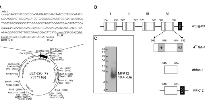
저자들은 βig-h3의 네 번째 fas-1 도메인의 YH18 모티프 가 류마티스관절염 조직에서 분리한 활막세포에서 세포의 부착 및 이주 조절하는 기전을 규명한 바 있다 (15). 그러 나 RGD 모티프와 YH18 모티프는 세포 부착과 이주의 기 본단위로서 효율적인 기능 조절을 위해서는 펩타이드를 증가시킬 필요가 있을 것으로 추정된다. 본 연구에서는 이 를 위해 βig-h3의 네 번째 fas-1 도메인에서 H1 및 H2 서 열을 제거한 dhfas-1과 RGD 펩타이드를 연결한 재조합 단 백(MFK12)를 클로닝하였다(그림 1A, B). 순분리한 단백을 면역블롯팅으로 조사하여 예상 크기인 10.4 kDa의 재조합 MFK12를 확인하였다(그림 1C).
Figure 1. Design of the recombinant MFK12 peptide. (A) A schematic representation of pET-29b(+) containing MFK12 sequence. (B) MFK12 peptide was produced by combination of dhfas-1 (amino acid 502-632) with RGD motif. (C) Western blot analysis of purified recombinant MFK12.
Figure 2. Effective inhibition of βig-h3-mediated adhesion and migration by MFK12. To elucidate the efficiency of MFK12 in the regulation of cell function, MFK12 was incubated with NIH3T3 cell line in different concentrations before performing adhesion (A) and migration assay (B). Data demonstrates the relative percentage of inhibition by MFK12 compared with that by the control. Results are the mean and SEM of 3 independent experiments (*p<0.05 versus control).
NIH3T3 세포를 이용한 MFK12 기능 억제조사
YH18 모티프는 활막세포의 부착을 비교적 고농도(100 μM 이상)로 처리하였을 때 억제할 수 있으며 (14), 섬유아세포 (MRC-5)나 혈관내피세포 부착억제들과도 유사하였다 (9,17).
본 연구에서는 마우스 섬유아세포주인 NIH3T3 세포의 β ig-h3-매개 부착 및 이주에 대한 MFK12의 효과를 확인하였 다. MFK12를 전처리한 결과, 세포 부착억제 효과는 1 μM에 서부터 뚜렷하게 관찰되었으며 대조군보다 80% 이상 억제 하였다(그림 2A). 부착 후 βig-h3에 의해 세포 이주가 촉진
되는 지 확인하기 위해 2-compartment transwell system을 이 용하여 이주시험을 하였으며, NIH3T3 세포는 βig-h3에 의해 농도의존적으로 이주하였다(data not shown). MFK12는 저농 도(1 μM)에서도 세포의 이주를 억제하였으며, 고농도(10 μ M)에서는 80%이상의 억제를 나타내었다(그림 2B).
CIA 마우스에서 MFK12의 치료효과
MFK12가 세포의 βig-h3-매개 부착 및 이주 저해에 관여 한다는 in vitro 실험결과를 바탕으로 마우스 CIA 모델을
Figure 3. MFK12 induces amelioration of collagen-induced arthritis (CIA) where βig-h3 is unregulated. (A) Immunohistochemical staining of F4/80 and βig-h3 in joint tissues from normal and CIA mice. Expression of F4/80-expressing macrophages and βig-h3 in serial sections of joint tissues (×200). (B&C) Development of arthritis in MFK12-treated mice. From day 22 following the first immunization, mice were treated with MFK12 (0, 10, and 30mg/kg/day, intraperitoneally). (B) Clinical arthritis index was recorded daily.
Values are the mean and SEM. (C) Incidence of arthritis among the four paws was recorded at day 30, 40, and 50 after the first immunization. Values are the mean and SEM. *p<0.05 versus the control.
이용하여 MFK12의 관절염 치료효과를 평가하였다. CIA 마우스의 관절조직에 βig-h3의 발현을 확인하였으며, 류 마티스관절염 활막조직과 마찬가지로 (14) βig-h3가 과발 현되어 있었다(그림 3A). 제 2형 콜라겐으로 마우스를 2차 면역화하였으며, 1차 면역화 후 22일째부터 매일 MFK12를 복강투여(10 mg/kg 및 30 mg/kg)한 치료군과 PBS만 투여한 대조군의 관절염 중증도를 평가하였다. MFK12 10 mg/kg 치료군에서는 1차 면역화 후 39일까지 CAI가 낮은 상태를 유지하였으나 40일 이후부터 급격하게 증가하였다. 이에 비해 MFK12 30 mg/kg 치료군에서는 치료기간 동안 CAI가 지속적으로 낮은 상태를 유지하였다(그림 3B).
관절염 침범 발의 빈도는 1차 면역화 후 50일째까지 10일 마다 관찰하였으며, 저용량 치료군에서는 대조군에 비해
CAI의 차이는 있었으나 발병률은 유사하였다. 고용량 치 료군에서는 치료 30일 동안 관절염의 발병률이 대조군의 20% 정도로 통계적으로 유의하게 낮았다(그림 3C). 이 결 과는 MFK12가 YH18 모티프에 비해 치료효과를 극대화할 수 있는 치료제로서의 가능성이 있음을 보여준다.
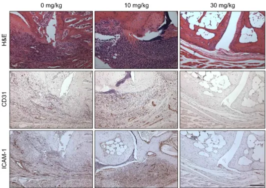
MFK12 치료 후 염증세포 침윤 및 매개물질 발현 변화 MFK12를 치료한 관절조직에서 조직학적 차이를 조사하 였으며, 저용량 치료군에서는 염증세포의 침윤과 연골 및 뼈의 파괴의 소견이 뚜렷이 발생하였으나 고용량 치료군에 서는 이런 소견이 거의 관찰되지 않았다(그림 4). MFK12로 치료한 관절조직에서 염증억제기전을 확인하기 위해 면역 화학염색으로 신생혈관 및 ICAM-1 분포를 확인하였다. 그
Figure 4. Histologies and immu- nohistologies of joints tissues from control and MFK-12 treated (10 and 30 mg/kg) CIA mice. Expre- ssion of CD31 and ICAM-1 was detected in CIA mice. (Hematoxy- lin counter stained for immunohis- tochemical stain, ×200).
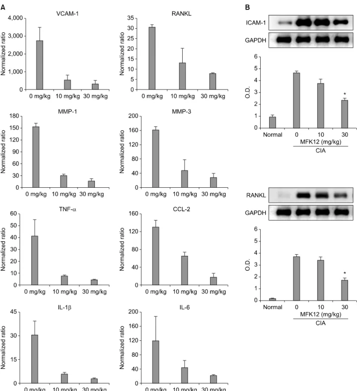
결과 대조군과 저용량 치료군은 신생혈관의 생성(CD31 양 성)이 많고 ICAM-1 발현도 높게 나타났으나, 고용량 치료 군에서는 신생혈관이 매우 드물었고 ICAM-1도 혈관벽에 미량 발현되어 있었다(그림 4). MFK12 치료군과 대조군의 관절조직을 채취하여 균질화한 후 총 RNA 및 단백을 추출 하여 염증매개물질의 발현 정도를 확인하였다. 반정량 RT-PCR을 통해 염증매개물의 전사체 발현을 조사한 결과, TNF-α, IL-1β, IL-6, RANKL, CCL-2, VCAM-1, MMP-1, MMP-3의 전사체는 저용량 치료군에서도 감소하였으나 고 용량 치료군에서 보다 일관된 감소소견을 보였다(그림 5A).
하지만 ICAM-1과 RANKL 단백 발현은 저용량 치료군에는 약간의 감소만 관찰되었으나 고용량 치료군에서는 통계적 으로 유의한 감소를 보였다(그림 5B). 이 결과는 MFK12 치 료에 의해 염증매개물의 mRNA 발현과 단백 생산이 모두 감소되지만 염증매개물에 따라서는 발현양상에 차이가 있 음을 보여준다.
고 찰
본 연구 결과, βig-h3의 H1, H2부위를 제거한 네 번째 fas-1 도메인(dhfas-1)과 RGD 펩타이드로 재조합한 MFK12 는 NIH3T3 세포의 βig-h3-매개 부착 및 이주를 통계적으 로 유의하게 저해하였고, YH18 모티프에 비해 저농도에서 효과를 나타내었다. 마우스CIA 모델에서는 MFK12 치료 군에서 농도의존적으로 관절염에 대한 치료효과를 나타내 었다. 조직학적 소견으로는 활막조직의 과증식, 파누스의 형성, 연골 및 뼈의 파괴를 감소시키면서 혈관신생을 억제 하였고 다양한 염증매개물질의 발현을 단백 및 전사체 수 준에서 억제하였다.
류마티스관절염의 활막조직은 섬유아세포양 활막세포가 증식 및 활성화되어 있고, 활막표층 및 하층에 세포외기질 단백 발현이 증가되어 있는데 이는 미세섬유구성단백, glycosaminoglycan (GAG) 및 fibronectin과 같은 부착성 단 백으로 구성되어 있으며 (2), 활막조직 뿐 만 아니라 관절 액에서도 발현이 뚜렷하게 증가되어 있다 (18,19). 기질의 수용체인 인테그린 역시 활막조직에 과발현되어 있어 세 포와 세포외기질 간의 활발한 상호작용이 이루어지고 있 다 (20). βig-h3는 다른 세포외기질단백에 비해 상대적으 로 작은 크기(68 kDa)를 가지며, laminin과 제 1형 및 제 4형 콜라겐 및 fibronectin과 직접적인 원섬유를 형성함으 로써, 염증세포나 거주세포의 이주 시 어댑터의 역할을 담 당한다 (21,22).
혈관내피세포 및 폐 섬유아세포 연구에서 네 개의 도메 인에 있는 YH 모티프들은 정도의 차이가 있으나 모두 세 포 부착을 저해할 수 있었다 (6,9,17). 하지만 일부 세포에 서는 두 번째와 네 번째 fas-1 도메인이 주로 부착 및 이주 조절을 하는 것으로 밝혀졌다 (8,9,23-25). 각각의 fas-1 도 메인 내에 있는 YH18 모티프의 아미노산 서열은 유사성 을 나타내지만 조금씩 차이가 있는데, 두 번째 도메인의 티로신은 아르기닌으로 치환되어 있으며, 네 번째 도메인 의 YH18 모티프와 유사한 정도의 소수성을 나타낸다 (17).
활막세포에서는 두 번째 fas-1 도메인보다 네 번째 fas-1 도 메인에 있는 YH18 모티프에 의해 부착이 더 효율적으로 저해되는 것을 확인하였다 (15). 한편 RGD 펩타이드는 α 5β1, αvβ2, αvβ3 혹은 αvβ5 등의 다양한 인테그린 과 결합함으로써 세포에 따라 부착, 이주, 성장, 분화 및 세포사멸 작용을 조절한다 (23,26,27). 본 연구결과 MFK12
Figure 5. Effect of MFK12 treatment on the expression of inflammatory mediators in CIA mice. (A) Semi-quantitative measurement of transcripts of inflammatory mediators from joint tissues. Transcripts were calculated as target gene/reference gene (18s ribosome RNA) ratios and normalized to the relative expression level of non-arthritis mice. (B) Immunoblotting analysis of ICAM-1 and RANKL for proteins extracted from joint tissues of representative paws. Graphs show the mean and SD optical density (O.D.) from at least 3 independent experiments. The results are means±SD (*p<0.05 versus 0 mg/kg).
는 마우스 섬유아세포의 부착 및 이주를 저농도에서도 매 우 효과적으로 억제하였다. 이는 네 번째 dhfas-1과 RGD 펩타이드 각각의 인테그린 결합능으로 인해 상대적으로 소량에 의해서도 세포의 인테그린에 대한 결합 포화도를
높일 수 있었기 때문으로 추정된다. 뿐만 아니라, 네 번째 dhfas-1과 RGD 펩타이드에 의한 인테그린 클러스터링은 야생형 βig-h3의 4개의 상동도메인에 의한 다중결합에 비 해 효율이 훨씬 낮아서 세포신호전달 경로를 약화시킬 수
있으며, 이를 통해 세포와 기질간의 상호작용을 조절하는 것으로 추정할 수 있다. 향후MFK12 단백에 의한 활막세 포, 내피세포, 림프구 및 대식세포 등의 기능조절 기전규 명을 위한 추가 연구가 필요할 것이다.
세포외기질단백을 기반으로 추출한 펩타이드는 인테그린 결합에 의한 세포 활성화 기능조절을 이용하여 질병치료제 로서 연구되어 왔다. 제 18형 콜라겐에서 추출한 endostatin (28)은 인테그린 α5β1 과, 그리고 제 4형 콜라겐에서 추출 한 tumstatin (29)은 인테그린 αvβ3와의 결합을 통해 내피 세포의 증식, 이주 및 혈관 신생을 저해할 수 있으며 (30), plasminogen에서 추출한 greenstatin과 angiostatin은 VEGF 발 현을 조절함으로써 종양의 진행과정에서 혈관신생을 억제 한다 (31). 또한 관절염 모델에 angiostatin 발현 유전자를 주 입하였을 때 관절염의 치료효과가 있었다(32). βig-h3에서 추출한 fastatin도 인테그린과의 결합을 통해 혈관신생을 억 제하고 단핵구의 부착 및 이주를 저해하는 것으로 보고되 었다 (10,33). 그러나 류마티스관절염에서 세포외기질단백 추출 펩타이드를 이용한 관절염 치료제 연구는 세계적으로 매우 부족한 상태며 향후 다양한 기질단백을 기반으로 하 는 펩타이드 약제의 개발연구가 필요하다.
새로운 작용기전의 관절염 치료제가 지속적으로 개발되 고 있지만 류마티스관절염의 발병에 있어 아직까지 밝혀 지지 않은 기전이 많이 남아있다. 그리고 단일 표적을 차단 하는 치료제뿐 만 아니라 좀 더 작용범위가 넓은 약제의 개발이 필요하다. 당뇨병 및 암과 같은 질환에 있어 βig-h3 를 이용한 다양한 기전 연구가 발표되었으나 (10,34,35), 류 마티스관절염에서 뚜렷하게 증가되어 있는 (14,15,36) β ig-h3를 이용한 연구의 진행은 부족한 상황이다. 따라서 향 후 본 연구에서 규명한 MFK12를 바탕으로 좀 더 낮은 농 도에서 보다 효과적인 작용을 나타낼 수 있는 새로운 구조 의 βig-h3 기반 재조합 단백에 대한 연구가 필요하다.
결 론
류마티스관절염의 진행에 중요한 역할을 담당하는 섬유 아세포양 활막세포는 세포외 기질단백과 결합하여 활막조 직내에서의 이주 및 활성화가 일어난다. βig-h3 기반 재조 합단백인 MFK12는 NIH3T3 세포의 부착 및 이주를 저농 도에서도 효과적으로 저해하는 것으로 나타났으며, 관절 염 동물 모델에서 관절의 파괴 및 변형, 활막조직의 과증 식, 파누스의 형성 등을 억제하였고, 또한 염증매개물질의 발현 역시 현저하게 감소시키는 것으로 확인하였다. 본 연 구로 βig-h3 네 번째 fas-1 도메인과 RGD 펩타이드가 류 마티스관절염에 있어서 효과적인 치료제로 사용될 가능성 을 제시하였다.
감사의 글
이 논문 또는 저서는 2006년 정부(교육인적자원부)의 재 원으로 한국학술진흥재단의 지원을 받아 수행된 연구임
(KRF-2006-311-E00313).
참고문헌
1. Knedla A, Neumann E, Müller-Ladner U. Developments in the synovial biology field 2006. Arthritis Res Ther 2007;9:209.
2. Firestein GS. Invasive fibroblast-like synoviocytes in rheumatoid arthritis. Passive responders or transformed aggressors? Arthritis Rheum 1996;39:1781-90.
3. Kim JM, Kim KH. Pathogenesis of rheumatoid arthritis.
J Korean Med Assoc 2010;53:853-61.
4. Barilla ML, Carsons SE. Fibronectin fragments and their role in inflammatory arthritis. Semin Arthritis Rheum 2000;29:252-65.
5. Tomasini-Johansson BR, Milbrink J, Pejler G. Vitronectin expression in rheumatoid arthritic synovia--inhibition of plasmin generation by vitronectin produced in vitro. Br J Rheumatol 1998;37:620-9.
6. Kim JE, Kim SJ, Lee BH, Park RW, Kim KS, Kim IS.
Identification of motifs for cell adhesion within the re- peated domains of transforming growth factor-β-induced gene, βig-h3. J Biol Chem 2000;275:30907-15.
7. Skonier J, Bennett K, Rothwell V, Kosowski S, Plowman G, Wallace P, et al. β ig-h3: a transforming growth fac- tor-β-responsive gene encoding a secreted protein that inhibits cell attachment in vitro and suppresses the growth of CHO cells in nude mice. DNA Cell Biol 1994;13:
571-84.
8. Park SW, Bae JS, Kim KS, Park SH, Lee BH, Choi JY, et al. βig-h3 promotes renal proximal tubular epithelial cell adhesion, migration and proliferation through the inter- action with α3β1 integrin. Exp Mol Med 2004;36:211-9.
9. Nam JO, Kim JE, Jeong HW, Lee SJ, Lee BH, Choi JY, et al. Identification of the αvβ3 integrin-interacting mo- tif of βig-h3 and its anti-angiogenic effect. J Biol Chem 2003;278:25902-9.
10. Nam JO, Jeong HW, Lee BH, Park RW, Kim IS.
Regulation of tumor angiogenesis by fastatin, the fourth FAS1 domain of βig-h3, via αvβ3 integrin. Cancer Res 2005;65:4153-61.
11. Kim JE, Kim SJ, Jeong HW, Lee BH, Choi JY, Park RW, et al. RGD peptides released from βig-h3, a TGF-β-in- duced cell-adhesive molecule, mediate apoptosis. Oncogene 2003;22:2045-53.
12. Ruoslahti E. RGD and other recognition sequences for integrins. Annu Rev Cell Dev Biol 1996;12:697-715.
13. Trabocchi A, Menchi G, Cini N, Bianchini F, Raspanti S, Bottoncetti A, et al. Click-chemistry-derived triazole li- gands of arginine-glycine-aspartate (RGD) integrins with a broad capacity to inhibit adhesion of melanoma cells and both in vitro and in vivo angiogenesis. J Med Chem 2010;53:7119-28.
14. Nam EJ, Sa KH, You DW, Cho JH, Seo JS, Han SW, et al. Up-regulated transforming growth factor β-inducible gene h3 in rheumatoid arthritis mediates adhesion and mi- gration of synoviocytes through αvβ3 integrin: Regulation by cytokines. Arthritis Rheum 2006;54: 2734-44.
15. Nam, EJ, Song EJ, Kim JM, Seo JS, Sa KH, Cho HJ, et al. βig-h3-Mediated Adhesion of Fibroblast-Like Synoviocytes in Rheumatoid Arthritis. J Korean Rheum Assoc 2008;15:222-9.
16. Brownlie RJ, Myers LK, Wooley PH, Corrigall VM, Bodman-Smith MD, Panayi GS, et al. Treatment of murine collagen-induced arthritis by the stress protein BiP via in- terleukin-4-producing regulatory T cells: a novel function for an ancient protein. Arthritis Rheum 2006;54:854-63.
17. Kim JE, Jeong HW, Nam JO, Lee BH, Choi JY, Park RW, et al. Identification of motifs in the fasciclin domains of the transforming growth factor-β-induced matrix pro- tein βig-h3 that interact with the αvβ5 integrin. J Biol Chem 2002;277:46159-65.
18. Clemmensen I, Hølund B, Andersen RB. Fibrin and fi- bronectin in rheumatoid synovial membrane and rheuma- toid synovial fluid. Arthritis Rheum 1983;26:479-85.
19. Vartio T, Vaheri A, Von Essen R, Isomäki H, Stenman S. Fibronectin in synovial fluid and tissue in rheumatoid arthritis. Eur J Clin Invest 1981;11:207-12.
20. Nikkari L, Aho H, Yli-Jama T, Larjava H, Jalkanen M, Heino J. Expression of integrin family of cell adhesion receptors in rheumatoid synovium. α 6 integrin subunit in normal and hyperplastic synovial lining cell layer. Am J Pathol 1993;142:1019-27.
21. Kim JE, Park RW, Choi JY, Bae YC, Kim KS, Joo CK, et al. Molecular properties of wild-type and mutant βIG-H3 proteins. Invest Ophthalmol Vis Sci 2002;43:656-61.
22. Sarkissian M, Lafyatis R. Integrin engagement regulates proliferation and collagenase expression of rheumatoid synovial fibroblasts. J Immunol 1999;162:1772-9.
23. LeBaron RG, Bezverkov KI, Zimber MP, Pavelec R, Skonier J, Purchio AF. βIG-H3, a novel secretory protein inducible by transforming growth factor-β, is present in normal skin and promotes the adhesion and spreading of dermal fibro- blasts in vitro. J Invest Dermatol 1995;104:844-9.
24. Kim JE, Kim EH, Han EH, Park RW, Park IH, Jun SH, et al. A TGF-β-inducible cell adhesion molecule, β ig-h3, is downregulated in melorheostosis and involved in osteogenesis. J Cell Biochem 2000;77:169-78.
25. Lee BH, Bae JS, Park RW, Kim JE, Park JY, Kim IS.
βig-h3 triggers signaling pathways mediating adhesion and migration of vascular smooth muscle cells through α vβ5 integrin. Exp Mol Med 2006;38:153-61.
26. Lutz R, Pataky K, Gadhari N, Marelli M, Brugger J, Chiquet M. Nano-stenciled RGD-gold patterns that inhibit
focal contact maturation induce lamellipodia formation in fibroblasts. PLoS One 2011;6:e25459.
27. Yoon SH, Mofrad MR. Cell adhesion and detachment on gold surfaces modified with a thiol-functionalized RGD peptide. Biomaterials 2011;32:7286-96.
28. Thomas JP, Arzoomanian RZ, Alberti D, Marnocha R, Lee F, Friedl A, et al. Phase I pharmacokinetic and phar- macodynamic study of recombinant human endostatin in patients with advanced solid tumors. J Clin Oncol 2003;
21:223-31.
29. Maeshima Y, Yerramalla UL, Dhanabal M, Holthaus KA, Barbashov S, Kharbanda S, et al. Extracellular ma- trix-derived peptide binds to α(v)β(3) integrin and in- hibits angiogenesis. J Biol Chem 2001;276:31959-68.
30. Sudhakar A, Sugimoto H, Yang C, Lively J, Zeisberg M, Kalluri R. Human tumstatin and human endostatin exhibit distinct antiangiogenic activities mediated by αvβ3 and α5β1 integrins. Proc Natl Acad Sci USA 2003;100:4766-71.
31. Hajitou A, Grignet C, Devy L, Berndt S, Blacher S, Deroanne CF, et al. The antitumoral effect of endostatin and angiostatin is associated with a down-regulation of vascular endothelial growth factor expression in tumor cells. FASEB J 2002;16:1802-4.
32. Kato K, Miyake K, Igarashi T, Yoshino S, Shimada T.
Human immunodeficiency virus vector-mediated in- tra-articular expression of angiostatin inhibits progression of collagen-induced arthritis in mice. Rheumatol Int 2005;25:522-9.
33. Kim HJ, Kim IS. Transforming growth factor-β-induced gene product, as a novel ligand of integrin αMβ2, pro- motes monocytes adhesion, migration and chemotaxis. Int J Biochem Cell Biol 2008;40:991-1004.
34. Cha DR, Kim IS, Kang YS, Han SY, Han KH, Shin C, et al. Urinary concentration of transforming growth fac- tor-β-inducible gene-h3(βig-h3) in patients with Type 2 diabetes mellitus. Diabet Med 2005;22:14-20.
35. Lee SH, Bae JS, Park SH, Lee BH, Park RW, Choi JY, et al. Expression of TGF-β-induced matrix protein β ig-h3 is up-regulated in the diabetic rat kidney and human proximal tubular epithelial cells treated with high glucose.
Kidney Int 2003;64:1012-21.
36. Kang YM, Kim SI, Kim JS, You DW, Sa KH, Park EJ, et al. Regulation of βig-h3 production in rheumatoid syn- ovitis by inflammatory madiators. J Korean Rheum Assoc 2005;12:73-82.