| Abstract |
1)PURPOSE: The purpose of this study was to determine the effect of different frequencies (4㎐ and 100㎐) of transcutaneous electrical nerve simulation (TENS) on pain relief using c-fos expression in the spinal cord of rat osteoarthritis to investigate the appropriate frequency for pain relief.
METHODS: Total of 30 Sprague-Dawley rats was used and randomly divided 2 groups according TENS frequency and applicate the TENS during 3 period (3 days, 7 days, 10 days). The induction of osteoarthritis by 3mg monosodium iodoacetat was injected into the right knee joint of rats. Three days later, commercially available TENS unit was used for stimulation was set to 20minutes on 3, 7, 10 days after surgery.
Western blot analysis system was used to detect immunoreactive proteins. The thickness of the bands were photographically measured by Scion Image.
RESULTS: When investigating the c-fos expression of
†Corresponding Author : [email protected]
This is an Open Access article distributed under the terms of the Creative Commons Attribution Non-Commercial License (http://creativecommons.org/licenses/by-nc/3.0) which permits unrestricted non-commercial use, distribution, and reproduction in any medium, provided the original work is properly cited.
TENS on spinal cord in OA knee over 10 days, between-groups differences in c-fos expression reached a significant level by day 10. For within-groups comparisons, the c-fos expression decreased significantly across days in low- and high-frequency TENS groups.
CONCLUSION: Whether at low- and high-frequency, the TENS as a therapy obtained beneficial effects of pain relief and TNES at high-frequency is more beneficial effects on the pain relief when TENS applied at injury site.
Key Words: Frequencies, Osteoarthritis, Transcutaneous electrical nerve stimulation, Pain relief
Ⅰ. Introduction
Osteoarthritis (OA) is the most common degenerative joint musculoskeletal disease that causes physical disability (Hinton et al., 2002). Mechanical stress and inflammation are involved in OA onset and progression. Disease management of OA is benefited in the early stage by therapies such as exercise, diet, anti-inflammatory medicines, and injections that delay the disease’s progression and pain.
Research Article Open Access
The Short-Term Effects of Difference Frequency of Transcutaneous Electrical Nerve Stimulation on Pain Relief using c-fos Expression in Spinal Cord with Knee
Osteoarthritis Rats
Hyun-Mo Koo, PT, PhD⋅Sang-Su Na, PT, PhD
1†Department of Physical Therapy, College of Science, Kyungsung University
1
Department of Physical Therapy, Daegu University Received: July 29, 2016 / Revised: July 29, 2016 / Accepted: August 23, 2016
ⓒ 2016 J Korean Soc Phys Med
Pain is one of the major factors in the interruption of movement, so pain relief is important for the quality of life for OA patients. Reports of pain in humans and animals have demonstrated that nociceptive stimuli can facilitate metabolic changes on the dorsal horn of the spinal cord (Klein et al., 2004; Schadrack et al., 1999). Met- and leu-enkephalin peptides are found on primary afferent terminals in the spinal cords in rodents (Wang et al., 2010).
The encephalin can reduce the responses of primary afferent stimulation, particularly of activated neurons, by noxious stimuli located in the dorsal horn (Duggan et al., 1977).
This is caused by inhibiting transmitter release by the nociceptive primary afferents (Beaudry et al., 2011). Thus, spinal nociception may modulate encephalin by inhibiting the primary afferents and subsequently by inhibiting the spinal nociceptive neurons. C-fos is a general pain marker in the spinal cord and is known as the suppressor of encephalin. Previous studies showed that c-fos activated neurons containing preproenkephalin (PPE), which produces encephalin and encodes for the proenkephalin polypeptide that yields methionine (met) and leucine (leu-) encephalin peptides after different types of nociceptive inputs (Hossaini et al., 2014).
Transcutaneous electrical nerve stimulation (TENS) is one of the physical modalities most often used clinically for the treatment of many types of pain, including that caused by OA of the knee. TENS is well documented in relieving pain, and various simulation parameters have been adopted, from 4㎐ to 100㎐ frequencies (Law and Cheing, 2004; Lee and Song, 2013). Previous studies found that both high-frequency TENS (100㎐) and low-frequency TENS (4㎐) affected hyperalgesia after treatment, and biochemical studies have demonstrated that different frequencies of TENS activate different endogenous opioid systems in the central nervous system (CNS) (Andersson et al., 1977; Han et al., 1991; Sluka et al., 1998). High-frequency stimulation accelerates the release of dynorphin, while low-frequency stimulation
releases enkephalins, B-endorphins, and endomorphins, which act on different receptors in the CNS. However, there is conflicting evidence regarding the effect of TENS frequencies. Previous study have demonstrated that high-frequency TENS produces more effective analgesia and a greater decrease in the activity of dorsal horn neurons than does low-frequency TENS (Willer et al., 1982).
However, evidence that low-frequency TENS provides a greater reduction of the activity of spinothalamic tract cells has also been reported (Garrison and Foreman, 1994; Lee et al., 1985).
Thus, the purpose of this study was to determine the effect of different frequencies (4㎐ and 100㎐) of TENS on pain relief using c-fos expression in the spinal cord of rat osteoarthritis models.
Ⅱ. Methods
1. Experimental animals and Procedures
In this study, total 30 adult male Wistar rats (8 weeks
of age) weighting between 250 and 300g. The induction
of osteoarthritis was performed as previously reported
(Jekal et al., 2014). In brief, 3mg monosodium iodoacetat
(MIA, Sigma, St Louis, MO, America) was injected into
the right knee joint of rats. Three days later, commercially
available TENS unit was used for stimulation was set to
20 minutes per day for 3, 7, 10 days after surgery. Two
frequencies were used, a high-frequency (100㎐; 20-40㎃)
and a low-frequency (4㎐; 10-20㎃) using pairs of rubber
electrodes placed on right knee. Pulse duration was either
100μsec or 250μsec. Injected animals were divided 2
groups randomly according to TENS (Endomed,
Enraf-nonius, Netherlands) frequency and applicate the
TENS during 3 period: high-frequency TENS (100㎐) for
3 days after surgery, high-frequency TENS (100㎐) for
7 days after surgery, high-frequency TENS (100㎐) for
10 days after surgery, low-frequency TENS (4㎐) for 3
days after surgery, low-frequency TENS (4㎐) for 7 days after surgery, low-frequency TENS (4㎐) for 10 days after surgery.
2. Western blot analysis
After the animals were anesthetized and sacrificed, the spinal cord of each group were collected and then homogenized and lysed with RIPA buffer. Equal amount of protein (20㎍) were resolve via 12% sodium dodecyl sulfate-polyacrylamide gel electrophoresis (SDS-PAGE) and transferred to nitrocellulose membranes. The blots was washed with TBS-T, blocked with 5% skim milk for 1 hour, then incubated with anti c-fos (1:1000) antibody at the dilutions recommended by the suppliers. The membranes was washed and the primary antibody was detected using horseradish peroxidase-conjugated goat anti-mouse IgG (1:3000). Western blot analysis system was used to detect immunoreactive proteins. The thickness of the bands were photographically measured by Scion Image.
3. Statistical analysis
PASW 18 was used for the analysis and significance level was set at .05. Repeated two-way ANOVA was used to analyze the effects of the group and period on the c-fos expression level, and Fisher’s LSD test was used for post
hoc evaluations.Ⅲ. Results
1. Comparison of c-fos expression level between low- and high-frequency TENS group The c-fos expression level of frequency and period in each group was two-way repeated ANOVA and LSD test was used for the post hoc evaluations. When investigating the c-fos expression of TENS on spinal cord in OA knee pain over 10 days, significant interaction between the
‘frequency’ and ‘period’ was observed (p=.004) (Table 1).
The between-groups differences in c-fos expression reached
Subjects SS Df MS F P
Frequency 5400.40 2 2700.70 569.26 .00*
Period 145.53 1 145.53 30.71 .00*
Frequency / period 68.27 2 34.63 7.21 .00*
*P<.05
Table 1. The statistical analysis of c-fos expression level in each group
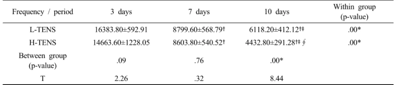
Frequency / period 3 days 7 days 10 days Within group
(p-value)
L-TENS 16383.80±592.91 8799.60±568.79† 6118.20±412.12†‡ .00*
H-TENS 14663.60±1228.05 8603.80±540.52† 4432.80±291.28†‡∮ .00*
Between group
(p-value) .09 .76 .00*
T 2.26 .32 8.44
Mean±SD; mean±standard deviation
†= significant difference from 3day P<.05
‡= significant difference from 7day P<.05
∮= significant difference from L-TENS P<.05
L-TENS: low-intensity transcutaneous electrical nerve simulation H-TENS: high-intensity transcutaneous electrical nerve simulation
Table 2. Changes of c-fos expression level on the 2 groups across period
a significant level by day 10 (p=.001). However, the c-fos expression level were not significantly different among the low- and high-frequency groups in day 3, day 7 (Table 2).
2. Comparison of c-fos expression level within- groups
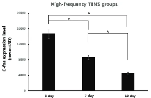
For within-groups comparisons, the c-fos expression decreased significantly across days in high-and low- frequency TENS groups. From day 3 to day 10, there was 63.7% decrease in low-frequency TNES group (Fig. 1), 69.8% decrease in high-frequency TENS groups (Fig. 2).
Ⅳ. Discussion
A is a chronic disease that is associated with reduced muscle strength, disability, and a progressive loss of function due to pain (Hinman et al., 2002). The progression of pain is not fully understood, but recent studies suggest that the central and peripheral nervous systems may be involved in chronic pain due to changes in the excitability of the spinal pathway (Imamura et al., 2008). Thus, the control of pain could influence muscle activation and normal movement for patients with OA. This study investigated the pain levels using c-fos in the spinal cord using Western blot analysis.
From the study results, it is evident that high- and
low-frequency TENS reduces c-fos expression in the spinal cords of rats with osteoarthritis after treatment. TENS is often used clinically to treat pain and is widely applied to patients with osteoarthritis. Many studies have been done to find the most effective parameters, including the frequency of TENS, but these parameters have yet to be identified. Ericksson and colleagues showed that low frequencies and high frequencies are more effective for the relief of pain compared with conventional TENS, but a different study demonstrated that there were no significant differences in pain levels (Eriksson et al., 1984; Jensen et al., 1991). Mechanical studies for the frequency of TENS showed various analgesic mechanisms in which low- frequency stimulation increases met-enkephalin-Arg- Phe (MEAP), while high-frequency stimulation accelerates the dynorphin (Han et al., 1991). Another study showed that alternating the stimulation frequency of TENS between low (2㎐) and high (100㎐) frequencies produces a synergistic interaction of dynorphin and encephalin and that it has more analgesic effect than a fixed frequency (Xiao-Hong et al., 1994). Following these results, different frequencies of TENS seem to relate to different analgesic mechanisms.
In this study, repeated application of low- and high-frequency TENS showed a significant decrease of c-fos expression. This is supported by the findings of the previous study, in which the application of TENS was useful for pain relief (Hossaini et al., 2014). The findings
Fig. 1. C-fos expression level of low-frequencyTENS group
Fig. 2. C-fos expression level of high-frequency TENS group