171
Clinical Practice Using Allograft (cadaver) Skin and Cultured Epithelial Autografts in Massive Burns
Kook Hyeun Jung, M.D., Do-hern Kim, M.D., Jun Her, M.D., Wook Chun, M.D., Jong Hyun Kim, M.D., Young Chul Jang, M.D.
1and Sae Wha Jeon
2, Ph.D.
Departments of Surgery and
1Plastic Surgery, Hangang Sacred Heart Hospital, College of Medicine, Hallym University,
2Tego Science, Seoul, Korea
Backgrounds: The procedure using allograft skin from cadavers has been the safest standard treatment for patients with extensive burns, but the results of the sandwich grafting technique using cadaver skin were not always successful. In massive burns, graft failure is crucial, so we have attempted to modify the sandwich grafting technique using CEAs (cultured epithelial autografts, Holoderm, Tego Science) instead of cadaver skin.
Methods: 1. Early selective escharectomy was performed. 2. Allograft skin (cadaver) was grafted in excision sites. 3. At the same time, a small skin biopsy (1 cm
2) was performed for CEAs (Holoderm) production. 4. A modified sandwich grafting technique (1: 3∼6 meshed autog- raft with CEAs) was performed for areas with formation of granulation after the allograft skin had come away. 5. Acellular dermal substitute (AlloDerm, Life Cell) was concurrently used in some cases. 6. In accordance with the wound conditions, we removed the CEAs after 4∼8 days.
If possible, we tried to leave them intact.
Results: Twelve subjects were included in this investigation. The mean age was 25.8 (1∼51) years old. The average burn area was 50.8 (25∼75) %TBSA. There were 7 cases of flame burns and 2 more cases each of chemical burns and electrical burns and 1 case of scalds. A 1:3-6 meshed autograft was used with CEAs. The mean area of the modified sandwich grafting technique used was 2,291.3 (1,120∼4,816) cm
2. All of these areas were successfully covered with epithelial cells. In 6 cases, AlloDerm was simultaneously used. In some cases, for infection control, Acticoat (Smith & Nephew) was used in the boundary of the unclean wound.
동종피부(사체피부)이식과 자기유래 배양피부를 이용한 광범위 화상의 치료
한림대학교 의과대학 한강성심병원 외과학교실, 1성형외과학교실, 2테고사이언스
정국현․김도헌․허 준․전 욱․김종현․장영철1․전세화2
책임저자:전 욱, 서울시 영등포구 영등포동 2가 94-200, 우편번호: 150-719, 한강성심병원 외과 Tel: 02-2639-5793, Fax: 02-2678-4386, E-mail: [email protected]
서 론
동종피부이식을 이용한 광범위화상의 치료는 가 장 안전한 표준 치료법이다. 조기 가피절제술을 시 행하고 그 부위에 사체피부를 이식함으로써 감염 을 예방하고 신선한 육아조직이 잘 자라게 도와준 다. 하지만 사체피부를 이용한 샌드위치 이식방법 은 항상 성공적이지는 않았다. 피부 공여부가 한정 된 광범위화상에서 자가 피부이식 실패는 치명적 이 될 수 있기에, 저자는 사체피부이식을 통하여 육아조직을 만들고 그 부위에 자기유래 배양피부
(CEA, Cultured Epithelial Autograft, Holoderm)를 이 용한 변형된 샌드위치 이식방법을 사용하여 성공 적으로 이들을 치료하였기에 보고하는 바이다.
대상 및 방법
2004년 9월부터 2005년 10월까지 한림대학교 한 강성심병원 화상센터에 입원한 12명의 광범위화상 환자를 대상으로 하였다.
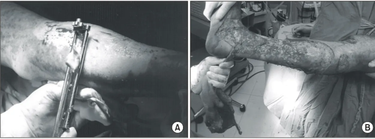
1. 조기 선택적 가피절제술을 시행한다(Fig. 1).
2. 사체피부를 절제부위에 이식하며, 동시에 자 기유래 배양피부(홀로덤, 테고사이언스) 생산을 위 Conclusions: CEAs were created from a full thickness biopsy of the patient's own skin and required 3 weeks to grow. The grafted allograft skin usually came away 3 weeks later. CEAs could help wide meshed autografts to be used safely in massive burn patients. Early selective escharectomy, allograft skin coverage, acellular dermal substitutes and wide meshed autograft with CEA application would be immensely helpful techniques in patients with extensive burns. If we can reduce CEA production time to 14 days, the allodermis of cadaver skin will be more useful in the new modified sandwich grafting technique
ꠏꠏꠏꠏꠏꠏꠏꠏꠏꠏꠏꠏꠏꠏꠏꠏꠏꠏꠏꠏꠏꠏꠏꠏꠏꠏꠏꠏꠏꠏꠏꠏꠏꠏꠏꠏꠏꠏꠏꠏꠏꠏꠏꠏꠏꠏꠏꠏꠏꠏꠏꠏꠏꠏꠏꠏꠏꠏꠏꠏꠏꠏꠏꠏꠏꠏꠏꠏꠏꠏꠏꠏꠏꠏꠏꠏꠏꠏꠏꠏꠏꠏ Key Words: Major burns, Cultured epithelial autograft
Fig. 1. Selective Escharectomy was performed for full thickness burn areas and, if possible, within 72 hours of the actual burn. (A) Tangential excision, (B) fascial level excision.
A B
해 1 cm 의 피부를 생검한다(Fig. 2, 3).
3. 3주 뒤 사체피부가 떨어지고 육아조직이 자란 부위에 자기유래 배양피부를 이용한 변형된 샌드
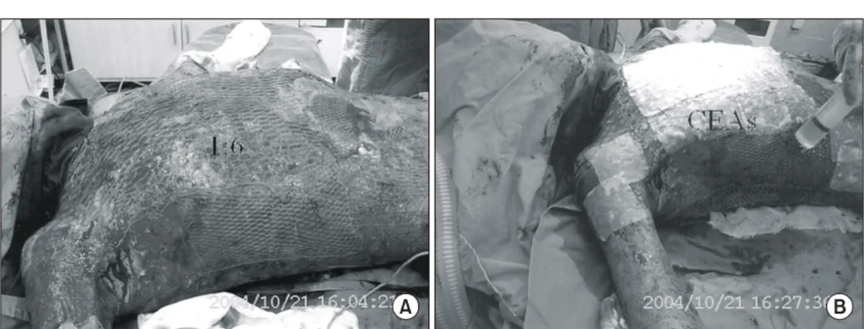
위치 이식방법(wide meshed autograft with CEAs)으 로 피부이식을 시행한다. 홀로덤은 fibrin glue를 분 사한 후 적용하였다(Fig. 4).
4. 경우에 따라 관절부위 수축을 예방하기위해 서 무세포성 진피대체물(AlloDerm, Life Cell사)을 동시에 사용한다(Fig. 5).
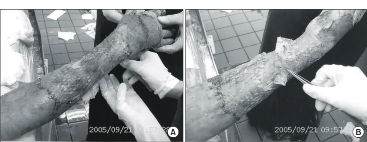
5. 시술부위 상태에 따라 홀로덤은 수술 후 4∼8 일 후에 제거한다. 하지만 가능한 한 오랫동안 둔 다(Fig. 6).
자기유래 배양피부를 사용한 후 드레싱은 일반 적인 피부이식술의 경우와 같이 Wet to Dry 드레 싱을 사용하였다.
결 과
12명의 광범위 화상 환자를 대상으로 하였다. 평 균 연령은 25.8 (1∼51)세이고 평균 화상면적은 Fig. 2. Allograft(cadaver) skin was grafted in excision
sites.
A
C
B
Fig. 3. (A, B, C) At the same time, a small skin
biopsy (1 cm
2) was performed for CEAs (Holoderm,
Tego Science) production.
ꠏꠏꠏꠏꠏꠏꠏꠏꠏꠏꠏꠏꠏꠏꠏꠏꠏꠏꠏꠏꠏꠏꠏꠏꠏꠏꠏꠏꠏꠏꠏꠏꠏꠏꠏꠏꠏꠏꠏꠏꠏꠏꠏꠏꠏꠏꠏꠏꠏꠏꠏꠏꠏꠏꠏꠏꠏꠏꠏꠏꠏꠏꠏꠏꠏꠏꠏꠏꠏꠏꠏꠏꠏꠏꠏꠏꠏꠏꠏꠏꠏꠏꠏꠏꠏꠏꠏꠏꠏꠏꠏꠏꠏꠏꠏꠏꠏꠏꠏꠏ
50.8 (25∼75) %TBSA (total body surface area)이었 다. 변형된 샌드위치 이식방법을 사용한 평균 면적 은 2,291.3 (1,120∼4,816) cm
2였다. 변형된 샌드위 치 이식을 위해 1:3∼6배의 자가 그물이식편이 사용되었으며, 모든 경우 자가이식편의 초기 생착 이 성공적으로 이루어졌다. 이후 거의 대부분의 부 위에 재상피화가 성공적으로 일어났으며, 6예에서 는 수술 후 관절구축을 예방하기 위해 관절부위에 진피대체물인 AlloDerm을 동시에 사용하였다. 수 술부위의 오염을 예방하기 위해 경계부위에 Acti- coat (Smith & Nephew)를 선택적으로 사용하였다
(Table 1).
고 찰
저자는 2003년 12월 이후 9명의 광범위 소아 화 상환자에서 부모로부터 얇은 피부(신선 동종피부) 를 얻어 사용함으로써 이들을 모두 생존시켰을 뿐 만 아니라, 신선 동종피부의 효과에 대해서 많은 것을 배울 수 있었다. 아주 얇은 동종피부 이식편 이지만 이들은 가피절제 부위와의 빠른 혈관 연결 을 통해 수술부위 감염을 예방하여, 진피가 남아있 Fig. 4. (A, B) A modified sandwich grafting technique (1:4∼6 meshed autograft with CEAs) was performed for areas with formation of granulation after the allograft skin had come away.
A B
Fig. 5. (A, B) Acellular dermal substitute (AlloDerm) was concurrently used in 6 cases to prevent scar contraction on the joint areas.
A B
는 부위는 스스로 재상피화가 일어나게 하였고, 3 도 화상부위에는 육아조직이 자라게 하였다. 수술 후 7∼8일 경에 동종피부이식편에 대한 혈관 연결 이 차단되게 되어 12일경에 탈락되지만 그 동안 환자를 확실하게 보호해 주었다. 경험이 늘면서 저 자는 접면괴사조직절제술 시에 지방층이 노출된 경우에는 환아의 피부를 조금 떼어내어 6배의 그 물이식편을 만들어 이식을 하고 그 위에 신선 동
종피부를 이식하는 샌드위치 이식방법을 사용하였 다. 100% 재상피화를 이루었으며, 따라서 대부분 한두 번의 수술로 치료를 끝낼 수 있었다. 하지만 샌드위치 이식방법을 사용한 부위는 경우에 따라 심한 수축을 보였다. 2004년 9월경 저자는 외국으 로부터 수입된 사체피부를 사용하기 시작하였는 데, 광범위화상환자에서 이 들을 이용한 샌드위치 이식방법은 결과가 그리 좋지는 않았다. 이는 아마 도 사체피부의 신선도가 떨어지고 접착력이 떨어 지기 때문이라 생각하였다. 피부공여부가 절대적 으로 부족한 광범위화상에서 이식 실패는 치명적 일 수도 있다. 사체피부는 이식 후 21일 전후로 탈 락되었다. 또한 사체피부를 이용함으로써 아주 높 은 범위의 광범위 화상 환자도 생존 기간이 연장 됨으로써, 이제는 그 넓은 화상부위를 덮을 수 있 는 방법을 생각해야 했다. 그러던 중 자기유래 배 양피부를 국내에서 사용할 수 있음과 배양 기간에 21일이 걸린다는 사실을 알게 되었다.
1)즉, 자기유 래 배양피부가 배양되는 3주간의 시간을 사체피부 를 이식함으로써 버틸 수 있다고 생각하였다. 더군 다나 육아조직이란 대부분의 구성성분이 신생혈관 이 풍부한 콜라겐 덩어리며, 혈액 공급이 좋기에 성공률을 더욱 높일 수 있다. 하지만 자기유래 배 양피부(Cultured Epithelial Autograft, 이하 CEA)를 사용함에 있어서 육아조직 위에 단순히 올려놓는 Fig. 6. (A, B) In accordance with the wound conditions, we removed the CEAs after 4∼8 days. If possible, we tried to leave them intact. 7 days later after modified sandwich grafting method.
A B
Table 1. Summary of Results
ꠚꠚꠚꠚꠚꠚꠚꠚꠚꠚꠚꠚꠚꠚꠚꠚꠚꠚꠚꠚꠚꠚꠚꠚꠚꠚꠚꠚꠚꠚꠚꠚꠚꠚꠚꠚꠚꠚꠚꠚꠚꠚꠚꠚꠚꠚꠚꠚ
Age/ Burn % Allograft CEAs
Case Mode
Sex of TBSA (%TBSA) (cm
2)
ꠏꠏꠏꠏꠏꠏꠏꠏꠏꠏꠏꠏꠏꠏꠏꠏꠏꠏꠏꠏꠏꠏꠏꠏꠏꠏꠏꠏꠏꠏꠏꠏꠏꠏꠏꠏꠏꠏꠏꠏꠏꠏꠏꠏꠏꠏꠏꠏ
1 42/M FB 42 40 1,960
2 4/F FB 35 35 1,960
3 35/M EB 43 35 1,120
4 38/F FB 34 30 1,120
5 24/M EB 65 35 1,120
6 4/F FB 75 70 2,240
7 4/F FB 25 25 1,120
8 21/M CB 70 45 4,816
9 51/M CB 60 35 3,360
10 41/F FB 37 35 1,960
11 1/M SB 70 67 2,240
12 44/M FB 53 48 4,480
ꠏꠏꠏꠏꠏꠏꠏꠏꠏꠏꠏꠏꠏꠏꠏꠏꠏꠏꠏꠏꠏꠏꠏꠏꠏꠏꠏꠏꠏꠏꠏꠏꠏꠏꠏꠏꠏꠏꠏꠏꠏꠏꠏꠏꠏꠏꠏꠏ