Usefulness of Quantitative Endotracheal Aspirate Cultures in Intensive Care Unit Patients with Suspected Pneumonia
It is difficult to differentiate pathogens responsible for pneumonia or colonization in patients with an endotracheal tube or in patients that have undergone tracheostomy. We evaluated the clinical usefulness of quantitative endotracheal aspirates cultures and sought to determine the result threshold level for positivity. The authors performed this
retrospective cohort study between December 1, 2004 and January 31, 2006. Forty-five suspected pneumonia patients admitted to an intensive care unit (ICU) with quantitative bronchoalveolar lavage (BAL) and endotracheal aspirate (EA) culture results were enrolled.
Using a threshold of 105 cfu/mL, 10 of the 45 (22.2%) quantitative EA cultures were positive, as compared with 7 (15.6%) BAL cultures. When BAL culture findings were used as the reference, the sensitivity and specificity of quantitative EA cultures were 85.7% and 89.5%, respectively, at a threshold of 105 cfu/mL, and 85.7% and 94.7%, respectively, at a threshold of 106 cfu/mL. Of the 10 EA culture positive patients, 2 patients with a result of -105 cfu/mL were BAL culture negative. The quantitative EA culture is a useful non-invasive tool for the diagnosis of pneumonia pathogens. It is suggested that a threshold level of 106 cfu/mL is appropriate.
Key Words: Quantitative Culture; Endotracheal Aspirate; Pneumonia Yoon Mi Shin1, Yeon-Mok Oh2,
Mi Na Kim3, Tae Sun Shim2, Chae-Man Lim2, Sang Do Lee2, Younsuck Koh2, Woo Sung Kim2, Dong Soon Kim2 and Sang-Bum Hong2
1Division of Pulmonary & Critical Care Medicine, Department of Internal Medicine, Cheongju St.
Mary Hospital, Cheongju; 2Division of Pulmonary &
Critical Care Medicine, Department of Internal Medicine; 3Department of Laboratory Medicine, University of Ulsan College of Medicine, Asan Medical Center, Seoul, Korea
Received: 13 January 2011 Accepted: 21 April 2011 Address for Correspondence:
Sang-Bum Hong, MD
Division of Pulmonary & Critical Care Medicine, Department of Internal Medicine, University of Ulsan College of Medicine, Asan Medical Center, 86 Asanbyeongwon-gil, Songpa-gu, Seoul 138-736, Korea
Tel: +82.2-3010-3893, Fax: +82.2-3010-6968 E-mail: [email protected]
DOI: 10.3346/jkms.2011.26.7.865 • J Korean Med Sci 2011; 26: 865-869
INTRODUCTION
The accurate diagnosis of newly developed pneumonia is diffi- cult in patients with an endotracheal tube or tracheostomy (1), since many other conditions, such as, tracheobronchitis (2), pul- monary edema, and atelectasis, can mimic pneumonia. There- fore bacteriologic tests are necessary to confirm pneumonia;
Infectious Diseases Society of America/American Thoracic So- ciety (IDSA/ATS) guideline is that samples of lower respiratory tract secretions should be obtained from all patients with sus- pected hospital acquired pneumonia (HAP) which should be collected before antibiotic changes. Samples can include an en- dotracheal aspirate, bronchoalveolar lavage sample, or protect- ed specimen brush sample (2).
But qualitative endotracheal aspirate (EA) culture cannot dif- ferentiate colonization from infection (3, 4). Although quantita- tive culture of bronchoalveolar lavage (BAL) fluid obtained by bronchoscopy can accurately diagnose pneumonia, this proce- dure is invasive and cannot be utilized in some patients, espe- cially in those who are critical (5-12).
Quantitative EA culture is non-invasive, easily learnt, and cheaper than quantitative BAL fluid culture (4, 13); and previ-
ous results have suggested that EA can be used as a substitute for BAL in quantitative cultures (4). However, this issue is con- troversial and result thresholds have not been determined. Fur- thermore, little data is available on quantitative EA cultures in Korea, or on the clinical applications of this methodology. Ac- cordingly, we evaluated the clinical usefulness of quantitative EA cultures in intensive care unit (ICU) patients with pneumo- nia, and sought to determine the result threshold level for posi- tivity.
MATERIALS AND METHODS
We performed this retrospective cohort study between Decem- ber 1, 2004 and January 31, 2006. Patients with an endotracheal tube or tracheostomy suspected of having pneumonia admitted to medical ICU at the Asan Medical Center were enrolled whose EA sample was collected within 2 days of a BAL sample. Patients with community acquired, hospital acquired, and health care associated pneumonia were included, as were immunocom- promised or immunocompetent patients, who had previously received antibiotics. However, patients a with lower clinical pul- monary infection score (CPIS) of < 6 or with an interval between
BAL fluid and EA sampling of > 48 hr were excluded.
We evaluated patient demographic data, hospital courses, microbiologic results, the use of antibiotics, and radiographic changes. Acute physiology and chronic health evaluation (APACHE) II scores were determined at ICU admission, and CPIS scores on the first and third days after the onset of hospital acquired pneumonia.
EA samples with > 10 epithelial cells per low power field on a Gram stained slide of a direct smear were rejected as inadequate for culture. EA cultures were quantified using calibrated loops.
Briefly, each 1 microliter of EA itself and 100-fold diluted EA were evenly streaked with 1 microliter-disposable plastic loop on en- tire surface of a chocolate agar plate, a sheep blood agar plate, and a MacConkey agar plate. Plates are incubated overnight in a 5% CO2 atmosphere at 35°C. Colonies were then counted and bacterial concentrations (cfu/mL) were calculated. Microorgan- isms with counts > 104 cfu/mL were submitted for identification and antimicrobial susceptibility testing. If no growth was detect- ed on any plate, the incubation was extended for 24 hr.
Statistical analysis was performed using SPSS version 10.0 (SPSS, Chicago, IL, USA). Results are expressed as means ± SDs.
The chi-square test was used to compare proportions. Threshold levels of 105, 106, and 107 cfu/mL were used to evaluate quanti-
tative EA culture positivity, but quantitative BAL culture positiv- ity was defined as 104 cfu/mL, which is an established threshold level. Sensitivity, specificity, and the predictive values of quan- titative EA cultures were calculated at each of the three thresh- old levels. Kappa analysis was used to determine concordance rates between quantitative EA and BAL culture results.
Ethics statement
This study protocol was approved by the institutional review board (IRB) of Asan Medical Center (Approval number: 2011- 0170). This study was exempted from written informed consent and all the data collected from this study was kept confidential.
RESULTS
A total of 45 patients (32 men, 13 women, mean age 63.4 ± 13.2 yr) were enrolled. Patient demographic data is provided in Table 1. Twenty-eight patients had hospital acquired pneumonia and 17 had community acquired pneumonia (CAP). Their underly- ing diseases included malignancy, chronic lung disease, rheu- matologic disease, and others. The majority of patients (n = 36) were immunocompromised, and 17 of these had previously re- ceived chemotherapeutic agents, corticosteroids, or immuno- suppressive agents. The major cause of ICU admission was re- spiratory failure and hypoxemia. Total patients’ mean APACHE II score at ICU admission was 29.4 ± 7.3. In patients with hospi- tal acquired pneumonia, mean CPIS scores on days 1 and 3 af- ter pneumonia onset were 8.7 ± 2.0 and 8.5 ± 2.0, respectively.
Table 2 shows the hospital courses of the patients. Mean length of ICU stay was 29.8 ± 26.8 days and mean duration of mechan- ical ventilation was 26.4 ± 25.3 days. The average time between BAL and EA sampling was 0.2 ± 0.9 days. Using a threshold of 105 cfu/mL, 10 (22.2%) quantitative EA cultures were positive, as compared with 7 (15.6%) BAL cultures (Table 3). Six of the 7 patients with a positive BAL cultures had a positive EA culture.
Of the 10 EA culture (+) patients, 2 patients with 105 cfu/mL had all BAL culture (-), and of the 8 EA culture (+) patients (at a thresh- Table 1. Patient demographics and clinical characteristics at baseline
Clinical characteristics Data
Total number 45
Age (yr) 63.4 ± 13.2
Sex (M/F) 32/13
Pneumonia HAP CAP
28 17 Underlying disease
Malignancy Chronic lung disease Transplantation recipient Rheumatologic disease Gastrointestinal disease History of malignancy Stroke
Other
19 8 2 4 4 3 3 2 Immune status
Immuno-competent
Immuno-compromised 9
36 Previous medication
Steroid
Chemotherapeutic agent Immunosuppressant None
5 8 4 28 Previous antibiotics
Yes
No 29
16 Cause of ICU care
Respiratory failure and hypoxemia Shock
Close monitoring Post-operation
36 3 4 2 HAP, hospital acquired pneumonia; CAP, community acquired pneumonia.
Table 2. Patient characteristics at ICU admission, clinical pulmonary infection score (CPIS) on days 1 and 3 after pneumonia onset in patients with hospital acquired pneu- monia and hospital courses (days)
Parameters Mean ± SD
APACHE II score on ICU admission 29.4 ± 7.3
CPIS (n = 28) Day 1
Day 3 8.7 ± 2.0
8.5 ± 2.0 Hospital days
Duration of ICU stay
Duration of mechanical ventilation 29.8 ± 26.8 26.4 ± 25.3 Duration of mechanical ventilation before BAL (total)
In HAP In CAP
8.1 ± 14.0 11.8 ± 17.0 2.2 ± 2.3
Time between BAL and EA 0.2 ± 0.9
SD, standard deviation.
old of 106 cfu/mL), 6 were BAL culture (+).
We calculated the diagnostic efficiencies of quantitative EA culture at each threshold level. When BAL culture findings were used as reference, the sensitivity and specificity of quantitative EA culture at a threshold of 105 cfu/mL were 85.7% and 89.5%, respectively and at a threshold of 106 cfu/mL, these were 85.7%
and 94.7%, respectively (Table 4). Kappa analysis showed that the concordance rate of the two diagnostic methods was best at a quantitative EA culture threshold of 106 cfu/mL. The area un- der the ROC curve of quantitative EA culture at this threshold was 0.902 (Fig. 1).
After obtaining culture results, antibiotic regimens were al-
tered in 19 (42%) of the 45 patients. In 16 (36%) patients, antibi- otics were deescalated and these included 12 patients on van- comycin because of a lack of evidence of methicillin-resistant Staphylococcus aureus infection. When we evaluated positivity rates with respect to time of antibiotic use before BAL, we ob- served higher positivity rate in patients who had received anti- biotics for 7 days or longer (P = 0.017).
DISCUSSION
This study shows that quantitative endotracheal aspirate culture is a useful non-invasive tool for the diagnosis of pneumonia pathogens in critically ill patients. Our findings suggest that a threshold level of 106 cfu/mL is appropriate because at this level excellent concordance was found with quantitative BAL culture results.
Ventilator associated pneumonia (VAP) or severe CAP is diffi- cult to diagnosis in ICU patients with an endotracheal tube or a tracheostomy. It is associated with high mortality and morbidity rates (14). Thus, early and accurate diagnosis and appropriate empirical antibiotic treatment are important outcome variables (14).
Many clinical conditions mimic pneumonia in these patients, which in practice, means that microbiologic evaluations are re- quired for the diagnosis of pneumonia. However, it is difficult to obtain specimens from the lower respiratory tract without con- tamination by colonizing bacteria. Therefore culture results are invariably often difficult to interpret. Since qualitative cultures are unable to distinguish between pathogens and colonizing bac- teria, the concept of quantitative culture was developed based Table 4. Characteristics of quantitative EA cultures and the concordance between quantitative BAL and EA cultures
Threshold Sensitivity (%) Specificity (%) PPV (%) NPV (%) κ P value Concordance
≥ 105 85.7 89.5 60.0 97.1 0.64 0.00 Good
≥ 106 85.7 94.7 75.0 97.3 0.76 0.00 Excellent
≥ 107 14.3 100 100 86.4 0.22 0.018 Poor
PPV, positive predictive value; NPV, negative predictive value.
Sensitivity
1-Specificity (%)
107 105 106
0 100
100
0
Fig. 1. ROC curve of quantitative EA culture.
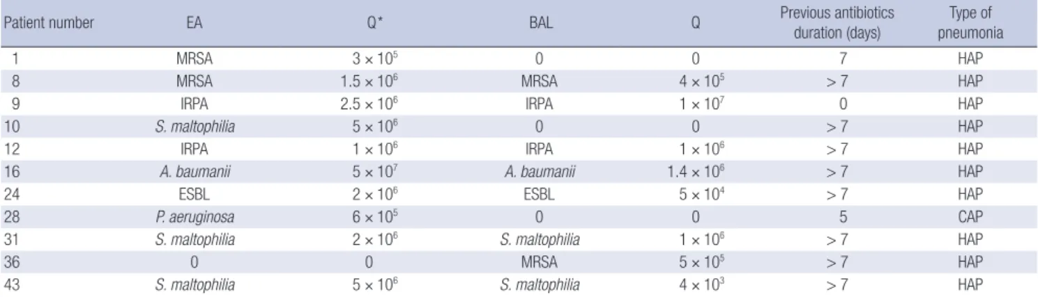
Table 3. Microbiologic results of quantitative cultures, previous antibiotics duration (days) before BAL and type of pneumonia in patients with positive culture
Patient number EA Q* BAL Q Previous antibiotics
duration (days) Type of pneumonia
1 MRSA 3 × 10⁵ 0 0 7 HAP
8 MRSA 1.5 × 10⁶ MRSA 4 × 10⁵ > 7 HAP
9 IRPA 2.5 × 10⁶ IRPA 1 × 10⁷ 0 HAP
10 S. maltophilia 5 × 10⁶ 0 0 > 7 HAP
12 IRPA 1 × 10⁶ IRPA 1 × 10⁶ > 7 HAP
16 A. baumanii 5 × 10⁷ A. baumanii 1.4 × 10⁶ > 7 HAP
24 ESBL 2 × 10⁶ ESBL 5 × 10⁴ > 7 HAP
28 P. aeruginosa 6 × 10⁵ 0 0 5 CAP
31 S. maltophilia 2 × 10⁶ S. maltophilia 1 × 10⁶ > 7 HAP
36 0 0 MRSA 5 × 10⁵ > 7 HAP
43 S. maltophilia 5 × 10⁶ S. maltophilia 4 × 10³ > 7 HAP
*Culture counts. MRSA, methicillin resistant Staphylococcus aureus; IRPA, imipenem resistant Pseudomonas aeruginosa; ESBL, extended-spectrum β-lactamases.
on BAL, protected specimen brush (PSB), blinded protected telescoping catheter, and EA cultures (15-21).
Quantitative EA cultures are straightforward, cheap, easily per- formed, and non-invasive and have been shown to be useful in Western countries (22-26). Previous studies included compara- tive evaluations of the accuracy of quantitative EA cultures for the diagnosis of VAP versus other diagnostic methods, such as, autopsy specimen, BAL, or PSB culture, or clinical methods (22, 24-28). In general, each of these methods is of clinical value, pri- marily because of the invasive nature of bronchoscopy (5-12), but their sensitivities and specificities vary. Additionally positiv- ity thresholds are controversial, 105 cfu/mL (22, 26, 29), or 106 cfu/mL (24, 25, 27).
No reference autopsy specimen was available for this study.
Therefore we used quantitative BAL cultures and clinical diag- nosis as diagnostic standards to confirm the presence of pneu- monia.
When we compared the sensitivity, specificity, and concor- dance of quantitative EA and quantitative BAL culture, we found that the optimal quantitative EA culture threshold level was 106 cfu/mL.
Furthermore, in patients with hospital acquired pneumonia, we also used CPIS (30). No patient with HAP had a CPIS score of < 6 on day 1 and 3 after pneumonia onset, so this study showed high specificity (94.7%) than previous studies that did not use it (24, 25, 27).
When 104 cfu/mL was used as a cutoff value for quantitative BAL cultures, we found that 15.6% of BAL cultures were positive, which is lower than previous studies. This may have been low because we do not routinely perform quantitative BAL culture in critically ill patients with suspected pneumonia. We performed quantitative BAL culture in ICU patients with a poor clinical re- sponse to a previous empirical regimen or atypical clinical symp- toms or signs. So we might enrolled many difficult patients to diagnose or patients suspected of having an atypical pathogens.
Furthermore, many of the cultured bacteria were drug resistant organisms. These may be partially explained by culture positiv- ity rate was high in patients with prolonged antibiotic use rather than short term antibiotic use. Only one CAP patient showed positive EA culture and the others were HAP patients. We had more HAP patients (HAP 28/CAP 17 patients) and most of CAP patients received antibiotics initiation before BAL. When we in- cluded in patients with HAP only, EA and BAL culture rates were 32.1% and 25% respectively.
In our cohort, the antibiotic regimen was changed in 19 pa- tients, including 12 maintained on antibiotics after vancomycin discontinuance, and these actions were taken based on our quan- titative culture results. Thus, quantitative EA culture is likely to reduce antibiotic use.
Several study limitations should be borne in mind. First, this was not a randomized controlled study. Besides BAL was not
performed routinely in all patients with pneumonia or in pa- tients that responded well to antibiotics due to its invasiveness.
Accordingly, this study was performed in patients who had con- tracted a difficult pathogen to diagnose. In addition, we also in- cluded many patients who had been previously treated with an- tibiotics, and we enrolled community and hospital acquired pneumonia cases.
Summarizing, quantitative EA cultures are useful non-inva- sive diagnostic tool in critically ill patients with an endotracheal tube or tracheostomy suspected of having pneumonia especial- ly in HAP. It is also suggested that the appropriate threshold lev- el for quantitative EA culture is 106 cfu/mL.
REFERENCES
1. Baughman RP. Diagnosis of ventilator-associated pneumonia. Microbes Infect 2005; 7: 262-7.
2. American Thoracic Society; Infectious Diseases Society of America.
Guidelines for the management of adults with hospital-acquired, venti- lator-associated, and healthcare-associated pneumonia. Am J Respir Crit Care Med 2005; 171: 388-416.
3. Lambotte O, Timsit JF, Garrouste-Orgeas M, Misset B, Benali A, Carlet J.
The significance of distal bronchial samples with commensals in ventilator- associated pneumonia: colonizer or pathogen? Chest 2002; 122: 1389-99.
4. Albert S, Kirchner J, Thomas H, Behne M, Schur J, Brade V. Role of quan- titative cultures and microscopic examinations of endotracheal aspirates in the diagnosis of pulmonary infections in ventilated patients. J Hosp Infect 1997; 37: 25-37.
5. Tsai SH, Cohen SS, Fenger EP. Bronchial perforation as a complication of bronchoscopy. Am Rev Tuberc 1958; 78: 106-10.
6. Prakash UB, Offord KP, Stubbs SE. Bronchoscopy in North America: the ACCP survey. Chest 1991; 100: 1668-75.
7. Jolliet P, Chevrolet JC. Bronchoscopy in the intensive care unit. Intensive Care Med 1992; 18: 160-9.
8. Valentine VG, Rizk NW, Hancock EW. A complication during bronchos- copy. Hosp Pract (Off Ed) 1993; 28: 22, 27.
9. Pereira W, Kovnat DM, Khan MA, Iacovino JR, Spivack ML, Snider GL.
Fever and pneumonia after flexible fiberoptic bronchoscopy. Am Rev Respir Dis 1975; 112: 59-64.
10. Kiss K, Pápai Z, Szima B, Kis S, Strausz J. Fiberoptic bronchoscopy in in- tensive care units. Orv Hetil 1996; 137: 1689-91.
11. Hammer DL, Aranda CP, Galati V, Adams FV. Massive intrabronchial aspiration of contents of pulmonary abscess after fiberoptic bronchosco- py. Chest 1978; 74: 306-7.
12. Friedman RL. Selective pneumothorax: a complication of bronchoscopy.
Dis Chest 1955; 27: 213-5.
13. Bergmans DC, Bonten MJ, De Leeuw PW, Stobberingh EE. Reproduc- ibility of quantitative cultures of endotracheal aspirates from mechani- cally ventilated patients. J Clin Microbiol 1997; 35: 796-8.
14. Chastre J, Fagon JY. Ventilator-associated pneumonia. Am J Respir Crit Care Med 2002; 165: 867-903.
15. Brun-Buisson C, Fartoukh M, Lechapt E, Honoré S, Zahar JR, Cerf C, Maitre B. Contribution of blinded, protected quantitative specimens to the diagnostic and therapeutic management of ventilator-associated
pneumonia. Chest 2005; 128: 533-44.
16. Clec’h C, Jauréguy F, Hamza L, Karoubi P, Fosse JP, Hamdi A, Vincent F, Gonzalez F, Cohen Y. Agreement between quantitative cultures of pos- tintubation tracheal aspiration and plugged telescoping catheter, pro- tected specimen brush, or BAL for the diagnosis of nosocomial pneumo- nia. Chest 2006; 130: 956-61.
17. Cook D, Mandell L. Endotracheal aspiration in the diagnosis of ventila- tor-associated pneumonia. Chest 2000; 117(4 Suppl 2): 195S-7S.
18. Elatrous S, Boukef R, Ouanes Besbes L, Marghli S, Nooman S, Nouira S, Abroug F. Diagnosis of ventilator-associated pneumonia: agreement be- tween quantitative cultures of endotracheal aspiration and plugged tele- scoping catheter. Intensive Care Med 2004; 30: 853-8.
19. Fangio P, Rouquette-Vincenti I, Rousseau JM, Soullié B, Brinquin L. Di- agnosis of ventilator-associated pneumonia: a prospective comparison of the telescoping plugged catheter with the endotracheal aspirate. Ann Fr Anesth Reanim 2002; 21: 184-92.
20. Fujitani S, Yu VL. Diagnosis of ventilator-associated pneumonia: focus on nonbronchoscopic techniques (nonbronchoscopic bronchoalveolar lavage, including mini-BAL, blinded protected specimen brush, and blind- ed bronchial sampling) and endotracheal aspirates. J Intensive Care Med 2006; 21: 17-21.
21. Papazian L, Thomas P, Garbe L, Guignon I, Thirion X, Charrel J, Bollet C, Fuentes P, Gouin F. Bronchoscopic or blind sampling techniques for the diagnosis of ventilator-associated pneumonia. Am J Respir Crit Care Med 1995; 152: 1982-91.
22. el-Ebiary M, Torres A, González J, de la Bellacasa JP, García C, Jiménez de Anta MT, Ferrer M, Rodriguez-Roisin R. Quantitative cultures of en- dotracheal aspirates for the diagnosis of ventilator-associated pneumo- nia. Am Rev Respir Dis 1993; 148: 1552-7.
23. Michel F, Franceschini B, Berger P, Arnal JM, Gainnier M, Sainty JM, Papazian L. Early antibiotic treatment for BAL-confirmed ventilator-as-
sociated pneumonia: a role for routine endotracheal aspirate cultures.
Chest 2005; 127: 589-97.
24. Marquette CH, Georges H, Wallet F, Ramon P, Saulnier F, Neviere R, Mathieu D, Rime A, Tonnel AB. Diagnostic efficiency of endotracheal aspirates with quantitative bacterial cultures in intubated patients with suspected pneumonia. Comparison with the protected specimen brush.
Am Rev Respir Dis 1993; 148: 138-44.
25. Torres A, Martos A, Puig de la Bellacasa J, Ferrer M, el-Ebiary M, González J, Gené A, Rodríguez-Roisin R. Specificity of endotracheal aspiration, protected specimen brush, and bronchoalveolar lavage in mechanically ventilated patients. Am Rev Respir Dis 1993; 147: 952-7.
26. Sauaia A, Moore FA, Moore EE, Haenel JB, Kaneer L, Read RA. Diagnos- ing pneumonia in mechanically ventilated trauma patients: endotrache- al aspirate versus bronchoalveolar lavage. J Trauma 1993; 35: 512-7.
27. Jourdain B, Novara A, Joly-Guillou ML, Dombret MC, Calvat S, Trouil- let JL, Gibert C, Chastre J. Role of quantitative cultures of endotracheal aspirates in the diagnosis of nosocomial pneumonia. Am J Respir Crit Care Med 1995; 152: 241-6.
28. Marquette CH, Copin MC, Wallet F, Neviere R, Saulnier F, Mathieu D, Durocher A, Ramon P, Tonnel AB. Diagnostic tests for pneumonia in ven- tilated patients: prospective evaluation of diagnostic accuracy using his- tology as a diagnostic gold standard. Am J Respir Crit Care Med 1995;
151: 1878-88.
29. Wu CL, Yang Dle, Wang NY, Kuo HT, Chen PZ. Quantitative culture of endotracheal aspirates in the diagnosis of ventilator-associated pneumo- nia in patients with treatment failure. Chest 2002; 122: 662-8.
30. Singh N, Rogers P, Atwood CW, Wagener MM, Yu VL. Short-course em- piric antibiotic therapy for patients with pulmonary infiltrates in the in- tensive care unit. A proposed solution for indiscriminate antibiotic pre- scription. Am J Respir Crit Care Med 2000; 162: 505-11.
AUTHOR SUMMARY
Usefulness of Quantitative Endotracheal Aspirate Cultures in Intensive Care Unit Patients with Suspected Pneumonia
Yoon Mi Shin, Yeon-Mok Oh, Mi Na Kim, Tae Sun Shim, Chae-Man Lim, Sang Do Lee, Younsuck Koh, Woo Sung Kim, Dong Soon Kim and Sang-Bum Hong
This study shows that quantitative endotracheal aspirate culture is a useful non-invasive tool for the diagnosis of pneumonia pathogens in critically ill patients. A threshold level of 106 cfu/mL is appropriate because excellent concordance with quantitative BAL culture results was found.