서 론
수질성유방암은합포체양상으로자라고, 단핵세포침윤등의특
징적인 조직학적 특징을 갖는 유방암의 하나이다. 1945년 Moore 와 Foote는 간질에 림프구침윤이 있으며 경계가 매우 좋은 유방 암으로 기술하였고, 발생률은 전체 유방암의 1-7% 정도로 보고 되고 있다.(1-3) 수질성유방암은 일반적으로 양호한 예후인자로 알려진 호르몬 수용체의 발현이 적고, 높은 핵등급과 조직학적 등 급을 갖는 것으로 알려져 있지만 예후에 있어서는 다른 침윤성유 방암에 비해 양호한 것으로 알려져 있다.(4-6) 하지만 Fisher 등 (7)과 Ellis 등(8)은 수질성유방암이 다른 침윤성유방암에 비해 예후에 있어 크게 유리하지 않다고 하였으며 이러한 차이는 수질 Purpose: Medullary carcinoma of the breast is a variant of
breast cancer characterized by the histologic appearance of poorly differentiated cells surrounded by a prominent lym- phoid stroma. Medullary carcinoma has been reported to carry a prognosis better than other invasive breast carcino- mas, but it is frequently overdiagnosed due to the difficulty in diagnosis. The aim of this study was to assess the clinical manifestations and outcome of medullary carcinoma of the breast. Methods: We reviewed the data of 91 patients diag- nosed with medullary carcinoma and 3,743 patients with invasive ductal carcinoma, not otherwise specified (NOS) from January 1980 to December 2005 at Yonsei University Severance Hospital. The clinicopathologic features, disease free survival (DFS) and overall survival (OS) for patients with medullary carcinoma were compared with those of the NOS patients. Results: With reviewing the pathologic slides, 69 (75.8%) patients had findings compatible with typical medu- llary carcinoma (TMC) and the remaining 22 (24.2%) patients were reclassified as atypical medullary carcinoma (AMC).
Early stage cancer was more frequent at medullary carcinoma and lymph node positive cancer was less frequent at medu-
llary carcinoma. The expression of ER/PR was positive in either the TMC (18.9%/16.2%) and AMC (15.0%/20.0%) as compared to the NOS (63.2%/57.2%), and the difference was significant (p<0.001). In contrast, the HER-2/neu expres- sion rate was significantly higher in the TMC (47.4%) and AMC (45.5%) than in the NOS (28.3%, p=0.001). The 10- year disease free survival and 10-year overall survival of the atypical medullary carcinoma patients (67.8%, 77.8%) were in fact similar to the NOS carcinoma patients (68.3%, 74.7%). There was significant difference in 10-year disease free survival and 10-year overall survival between the TMC (77.8%, 86.0%) and NOS carcinoma (68.3%, 74.7%) patients (p=0.002, p=0.006). Conclusion: The clinical outcome of typical medullary carcinoma is favorable in spite of its aggres- sive pathologic features and it differs from atypical medullary carcinoma. For precise prediction of prognosis of medullary cancer, we should apply strict criteria for the diagnosis of subtype with medullary features.
Key Words: Invasive ductal carcinoma, Medullary carcinoma of the breast 중심단어: 침윤성유관암, 수질성유방암
Clinical Analysis of Medullary Carcinoma of the Breast
Jae-Won Oh
1, Seho Park
1, Joo-Hee Kim
1, Ja-Seung Koo
2, Ho Hur
1, Woo-Ick Yang
2,3, Byeong-Woo Park
1,3, Kyong-Sik Lee
1Departments of 1Surgery and 2Pathology, 3Brain Korea 21 Project, Yonsei University College of Medicine, Seoul, Korea
Breast Cancer
O R I G I N A L A R T I C L E
오재원1ㆍ박세호1ㆍ김주희1ㆍ구자승2ㆍ허 호1ㆍ양우익2,3ㆍ박병우1,3ㆍ이경식1
1연세대학교 의과대학 외과학교실∙2병리학교실∙3BK21 project
수질성유방암의 임상병리학적 특성과 예후
책임저자: 박병우
120-752 서울시 서대문구 신촌동 134, 연세대학교 의과대학 외과 Tel: 02-2228-2100, Fax: 02-313-8289
E-mail: bwpark@yumc.yonsei.ac.kr
접수일: 2008년 6월 16일 게재승인일: 2008년 11월 13일
*본 논문은 2006년 춘계 한국유방암학회에서 구연되었음.
*본 연구는 Brain Korea 21 project의 지원 및 ㄜ동아제약, ㄜ노바티스, ㄜ아스트라제네카, ㄜ릴리의 지원으로 이루어졌음.
47
성유방암의 진단기준의 차이에 기인한 것으로 생각된다. 따라서 예후의 정확한 예측을 위해 수질암에 대한 엄격한 진단기준의 중 요성이 강조되게 되었다.(9,10)
유방암의 조직학적 진단은 재현성이 높고 예후에 있어서 동일 한 결과를 나타낼 수 있어야 하지만 초기 수질성유방암의 진단에 있어 연구자에 따라 차이가 있었으며, Fisher 등(11)과 Ridolfi 등(3)의 진단기준을 사용했을 때 검사자 간 진단의 재현성이 일치 하지 않는 문제점이 있어,(12) 보다 단순화된 진단기준들이 제시 되기도 하였지만,(9,10,13) 현재까지도 예후를 가장 잘 대변하는 것으로 알려진 1977년 Ridolfi 등(3)에 의해 제안된 진단기준이 널리 사용되고 있다. 저자들은 과거 수질성유방암으로 진단된 환 자의 병리조직 슬라이드를 Ridolfi 등(3)의 진단기준에 의거하여 전형적 수질성유방암과, 비전형적 수질성유방암으로 구분하여 각 각의 임상병리학적 특성과 예후를 침윤성유관암(Not Other- wise Specified, NOS)과 비교하였다.
방 법
1. 대상환자
1980년 1월에서 2005년 12월까지 연세대학교 의과대학 세브 란스병원에서 유방암으로 수술받은 환자는 4,601명이었으며 이 들을 침윤성유관암(NOS, 3743), 수질성유방암과 기타(남성유방 암, 비침윤성유방암, 특수형유방암 등)로 분류하여 기타를 제외 하고 연구를 시행하였다. 대상환자 중 수질성유방암으로 진단받 은 환자는 총 91명이었으며 전체의 2.4%에 해당하였다. 전형적 수질성유방암과 비전형적 수질성유방암의 분류는 Ridolfi 등(3) 의 진단기준(Table 1)에 근거하였다. 평균추적관찰 기간은 73개 월이었으며 의무기록을 토대로 진단 당시 나이, 종괴의 크기, TNM 병기, 액와림프절 전이, 수술방법, 호르몬수용체 발현 유 무, HER2/neu 발현, 수술 후 보조치료 방법 및 생존율에 대해 후향적으로 연구를 시행하였으며 병기 IV인 침윤성유관암 36명 은 생존율 분석에서 제외하였다.
2. 면역조직화학적염색의판정
호르몬수용체 발현 유무의 판정은 면역조직화학적 염색상 1- 10% 염색 정도를 보이는 등급 2 이상을 양성으로 정하였으며, Ligand-binding assay가 시행되었던 1994년 이전의 결과는 3 fmol/mg protein 이상일 경우를 양성으로 정하였다. HER2/
neu의 양성판정은 면역조직화학적 염색에 의한 염색 정도의 점 수가 3+인 경우만을 양성으로 정하였다.
3. 통계학적분석
분석처리는 SPSS (version 12.0)를 사용하였으며, t-test와 chi-square test를 이용하여 유의성을 분석하였고, 생존율의 분 석은 Kaplan-Meier curve을 이용하였고 log-rank method를 이용하여 유의확률을 비교하였다. p-value가 0.05 미만일 경우 통계학적 의미가 있는 것으로 정의하였다.
결 과
1. 병리학적재검
수질성유방암으로 진단되었던 환자의 조직 슬라이드를 Ridolfi 등(3)의 진단기준(Table 1)에 기준하여 재검하였고 전형적 수질 성유방암은 총 91예 중 69명(75.8%)이었으며, 비전형적 수질성 유방암은 22예(24.2%)였다. 합포체 형성이 75% 미만인 핵분화 도가 나쁜 저분화성 침윤성유관암을 수질성유방암으로 진단한 예는 없었다.
2. 임상및병리학적특성
전형적 수질성유방암, 비전형적 수질성유방암, 침윤성유관암 세 군 간의 연령은 침윤성유관암군에서 47.6세로 가장 높았지만 통계학적으로 유의한 차이는 없었다. 종양의 크기와, 수술방법에 있어서도 세 군 간에 차이가 없었다. 유방암에서 림프절 전이의 빈 도는 전형적 수질성유방암에서 21.7%, 비전형적 수질성유방암에 서 31.8%, 침윤성유관암에서 48.1%로 유의한 차이가 있었다(p<
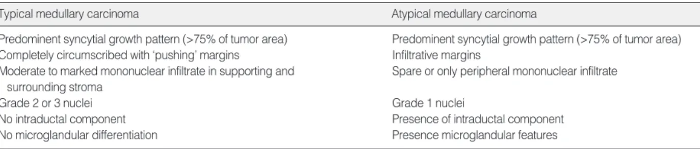
Table 1. Histologic criteria for classification of medullary carcinoma of the breast*
Typical medullary carcinoma Atypical medullary carcinoma
Predominent syncytial growth pattern (>75% of tumor area) Predominent syncytial growth pattern (>75% of tumor area) Completely circumscribed with ‘pushing’ margins Infiltrative margins
Moderate to marked mononuclear infiltrate in supporting and Spare or only peripheral mononuclear infiltrate surrounding stroma
Grade 2 or 3 nuclei Grade 1 nuclei
No intraductal component Presence of intraductal component
No microglandular differentiation Presence microglandular features
*adapted from Ridolfi et al.(3)
0.001). TNM 병기는 수질성유방암에서 1기 2기 조기유방암이 침 윤성유관암에 비해 많았다(p=0.002). 에스트로겐수용체와 프로 게스테론수용체의 경우 전형적 수질성유방암에서 18.9%와 16.2%
였으며, 비전형적 수질성유방암에서 15.0%, 20.0%의 양성률을 보였고, 침윤성유관암에서 63.2%, 57.2%로 나타나 수질성유방 암과 침윤성유관암 사이에 의미 있는 차이가 있었다(p<0.001).
HER2/neu 발현은 전형적 수질성유방암과 비전형적 수질성유 방암에서 47.4%, 45.5%로 침윤성유관암에서의 28.3%에 비해
높게 나타났다(p=0.001). 수술 후 호르몬 치료는 호르몬수용체 양성이 상대적으로 많았던 침윤성유관암에서 많이 시행되었으며, 항암치료도 침윤성유관암군에서 좀더 많이 시행된 것으로 조사 되었다(p<0.001, Table 2).
3. 생존율분석 1) 무병생존율
5년, 10년 무병생존율은 전형적 수질성유방암이 85.4%, 77.8%
를 보였으며 비전형적 수질성유방암은 75.3%, 67.8%를 침윤성 유관암은 77.6%, 68.3%의 생존율을 각각 보여 전형적 수질성유 방암과 침윤성유관암 사이에는 생존율의 차이가 있었지만(p=
0.002), 비전형적 수질성유방암과 침윤성유관암과는 의미 있는 차이가 없었다(p=0.117, Figure 1).
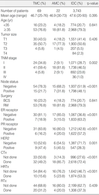
Table 2. Clinicopathologic characteristics
TMC (%) AMC (%) IDC (%) p-value
Number of patients 69 22 3,743
Mean age (range) 46.7 (25-76) 46.9 (30-73) 47.6 (20-93) 0.395 Age (yr)
<35 16 (23.2) 4 (18.2) 774 (20.7) 0.841
≥35 53 (76.8) 18 (81.8) 2,968 (79.3) Tumor size
T1 30 (43.5) 4 (18.2) 1,551 (41.4) 0.426 T2 35 (50.7) 17 (77.3) 1,900 (50.8)
T3 4 (5.8) 1 (4.5) 207 (5.5)
T4 84 (2.3)
TNM stage
I 24 (34.8) 2 (9.1) 1,071 (28.7) 0.002
II 41 (59.4) 18 (81.8) 1,738 (46.5)
III 4 (5.8) 2 (9.1) 892 (23.8)
IV 36 (1.0)
Node status
Negative 54 (78.3) 15 (68.2) 1,937 (51.9) <0.001 Positive 15 (21.7) 7 (31.8) 1,798 (48.1) Surgery
BCS 16 (23.2) 4 (18.2) 774 (20.7) 0.841 RM 53 (76.8) 18 (81.8) 2,968 (79.3) ER receptor
Negative 30 (81.1) 17 (85.0) 1,067 (36.8) <0.001 Positive 7 (18.9) 3 (15.0) 1,833 (63.2) PR receptor
Negative 31 (83.8) 16 (80.0) 1,212 (42.8) <0.001 Positive 6 (16.2) 4 (20.0) 1,622 (57.2) HER2
Negative 10 (52.6) 6 (54.5) 1,387 (71.7) 0.001 Positive 9 (47.4) 5 (45.5) 547 (28.3) CTx
Not done 33 (50.8) 3 (14.3) 996 (27.6) <0.001 Done 32 (49.2) 18 (85.7) 2,618 (72.4) HRTx
Not done 54 (84.4) 16 (76.2) 1,642 (46.7) <0.001 Done 10 (15.6) 5 (23.8) 1,874 (53.3) RTx
Not done 44 (68.8) 16 (80.0) 2,199 (62.7) 0.439 Done 20 (31.2) 4 (20.0) 1,306 (37.3) TMC=typical medullary carcinoma; AMC=atypical medullary carci- noma; IDC=invasive ductal carcinoma; ER=estrogen; PR=proges- terone; CTx=chemotherapy; HRTx=hormonal therapy; RTx=radiation therapy.
Cumulative survival
1.0
0.8
0.6
0.4
0.2
0.0
0 20 40 60 80 100 120
Months
p=0.0019 TMC (n=69) AMC (n=22) IDC (n=3,707)
Figure 1. Comparison of disease-free survival curve between medullary subtype and infiltrating ductal carcinoma.
TMC=typical medullary carcinoma; AMC=atypical medullary carcinoma; IDC=invasive ductal carcinoma.
Cumulative survival
1.0
0.8
0.6
0.4
0.2
0.0
0 20 40 60 80 100 120
Months
p=0.0059 TMC (n=69) AMC (n=22) IDC (n=3,707)
Figure 2. Comparison of overall survival curve between medullary subtype and infiltrating ductal carcinoma.
TMC=typical medullary carcinoma; AMC=atypical medullary carcinoma; IDC=invasive ductal carcinoma.
2) 전체생존율
5년, 10년 누적생존율은 전형적 수질성유방암이 90.7%, 86.0%
를 보였으며 비전형적 수질성유방암은 84.9%, 77.8%를 침윤성 유관암은 80.5%, 74.7%의 생존율을 각각 보여 전형적 수질성유 방암과 침윤성유관암 사이에는 생존율의 차이를 관찰할 수 있었 으나(p=0.006), 비전형적 수질성유방암과 침윤성유관암 사이에 는 차이가 없었다(p=0.314, Figure 2).
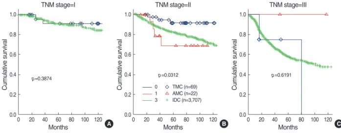
3) 병기별생존율의비교
TNM 병기 1기와 3기의 경우는 생존율에 있어 통계학적 차이 를 보이지 않았지만 병기 2기에서는 전형적 수질성유방암과 침윤 성유관암 사이에 의미 있는 무병생존율과 전체 생존율의 차이를 TMC=typical medullary carcinoma; DFS=disease free survival; OS=
overall survival; ER=estrogen receptor; PR=progesterone receptor.
Age (<35 vs. ≥35 ) 0.408 0.543
Tumor size (≤2 cm vs. >2 cm) 0.689 0.665
Nodal status (+/-) 0.042 0.045
ER (+/-) 0.587 0.597
PR (+/-) 0.511 0.588
Chemotherapy (Not done/done) 0.762 0.692 Hormonal therapy (Not done/done) 0.479 0.368 Radiation therapy (Not done/done) 0.516 0.495 Table 3. Prognostic significance of clinicopathologic variables of TMC
Variable p-value
DFS OS
Figure 3. Disease free survival curve according to TNM stage.
TMC=typical medullary carcinoma; AMC=atypical medullary carcinoma; IDC=invasive ductal carcinoma.
A
Cumulative survival
1.0
0.8
0.6
0.4
0.2
0.00 20 40 60 80 100 120 Months
TNM stage=I TNM stage=II TNM stage=III
p=0.3874
B
Cumulative survival
1.0
0.8
0.6
0.4
0.2
0.00 20 40 60 80 100 120 Months
p=0.0312
TMC (n=69) AMC (n=22) IDC (n=3,707) 0
1 3
C
Cumulative survival
1.0
0.8
0.6
0.4
0.2
0.00 20 40 60 80 100 120 Months
p=0.6191
Figure 4. Overall survival curve according to TNM stage.
TMC=typical medullary carcinoma; AMC=atypical medullary carcinoma; IDC=invasive ductal carcinoma.
A
Cumulative survival
1.0
0.8
0.6
0.4
0.2
0.00 20 40 60 80 100 120 Months
p=0.6417
B
Cumulative survival
1.0
0.8
0.6
0.4
0.2
0.00 20 40 60 80 100 120 Months
p=0.0418
TMC (n=69) AMC (n=22) IDC (n=3,707)
C
Cumulative survival
1.0
0.8
0.6
0.4
0.2
0.00 20 40 60 80 100 120 Months
p=0.5850
TNM stage=I TNM stage=II TNM stage=III
확인할 수 있었다(p=0.031, p=0.042, Figure 3, 4).
4. 전형적수질성유방암의예후인자분석
전형적 수질성유방암의 생존에 영향을 미치는 요인에 대한 단 변량분석 시 환자의 나이, 종양의 크기, 호르몬 수용체 발현 유무, 수술 후 보조요법의 종류와 유무는 통계학적 유의성을 확인할 수 없었으나 림프절 침범여부는 무병생존율과 전체생존율에 영향을 미치는 요인으로 나타났다(p=0.042, p=0.045, Table 3).
고 찰
1977년 Ridolfi 등(3)이 수질성유방암의 여섯 가지 엄격한 진 단기준을 제시한 이후 진단의 어려움으로 병리학자 간 혹은 동일 한 병리학자 내에서도 진단의 재현성이 떨어진다는 문제점이 제 시되었고, 그로 인해 단순화되고 재현성이 높은 진단기준이 계속 보고되었다.(9,10,13,14) 그러나 단순화된 진단기준들은 수질암 의 과잉진단과 이로 인해 일반적인 침윤성유방암에 가까운 비전 형적 수질성유방암과, 비수질성유방암을 수질성유방암으로 진단 하여 부적절한 치료와 예후의 예측에 있어 문제점을 만들게 되었 다.(14,15)
1997년 Jensen 등(5)은 진단기준에 따른 수질성유방암의 재 분류와 그에 따른 예후를 비교하여 어떤 진단기준이 예후를 가장 잘 반영하는가에 대한 연구를 시행하였다. Ridolfi 등(3)의 진단 기준보다 단순화된 Pedersen 등(12)의 진단기준이나, 더 엄격한 Tavassoli(6)의 진단기준 모두 예후의 차이를 잘 나타내지 못함 을 확인하였고, Ridolfi 등(3)의 진단기준에 의한 분류가 전형적 수질성유방암의 예후를 가장 잘 반영한다고 주장하였다. 본 연구 도 Ridolfi 등(3)의 진단기준에 따라 수질성유방암을 분류하여 진 행하였으며, 전형적 수질성유방암의 예후를 잘 반영하는 결과를 보였다.
유방암의 임상병리학적 특성 중 침윤성유방암에서 적용할 수 있 는 예후인자들이 수질성 유방암에서는 혼동을 야기시킬 수 있는 데 변형 Bloom-Richardson의 점수 계산법에 의한 핵등급이나, 호르몬 수용체발현, HER2/neu 발현 등은 다른 침윤성유방암과 는 반대되는 결과를 나타낸다. 수질성유방암의 임상양상을 살펴보 면 다른 침윤성암종에 비해 젊은 나이에 발병한다는 보고와,(10, 14) 비슷한 연령 때에 발병한다는 보고들도 있으나,(4,16,17) 본 연구결과는 수질성유방암과 다른 침윤성암종 사이에서 발병연령 의 차이는 없었다. 유방암의 예후인자로 가장 중요한 액와림프절 전이빈도는 수질성유방암에서 상대적으로 낮아 대부분의 연구에 서 전이 양성률을 낮게 보고하고 있다.(10,14,18) 저자들의 결과 도 전형적 수질성유방암에서 액와림프절 전이가 21.7%로 다른 침
윤성암의 48.1%에 비해 낮은 것으로 조사되었다. 호르몬수용체 양 성률은 수질성유방암에서 다른 침윤성암종에 비해 낮은 것으로 보 고되고 있다. Jensen 등(19)은 수질성유방암의 형태를 갖는 60명 의 대상 중 13명의 전형적 수질성유방암 환자에서 모두 호르몬수 용체가 발현되지 않았다고 보고하였고, Ponsky 등(4)도 수질성 유방암에서 에스트로겐수용체 양성률을 25%, 프로게스테론수용 체 양성률을 10%로 보고하였다. 본 연구의 결과에서 전형적 수질 성유방암과 비전형적 수질성유방암의 에스트로겐수용체 양성률 (18.9%, 15.0%)과 프로게스테론수용체 양성률(16.2%, 20.0%)이 다른 침윤성암종(63.2%, 57.2%)에 비해 낮은 것을 확인할 수 있 었다(p<0.001, Table 2). HER2/neu 유전자의 증폭의 경우, Xu 등(20)은 비전형적 수질성유방암에서 46%의 발현율을 보고하였 다. 저자들도 전형적 수질성유방암과 비전형적 수질성유방암에서 HER2/neu 발현이 각각 47.4%, 45.5%로 다른 침윤성유관암의 28.3%에 비해 높게 측정 되었지만 조사대상의 수가 적어 결과를 단정하기는 어렵다. Jacquemier 등(21)은 수질성유방암의 이해 와 진단을 향상시키기 위한 방법으로 tissue microarray (TMA) 를 이용하여 여러 표지자를 알아보았으며 P-cardherin, MIB1, ERBB2, p53과의 연관성을 언급하였다. 저자들도 유방암의 성 장과 진행에 관여하는 보다 많은 예후인자에 대한 조사가 필요할 것으로 생각된다. 이와 같이 수질성유방암은 낮은 호르몬수용체 발현과 높은 HER2/neu 발현 등의 분화가 나쁜 병리학적 특성을 보이면서 액와림프절 전이가 적은 생물학적 특성을 보인다.
전형적 수질성유방암의 예후에 있어 Fourquet 등(17)은 83%의 환자가 재발 없이 6년간 생존하였다고 보고하였고, Ridolfi 등(3) 도 수술적 치료 후 단핵구 침윤 정도에 따라 84-91%가 10년 생 존율을 보였으며, Krutz 등(22)과 Wargotz 등(14)은 각각 90%
와 94%의 5년 생존율을 보였다고 보고하였다. 하지만 비전형적 수질성유방암의 예후는 다른 침윤성유방암과 크게 다르지 않아 Rapin 등(10)은 10년 무병생존율을 각각 53%, 51%로 보고하였 다. 본 연구결과 역시 10년 무병생존율과 전체생존율이 전형적 수질성유방암(77.8%, 86.0%), 비전형적 수질성유방암(67.8%, 77.8%)에서 각각 조사되어 침윤성유관암(68.3%, 74.7%)과의 비교에서 전형적 수질성유방암의 예후는 침윤성유관암보다 양호 한 것으로 조사되었으며, 비전형적 수질성유방암과 침윤성유관 암 사이의 예후에 있어서는 차이가 없었다. 저자들은 수질성유방 암과 침윤성유관암의 병기별 생존율을 비교 분석하였으며 TNM 병기 1기 에서는 생존율의 의미 있는 차이를 발견하지 못하였으 며, 병기 3의 경우도 생존율의 차이를 확인할 수 없었는데 유효분 석 대상의 수가 적어 결과를 단정하기 어렵다. 병기 2의 경우는 무병생존율과 전체생존율에 있어 모두 전형적 수질성유방암이 침윤성유관암에 비해 예후가 좋은 것으로 나타났으며, 무병생존
율에 있어서는 전형적 수질/성유방암과 비전형적 수질성유방암 사이에도 전형적 수질성유방암이 양호한 예후를 보이는 것으로 나타났다(p=0.037).
전형적 수질성유방암의 생존에 영향을 주는 요인에 대한 분석 으로 Reinfuss 등(18)은 52명의 전형적 수질성유방암 환자에 대 한 조사에서 17명이 액와림프절 전이 양성이었으며 10년 무병생 존율과 관련된 인자는 단지 액와림프절 전이 유무라고 주장하였 고, Kim 등(23)도 수질성유방암에서 림프절 전이가 있거나 35세 이하의 젊은 연령인 경우 재발이 증가함을 확인하였다. 저자들도 단변량분석을 통한 전형적 수질성유방암의 생존에 영향을 주는 인자가 림프절 전이여부임을 확인하였지만, 발병연령은 생존율 에 영향을 미치는 요인으로 확인되지 않았다. 그 외 HER2/neu 에 대한 분석은 추가적인 환자 예와 대상기간을 다시 정하여 분석 하는 것이 필요할 것으로 사료된다.
결 론
전형적 수질성유방암은 병리학적 특성상 높은 핵등급과 조직 학적 등급, 호르몬 수용체 발현율의 감소 등 좋지 않은 예후인자 가 많음에도 불구하고 다른 침윤성유관암종에 비해 좋은 예후를 나타내지만, 비전형적 수질성유방암은 다른 침윤성유관암종과 비슷한 예후를 나타내는 것으로 조사되었다. 따라서 수질성유방 암의 진단은 전형적 수질성 유방암인지 비전형적 수질성 유방암 인지 아형의 분류에 따른 정확한 진단이 필요하며 이에 따라 추가 적 치료 방향을 설정하는 것이 치료 성적의 향상에 도움이 될 것 으로 생각된다.
참고문헌
1. Moore OS, Foote FW Jr. The relatively favorable prognosis of medu- llary carcinoma of the breast. Cancer 1949;2:635-42.
2. Dardick I, Yazdi HM, Brosko C, Rippstein P, Hickey NM. A quan- titative comparison of light and electron microscopic diagnoses in specimens obtained by fine-needle aspiration biopsy. Ultrastruct Pathol 1991;15:105-29.
3. Ridolfi RL, Rosen PP, Port A, Kinne D, Mike V. Medullary carci- noma of the breast: a clinicopathologic study with 10 year follow- up. Cancer 1977;40:1365-85.
4. Ponsky JL, Gliga L, Reynolds S. Medullary carcinoma of the breast:
an association with negative hormonal receptors. J Surg Oncol 1984;
25:76-8.
5. Jensen ML, Kiaer H, Andersen J, Jensen V, Melsen F. Prognostic
comparison of three classifications for medullary carcinomas of the breast. Histopathology 1997;30:523-32.
6. Tavassoli FA. Infiltrating carcinomas, common and familiar special types: medullary carcinoma. In: Schnitt ST, editor. Pathology the breast. Norwalk: Appleton & Lange; 1992. p.333-9.
7. Fisher ER, Kenny JP, Sass R, Dimitrov NV, Siderits RH, Fisher B.
Medullary cancer of the breast revisited. Breast Cancer Res Treat 1990;16:215-29.
8. Ellis IO, Galea M, Broughton N, Locker A, Blamey RW, Elston CW.
Pathological prognostic factors in breast cancer. II. Histological type.
Relationship with survival in a large study with long-term follow-up.
Histopathology 1992;20:479-89.
9. Pedersen L, Zedeler K, Holck S, Schiodt T, Mouridsen HT. Medu- llary carcinoma of the breast, proposal for a new simplified histopa- thological definition. Based on prognostic observations and observa- tions on inter- and intraobserver variability of 11 histopathological characteristics in 131 breast carcinomas with medullary features. Br J Cancer 1991;63:591-5.
10. Rapin V, Contesso G, Mouriesse H, Bertin F, Lacombe MJ, Piekar- ski JD, et al. Medullary breast carcinoma. A reevaluation of 95 cases of breast cancer with inflammatory stroma. Cancer 1988;61:2503-10.
11. Fisher ER, Gregorio RM, Fisher B, Redmond C, Vellios F, Sommers SC. The pathology of invasive breast cancer. A syllabus derived from findings of the National Surgical Adjuvant Breast Project (protocol no. 4). Cancer 1975;36:1-85.
12. Pedersen L, Holck S, Schiodt T, Zedeler K, Mouridsen HT. Inter- and intraobserver variability in the histopathological diagnosis of medullary carcinoma of the breast, and its prognostic implications.
Breast Cancer Res Treat 1989;14:91-9.
13. Pedersen L, Holck S, Schiodt T, Zedeler K, Mouridsen HT. Medu- llary carcinoma of the breast, prognostic importance of characteristic histopathological features evaluated in a multivariate Cox analysis.
Eur J Cancer 1994;30A:1792-7.
14. Wargotz ES, Silverberg SG. Medullary carcinoma of the breast: a clinicopathologic study with appraisal of current diagnostic criteria.
Hum Pathol 1988;19:1340-6.
15. Rubens JR, Lewandrowski KB, Kopans DB, Koerner FC, Hall DA, McCarthy KA. Medullary carcinoma of the breast. Overdiagnosis of a prognostically favorable neoplasm. Arch Surg 1990;125:601-4.
16. Black CL, Morris DM, Goldman LI, McDonald JC. The significance of lymph node involvement in patients with medullary carcinoma of the breast. Surg Gynecol Obstet 1983;157:497-9.
17. Fourquet A, Vilcoq JR, Zafrani B, Schlienger P, Jullien D, Campana F. Medullary breast carcinoma: the role of radiotherapy as primary treatment. Radiother Oncol 1987;10:1-6.
18. Reinfuss M, Stelmach A, Mitus J, Rys J, Duda K. Typical medullary carcinoma of the breast: a clinical and pathological analysis of 52 cases. J Surg Oncol 1995;60:89-94.
19. Jensen ML, Kiaer H, Melsen F. Medullary breast carcinoma vs. poorly differentiated ductal carcinoma: an immunohistochemical study with keratin 19 and oestrogen receptor staining. Histopathology 1996;29:
241-5.
20. Xu R, Feiner H, Li P, Yee H, Inghirami G, Delgado Y, et al. Differen- tial amplification and overexpression of HER-2/neu, p53, MIB1, and
estrogen receptor/progesterone receptor among medullary carcinoma, atypical medullary carcinoma, and high-grade invasive ductal car- cinoma of breast. Arch Pathol Lab Med 2003;127:1458-64.
21. Jacquemier J, Padovani L, Rabayrol L, Lakhani SR, Penault-Llorca F, Denoux Y, et al. Typical medullary breast carcinomas have a basal/
myoepithelial phenotype. J Pathol 2005;207:260-8.
22. Kurtz JM, Jacquemier J, Torhorst J, Spitalier JM, Amalric R, Hunig R, et al. Conservation therapy for breast cancers other than infiltrating ductal carcinoma. Cancer 1989;63:1630-5.
23. Kim SW, Kang HJ, Noh DY, Youn YK, Oh SK, Choe KJ. Compa- rison of the prognostic factors between medullary cancer and infiltra- ting ductal carcinoma in the breast. J Korean Surg Soc 2000;59:182-90.