ABSTRACT
Objective: Choice of hysterectomy and adjuvant treatment for International Federation of Gynecology and Obstetrics (FIGO) 2009 stage II endometrioid endometrial cancer (EEC) is still controversial. Aims of this study were to evaluate survival benefits and adverse effects of different hysterectomies with or without adjuvant radiotherapy (RT), and to identify prognostic factors.
Methods: The patients at 14 member hospitals of the Taiwanese Gynecologic Oncology Group from 1992 to 2013 were retrospectively investigated. Patients were divided into simple hysterectomy (SH) alone, SH with RT, radical hysterectomy (RH) alone, and RH with RT groups. Endpoints were recurrence-free survival (RFS), overall survival (OS), disease-specific survival (DSS), adverse effects and prognostic factors for survival.
Results: Total of 246 patients were enrolled. The 5-year RFS, OS, DSS and recurrence rates for the entire cohort were 89.5%, 94.3%, 96.2% and 10.2%, respectively. Patients receiving RH had more adverse effects including blood loss (p<0.001), recurrent urinary tract infections (p=0.013), and leg lymphedema (p=0.038). Age over 50-year (HR=9.2; 95% confidence
Original Article
Hung-Chun Fu ,1 Jen-Ruei Chen,2 Min-Yu Chen,3 Keng-Fu Hsu ,4 Wen-Fang Cheng ,5 An-Jen Chiang ,6 Yu-Min Ke ,7 Yu-Chieh Chen,8 Yin-Yi Chang,9 Chia-Yen Huang,10 Chieh-Yi Kang,11 Yuan-Yee Kan,12 Sheng-Mou Hsiao,13 Ming-Shyen Yen 14
1 Department of Obstetrics and Gynecology, Kaohsiung Chang Gung Memorial Hospital and Chang Gung University College of Medicine, Kaohsiung, Taiwan
2Department of Obstetrics and Gynecology, MacKay Memorial Hospital, Taipei, Taiwan
3Department of Obstetrics and Gynecology, Chang Gung Memorial Hospital, Linko, Taiwan
4 Department of Obstetrics and Gynecology, National Cheng Kung University Hospital, College of Medicine, National Cheng Kung University, Tainan, Taiwan
5Department of Obstetrics and Gynecology, College of Medicine, National Taiwan University, Taipei, Taiwan
6Department of Obstetrics and Gynecology, Kaohsiung Veterans General Hospital, Kaohsiung, Taiwan
7Department of Obstetrics and Gynecology, Taichung Veterans General Hospital, Taichung, Taiwan
8 Department of Obstetrics and Gynecology, Kaohsiung Medical University Chung-Ho Memorial Hospital, Kaohsiung, Taiwan
9Department of Obstetrics and Gynecology, China Medical University Hospital, Taichung, Taiwan
10 Gynecologic Cancer Center, Department of Obstetrics and Gynecology, Cathay General Hospital, Taipei, Taiwan
11 Center for Reproductive Medicine, Department of Obstetrics and Gynecology, Chi-Mei Medical Center, Tainan, Taiwan
12Department of Obstetrics and Gynecology, Yuan's General Hospital, Kaohsiung, Taiwan
13Department of Obstetrics and Gynecology, Far Eastern Memorial Hospital, Banqiao, New Taipei, Taiwan
14 Department of Obstetrics and Gynecology, Taipei Veterans General Hospital and Department of Obstetrics and Gynecology, National Yang-Ming University School of Medicine, Taipei, Taiwan
Treatment outcomes of patients
with stage II pure endometrioid-type endometrial cancer: a Taiwanese
Gynecologic Oncology Group (TGOG- 2006) retrospective cohort study
Received: Mar 30, 2018 Revised: Apr 24, 2018 Accepted: May 3, 2018 Correspondence to Ming-Shyen Yen
Department of Obstetrics and Gynecology, Taipei Veterans General Hospital and Department of Obstetrics and Gynecology, National Yang-Ming University School of Medicine, 201, Section 2, Shih-Pai Road, Taipei 112, Taiwan.
E-mail: [email protected] Copyright © 2018. Asian Society of Gynecologic Oncology, Korean Society of Gynecologic Oncology
This is an Open Access article distributed under the terms of the Creative Commons Attribution Non-Commercial License (https://
creativecommons.org/licenses/by-nc/4.0/) which permits unrestricted non-commercial use, distribution, and reproduction in any medium, provided the original work is properly cited.
ORCID iDs Hung-Chun Fu
https://orcid.org/0000-0002-0660-9349 Keng-Fu Hsu
https://orcid.org/0000-0002-2698-4971 Wen-Fang Cheng
https://orcid.org/0000-0002-3282-6304 An-Jen Chiang
https://orcid.org/0000-0001-6892-8559 Yu-Min Ke
https://orcid.org/0000-0003-2462-9244 Ming-Shyen Yen
https://orcid.org/0000-0002-7990-2837
interval [CI], 1.2–70.9) and grade 3 histology (HR=7.28; 95% CI, 1.45–36.6) were independent predictors of OS. Grade 3 histology was an independent predictor of RFS (HR=5.13; 95% CI, 1.38–19.1) and DSS (HR=5.97; 95% CI, 1.06–58.7). Patients receiving adjuvant RT had lower locoregional recurrence (p=0.046), but no impact on survival.
Conclusion: Different treatment modalities yield similar survival outcomes. Patients receiving SH with RT had lower locoregional recurrent with acceptable morbidity. Age and tumor grading remained significant predictors for survival among patients with FIGO 2009 stage II EEC.
Keywords: Uterine cancer; Endometrioid; General Surgery; Radiotherapy; Survival;
Recurrence
INTRODUCTION
Endometrial cancer (EC) is one of the most common gynecologic cancers worldwide [1]. In Taiwan, the incidence of EC has increased rapidly in the past decade [2]. The most common histology is the endometrioid type, and the majority of tumors are confined to the uterine corpus. The 2009 International Federation of Gynecology and Obstetrics (FIGO) staging defined stage II disease as pathologic involvement of the uterine cervix stroma [3]. The presence of glandular involvement was not included in the definition of stage II disease because the prognosis of these patients was not worse than those with stage I. According to previous reports, 7%–12% of ECs are stage II [4-6].
Total hysterectomy, bilateral salpingo-oophorectomy (BSO), and bilateral pelvic and para- aortic lymph node dissection are recommended as staging surgery for EC [7]. The optimal type of hysterectomy for patients with stage II EC remains controversial, and treatment options range from simple (extrafascial) hysterectomy (SH) to radical hysterectomy (RH).
RH has been recommended for stage II disease due to improvements in locoregional control and survival compared with SH [8,9]. However, other studies that evaluated the prognostic factors in patients with FIGO 2009 stage II did not find that the type of hysterectomy was a risk factor for survival or recurrence [10-14]. Therefore, the choice of hysterectomy for stage II EC is still controversial. Thus, we conducted a retrospective, multicenter study to evaluate survival benefits and adverse effects of different hysterectomies with or without adjuvant RT, and to identify prognostic factors among patients with FIGO stage II endometrioid endometrial cancer (EEC).
MATERIALS AND METHODS
1. Patients
The medical records of patients who underwent primary treatment between January 1992 and December 2013 at the member hospitals of the Taiwanese Gynecologic Oncology Group (TGOG) were retrospectively reviewed. The study protocol was approved by the Institutional Review Board of each hospital (approval number: 105-3816C). The inclusion criterion was post-operative pathology-proven FIGO 2009 stage II EEC. Patients with incomplete surgical staging (without BSO or lymphadenectomy), an inaccurate pathology report, incomplete medical records, or a lack of follow-up were excluded from the study. The detailed medical records were retrospectively evaluated until the end of the follow-up period (31 December 2014).
Presentation
The findings of the study had been presented at 2015 Asian Society of Gynecologic Oncology (ASGO) biennial meeting.
Funding
The work was supported by the research platform of the Taiwanese Gynecologic Oncology Group (TGOG), TR8-Taiwan Clinical Trial Consortium for Gynecologic Oncology V, supported by a grant from the National Science Council, Taiwan (NSC 103-2325-B- 195.002). The work was also supported by the grant (CMRPG8E0841) from Kaohsiung Chang Gung Memorial Hospital.
Conflict of Interest
No potential conflict of interest relevant to this article was reported.
Author Contributions
Conceptualization: F.H.C., Y.M.S.; Formal analysis: F.H.C.; Methodology: F.H.C., C.J.R., C.M.Y., H.K.F., C.W.F., C.A.J., K.Y.M., C.Y.C., C.Y.Y., H.C.Y., K.C.Y., K.Y.Y., H.S.M., Y.M.S.;
Resources: F.H.C., C.J.R., C.M.Y., H.K.F., C.W.F., C.A.J., K.Y.M., C.Y.C., C.Y.Y., H.C.Y., K.C.Y., K.Y.Y., H.S.M., Y.M.S.; Supervision: Y.M.S.; Validation:
Y.M.S.; Writing - original draft: F.H.C.; Writing - review & editing: C.J.R., C.M.Y., H.K.F., C.W.F., C.A.J., K.Y.M., C.Y.C., C.Y.Y., H.C.Y., K.C.Y., K.Y.Y., H.S.M., Y.M.S.
Demographic data, including age at the time of surgery, body mass index (BMI), parity, menopausal status, and history of medical diseases were obtained from the medical records.
The staging and histologic grading criteria were determined post-operatively and were based on the FIGO 2009 staging system [3]. Surgical staging procedures consisted of washing cytology, SH or RH, BSO, and lymph node dissection (pelvic with or without para-aortic).
The decision to give the patients adjuvant therapy was based on the extent of the disease, medical co-morbidities, and the institutional practices during that time. The clinical follow- up assessments of the disease consisted of pelvic physical examinations, vaginal cytology, determination of tumor markers, and imaging examinations when clinically indicated.
Grading of adverse events was judged by the Common Terminology Criteria for Adverse Events (CTCAE) v4.0 [15]. Chronic adverse events were defined as repeated hospitalizations with the same diagnosis for more than 6 months after surgery. The stage of leg edema was defined according to the Fifth World Health Organization Expert Committee on Filariasis.
Follow-up data, including sites of recurrence, date at the diagnosis of recurrence, and patient status at the last visit or the end of the follow-up period (31 December 2014).
The following pathologic factors were evaluated: FIGO grade (G); depth of myometrial invasion; lymphovascular space invasion (LVSI); size of the tumor; parametrium involvement;
and washing cytology results. A pathology review was conducted at each institution.
2. Statistical methods
Recurrence-free survival (RFS) and locoregional recurrence free survival (LRFS) was calculated from the date of staging surgery to the date of diagnosis of recurrence or last contact for the recurrence-free patients. Locoregional recurrence was defined as the vaginal stump, pelvic or vaginal with pelvic recurrence. Overall survival (OS) was defined as the period from the date of surgery to the date of death or last contact. Disease-specific survival (DSS) was defined as the date of surgery to the date of death from EC or last contact.
The χ2 test was used to analyze categorical variables and the Student's t-test was used for continuous variables. The Kaplan-Meier method was used to calculate patient survival distribution. The significance of the survival distribution in each group was tested using the log-rank test. Cox proportional hazard analysis was used to evaluate the prognostic factors for survival. Multivariate analysis using Cox stepwise forward regression was conducted for the covariates in univariate analysis with a p-value <0.05, hazard ratios (HRs) and 95%
confidence intervals (CIs) were calculated. A p-value <0.05 was considered to be statistically significant. All statistical calculations were performed using SPSS software for Windows version 20 (SPSS Inc., Chicago, IL, USA).
RESULTS
1. Characteristics of the patients
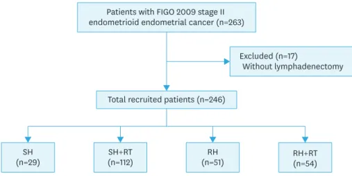
We identified 246 patients with stage II (FIGO 2009) EEC who were treated between 1992 and 2013 at the 14 TGOG member hospitals (Fig. 1). The median duration of follow-up was 78 months (range, 0.5–259 months). Table 1 shows the characteristics of the patients. The mean age was 53.1 years at the time EC was diagnosed. One hundred and forty-one (57.3%) of the patients underwent SHs and 105 patients (42.7%) underwent RHs. After surgery, 112 patients (79.4%) in the SH group and 54 patients (51.4%) in the RH group received adjuvant RT, while the other patients were managed with surveillance. Ten patients (4.1%) received
vaginal brachytherapy only, 44 patients (18.1%) received pelvic radiotherapy (RT), and 111 patients (45.7%) received both vaginal and pelvic irradiation. Thirty patients (12.4%) received adjuvant chemotherapy (CT; 3 [10.7%] in the SH group, 12 [10.8%] in the SH + RT group, 8 [16.0%] in the RH group, and 7 [13.2%] in the RH + RT group). Twenty-five patients (10.2%) had tumor recurrence during the follow-up period, including locoregional recurrence 14 and distant recurrence in10 patients, while only one patient had both locoregional and distal recurrences. The pathologic characteristics are shown in Table 1.
The clinic and pathologic characteristics of patients receiving different treatment modalities were showed in Supplementary Table 1. Patients with adjuvant RT after SH and RH had higher ratio of deep myometrium invasion and LVSI. Supplementary Table 2 showed the clinical and pathologic characteristics of patients receiving adjuvant RT or not. In adjuvant RT group, we found higher ratio of patients with deep myometrium invasion (RT vs. no RT:
56.1% vs. 23.8%, p<0.001), LVSI (RT vs. no RT: 27.9% vs. 10.0%, p=0.021) and SH (RT vs. no RT: 66.3% vs. 35.0%, p<0.001). Supplementary Table 3 showed the clinical and pathologic characteristics of patients receiving adjuvant CT or not. In adjuvant CT group, we found higher ratio of patients with grade 3 tumors (CT vs. no CT: 51.7% vs. 14.2%, p<0.001).
Supplementary Table 4 showed the clinical and pathologic characteristics of patients receiving SH or RH. In SH group, we found higher ratio of patients with adjuvant RT (SH vs.
RH: 78.0% vs. 53.3%, p<0.001) and lower ratio of chronic adverse effect (SH vs. RH: 15.2%
vs. 26.7%, p=0.028).
2. Survival pattern of the patients receiving different treatments
The 5-year RFS, OS, DSS, and LRFS, for the entire cohort were 89.5%, 94.3%, 96.2%, and 93.8%, respectively. RFS, LRFS, OS, and DSS curves of the patients receiving different treatment modalities are shown in Fig. 2, respectively. There were also no significant survival differences among the different treatment modalities.
3. Pattern of recurrence according to treatment
Twenty-five (10.2%) patients were diagnosed with tumor recurrences. There was a significant difference in the site of recurrence between the 4 groups (p=0.046; Table 2). The patients who received adjuvant RT had a lower locoregional recurrence rate (3.0% vs. 10.0%, p=0.021) regardless of the type of hysterectomy.
(n=29)SH SH+RT
(n=112)
Total recruited patients (n=246)
(n=51)RH RH+RT
(n=54) Patients with FIGO 2009 stage II
endometrioid endometrial cancer (n=263)
Excluded (n=17)
Without lymphadenectomy
Fig. 1. Flowchart of our retrospective study design.
FIGO, International Federation of Gynecology and Obstetrics; RH, radical hysterectomy; RT, radiotherapy; SH, simple hysterectomy.
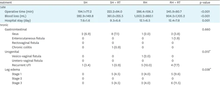
4. Acute and chronic adverse effects according to different treatments
Adverse effects according to the treatment modality are shown in Table 3. The patients who received RH had significantly longer operative time and hospital stay, and more surgical blood loss. Of the chronic adverse effects, vesicovaginal fistulas and recurrent urinary tract infections were more frequently observed in the RH with or without RT groups (p=0.013).The patients who received SH only had the lowest rate of lower limb lymphedema (p=0.038).
Lower limb lymphedema was most frequently observed in the RH with RT group.
5. Analysis of risk factors for recurrence and patient survival
The results of univariate (log-rank test) and multivariate (Cox proportional hazard) analyses of RFS, OS, DSS, and LRFS, are shown in Supplementary Tables 5-8, respectively. Based on the multivariate analysis (Table 4), age more than 50-year-old (HR=9.22; 95% CI=1.20–70.9) and Table 1. Patient characteristics (n=246)
Variable Values
Age (yr) 53.1±10.6
BMI (kg/m2) 25.2±4.5
Nulliparity 40 (16.3)
Menopause 124 (50.4)
Medical diseases 81 (32.9)
Hypertension 55 (22.4)
Diabetes mellitus 32 (13.0)
Co-existing history of malignancy 21 (8.5)
Breast cancer 3 (1.2)
Colon cancer 7 (2.8)
Adjuvant CT 30 (12.4)
Treatment type
SH 29 (11.8)
SH + RT 112 (45.5)
RH 51 (20.7)
RH + RT 54 (22.0)
Adjuvant RT 166 (67.5)
Vagina only 10 (4.1)
Pelvis only 44 (17.9)
Pelvis and vagina 111 (45.1)
Lymphadenectomy
Pelvic only 101 (41.1)
Pelvic and para-aortic 145 (58.9)
Tumor FIGO grade
Grade 1 92 (37.4)
Grade 2 105 (42.7)
Grade 3 46 (18.7)
Myometrium >1/2 111 (45.1)
LVSI 51 (20.7)
PM invasion 4 (1.6)
Positive washing cytology 6 (2.4)
Recurrence 25 (10.2)
Recurrent sites
Locoregional 14
Distant 10
Mixed 1
Cause of death
Disease-related 8 (3.3)
Non-disease related 6 (2.4)
Continuous data are presented as the mean±standard deviation or number (%).
BMI, body mass index; CT, chemotherapy; FIGO, International Federation of Gynecology and Obstetrics; LVSI, lymphovascular space invasion; PM, parametrium; RH, radical hysterectomy; RT, radiotherapy; SH, simple hysterectomy.
0.6
0.50 50 100
RFS
0.7 0.8 0.9 1.0
300
150 200 250
SH+RT SH
RH+RT RH
Time (mo) 0.6
0.50 50 100
LRFS
0.7 0.8 0.9 1.0
300
150 200 250
SH+RT SH
RH+RT RH
Time (mo)
A B
Time (mo) 0.6
0.50 50 100
OS
0.7 0.8 0.9 1.0
300
150 200 250
SH+RT SH
RH+RT RH
Time (mo) 0.6
0.50 50 100
DSS
0.7 0.8 0.9 1.0
300
150 200 250
SH+RT SH
RH+RT RH
C D
Fig. 2. Survival curves for the patients receiving different treatments. (A) RFS, (B) LRFS, (C) OS, (D) DSS, according to the treatment modality in 246 patients with FIGO 2009 stage II EEC. (A) Five-year RFS rates for SH alone, SH with RT, RH alone, and RH with RT were 86.9%, 90.1%, 87.3%, and 91.5%, respectively (p=0.706). (B) Five-year LRFS rates for SH alone, SH with RT, RH alone, and RH with RT were 88.9%, 96.4%, 88.2%, and 93.9%, respectively (p=0.141). (C) Five- year OS rate for SH alone, SH with RT, RH alone, and RH with RT were 94.7%, 93.0%, 95.7%, and 95.1%, respectively (p=0.863). (D) Five-year DSS rates for SH alone, SH with RT, RH alone, and RH with RT were 94.7%, 96.2%, 95.7%, and 97.4%, respectively (p=0.938).
DSS, disease-specific survival; LRFS, locoregional recurrence free survival; OS, overall survival; RFS, recurrence-free survival; RH, radical hysterectomy; RT, radiotherapy; SH, simple hysterectomy.
Table 2. Recurrence pattern according to different treatment groups
Treatment No. Recurrent site Total p-value
Locoregional Distant Mixed
SH 29 3 (10.3) 0 0 3 (11.1) 0.046
SH + RT 112 4 (3.6) 7 (6.2) 0 11 (9.8)
RH 51 6 (11.8) 1 (2.0) 0 7 (13.7)
RH + RT 54 1 (1.9) 2 (3.7) 1 (1.9) 4 (7.5)
Data are presented as the number (%).
RH, radical hysterectomy; RT, radiotherapy; SH, simple hysterectomy.
high tumor grade (HR=7.28; 95% CI=1.45–36.6) were significant poor predictors for OS. High tumor grade was the only significantly poor predictor for RFS (HR=5.13; 95% CI=1.38–19.1) and DSS (HR=5.97; 95% CI=1.06–58.7). High tumor grade (HR=5.57; 95% CI=1.58–19.5) and RT (HR=0.246; 95% CI=0.09–0.70) were significant predictors for locoregional recurrence.
The types of surgery were not significant predictors for OS, RFS, DSS, or LRFS.
DISCUSSION
This study demonstrated that the type of surgery was not associated with RFS, DSS, or OS for patients with FIGO 2009 stage II EEC regardless of whether or not they received RT. Adjuvant RT significantly improved the locoregional control rate, but not survivals. High tumor grade was an independent predictor of RFS, OS, DSS and LRFS. Age (≥50-years) was also an independent predictor of OS (Table 4).
In our series, the 5-year RFS, OS, and DSS rates for all of the patients were 89.5%, 94.3%, and 96.2%, respectively. The survival rates were better than previous reports [10-13,16,17].
Table 3. Acute and chronic adverse effects according to different treatment groups
Treatment SH SH + RT RH RH + RT p-value
Acute
Operative time (min) 194.1±77.2 223.2±84.0 286.4±106.3 245.9±80.7 <0.001
Blood loss (mL) 282.9±149.8 361.0±355.5 1,003.2±860.1 904.2±1,105.2 <0.001
Hospital stay (day) 7.6±1.6 9.3±6.6 12.1±6.5 12.4±7.8 0.001
Chronic
Gastrointestinal 0.660
Ileus 2 (6.9) 8 (7.1) 1 (2.0) 2 (3.8)
Enterocutaneous fistula 0 0 0 1 (1.9)
Rectovaginal fistula 0 0 0 0
Chronic colitis 0 1 (0.9) 0 0
Urogenital 0.013*
Vesico-vaginal fistula 0 0 1 (2.0) 0
Uretero-vaginal fistula 0 0 0 0
Recurrent UTI 1 (3.4) 1 (0.9) 5 (10.0) 4 (7.7)
Leg edema 0.038*
Stage 1 0 5 (4.5) 2 (4.0) 5 (9.6)
Stage 2 0 0 0 0
Stage 3 0 5 (4.5) 2 (4.0) 6 (11.5)
Continuous data are presented as the mean±standard deviation. Categorical data are presented as number (%). Stage of leg edema was defined according to the Fifth World Health Organization Expert Committee on Filariasis.
RH, radical hysterectomy; RT, radiotherapy; SH, simple hysterectomy; UIT, urinary tract infection.
*p-value <0.05.
Table 4. The results of multivariate analysis for survival endpoints
Variable OS RFS DSS Locoregional recurrence
HR 95% CI of HR p-value HR 95% CI of HR p-value HR 95% CI of HR p-value HR 95% CI of HR p-value Age (≥50 vs. <50 yr) 9.22 1.20–70.90 0.033* 2.72 0.82–9.04 0.103 9.93 0.58–82.30 0.125 1.62 0.36–7.32 0.533 RT (yes vs. no) 0.56 0.13–2.54 0.455 0.32 0.10–1.01 0.051 0.31 0.03–3.59 0.348 0.246 0.09–0.70 0.009* Grade (2 vs. 1) 2.30 0.46–11.50 0.309 1.78 0.47–6.77 0.367 1.42 0.13–16.00 0.754 1.08 0.27–4.37 0.909 Grade (3 vs. 1) 7.28 1.45–36.60 0.016* 5.13 1.38–19.10 0.017* 5.97 1.06–58.70 0.048* 5.57 1.58–19.50 0.007* Adjuvant CT (yes vs. no) 1.33 0.28–6.22 0.721 1.50 0.51–4.37 0.459 2.44 0.30–19.70 0.405 0.27 0.02–3.29 0.302 Depth of invasion
(<1/2 vs. ≥1/2) 5.10 0.90–28.80 0.065 2.78 0.99–7.75 0.051 2.04 0.40–10.40 0.391 3.32 0.67–16.40 0.141 LVSI (yes vs. no) 1.13 0.31–4.22 0.851 1.44 0.58–3.56 0.433 1.37 0.17–10.90 0.767 1.20 0.27–5.40 0.811 CI, confidence interval; CT, chemotherapy; DSS, disease-specific survival; HR, hazard ratio; LVSI, lymphovascular space invasion; OS, overall survival; RFS, recurrence-free survival; RT, radiotherapy.
*p-value <0.05.
A possible explanation for this finding is that we only included patients with FIGO 2009 stage II EEC with complete staging surgery; patients with high-risk histologic subtypes (carcinosarcoma, serous and clear cell carcinoma) were excluded from this study. Another interesting finding is that our patients were much younger than previously reported [7]. Age is known as a risk factor for survival [11,12,16], and the median age at diagnosis was 52 years (range, 26–86 years) in this study. This is not due to patient selection bias, because in Taiwan the median age at the time of diagnosis of uterine corpus cancer is 55 years [24], which is also younger than in a previous report [7]. In Mahdi et al.'s study [17], Asian women are younger than non-Hispanic white women at the initial presentation of endometrial adenocarcinoma.
Further studies are required to investigate possible explanations for such racial differences.
Cohn et al. [18] and Sartori et al. [19] reported statistically significant improvements in 5-year survival in patients who underwent RHs; however, their results were not based on multivariate analysis. In addition, not all of the patients in the reports received adequate lymphadenectomies. Because cervical involvement is a risk factor for lymph node metastasis [20-22], these patients may have been assigned to a higher stage due to lymph node
metastasis. In contrast, other investigators have not reported a survival benefit based on the type of surgical procedure performed [11,12,14,16]. In our multivariate model, RH had no significant beneficial effect on OS, RFS, DSS, or LRFS. RH alone does not increase survival in stage II EC, as in cervical cancer, because the tumor behavior is different for EC with cervical involvement compared to primary cervical cancer. A lower parametrium invasion [23] and higher pelvic lymph node metastasis had been reported in patients with EC with cervical involvement [20]. RH is mostly beneficial in women with gross cervical involvement pre- operatively [16].
After surgery for EC, external beam or vaginal brachytherapy is recommended for women with stage II disease [8]. A systematic review and meta-analysis assessed the benefits of adjuvant RT in women with early EC [25,26]. The investigators found that adjuvant RT reduced the risk of locoregional recurrence, but did not impact DSS or OS [13,16,19,25,26]. In our study, we also found that adjuvant RT improved locoregional control, but did no impact on RFS, OS, or DSS. Even, the patients with adjuvant RT had poor histologic factors as deeper myometrium invasion and LVSI (Supplementary Table 2).
There were no significant differences in major acute side effects including major organ injuries or death between the SH and RH groups in this study. RH required a significantly longer operative time, was associated with greater intra-operative blood loss, and led to longer hospital stay. Chronic side effects, including recurrent urinary tract infections, and leg edema occurred more frequently in the RH group. Our patients receiving RH and adjuvant RT had the highest rate of grade 3 leg edema (11.5%). Other investigators had reported that complications occurred more frequently in RH patients [10,11].
In the present study, histologic grade was an independent prognostic factor for OS, RFS, DSS, and locoregional recurrence (Supplementary Fig. 1), which is consistent with previous studies [9,12,13,16,27]. Both locoregional and distant recurrence rate were much higher in the patients with G3 tumors than in those with G1/G2 tumors (p=0.007, Supplementary Table 9). This finding suggests that adjuvant CT may play a role in these patients. Susumu et al. [28]
reported a randomized control trial that compared pelvic RT and CT with cyclophosphamide, doxorubicin, and cisplatin in patients with stage IC-IIIC EC, and suggested a survival advantage with CT in the patients from the high-to-intermediate risk group (stage IC, >70 years of age,
grade 3, stage II, or positive cytology with >50% myometrial invasion). However, Maggi et al.
[29] failed to show any improvement in RFS or OS in patients with stage IC G3 or II G3 treated with adjuvant CT or RT. In addition, the Gynecologic Oncology Group (GOG) 249 trial did not show any benefit of adjuvant CT plus brachytherapy compared to external beam RT with respect to RFS and OS. In our series, of 46 patients with G3 EEC, six did not receive adjuvant treatment, four received adjuvant CT only, 24 received RT only and 12 received both of CT and RT. The results showed that only RT significantly improved locoregional control (p<0.001). We did not find any benefit in OS or RFS in the patients who received adjuvant CT. Taken together, the role of CT for stage II G3 EEC remains controversial.
The strengths of this study are that we only included patients with FIGO 2009 stage II EEC with complete staging surgery (SH or RH, BSO, and pelvic lymphadenectomy), patients from medical centers throughout Taiwan, the length of follow-up, comparisons of the survival with different treatment modalities, and the description of rates and recurrence patterns. In comparison, other studies have included stage II EC with or without pelvic lymphadenectomy [9,11,12,16,23]. This is also the largest series to date on women with FIGO 2009 stage II EEC with complete staging surgery.
The limitations of this study included the retrospective design and lack of central pathologic review. The retrospective nature of the current study and the potential for selection bias is the main limitation. We included patients who were treated over a long period of time (>20 years), and developments in RT technology were not considered in this study. In addition, the patients were treated at different academic institutions with different philosophies with regards to surgery and adjuvant treatments.
In conclusion, we demonstrated that different treatment modalities for FIGO stage II disease yielded similar survival outcomes. Adjuvant RT reduced locoregional recurrence, but did not have any impact on survival. If gross cervical involvement was found pre-operatively, RH may be beneficial. For small or subtle cervical lesions of patients with EC found by pelvic examination or imaging study before surgery, SH with RT appeared to be a suitable treatment strategy for FIGO 2009 stage II EEC due to acceptable morbidity and lower locoregional recurrence. Patients with poor prognostic factors including older age and high histologic grade (G3) may need aggressive adjuvant therapy such as RT or CT. Further well-designed studies are required to confirm our findings.
ACKNOWLEDGMENTS
We are grateful to Dr. How-Ran Guo of the Department of Environmental and Occupational Health, College of Medicine, National Cheng Kung University, for providing statistical consultation.
SUPPLEMENTARY MATERIALS
Supplementary Table 1
Clinic and pathologic characteristics of patients receiving different treatment modalities Click here to view
Supplementary Table 2
Clinic and pathologic characteristics of patients with/without adjuvant RT Click here to view
Supplementary Table 3
Clinic and pathologic characteristics of patients with/without adjuvant CT Click here to view
Supplementary Table 4
Clinic and pathologic characteristics of patients with SH and RH Click here to view
Supplementary Table 5
Risk factor association with RFS Click here to viewSupplementary Table 6
Risk factor association with OS Click here to viewSupplementary Table 7
Risk factor association with DSS Click here to viewSupplementary Table 8
Risk factor association with LRFS Click here to viewSupplementary Table 9
Recurrent pattern according to different histologic grade groups Click here to view
Supplementary Fig. 1
Survival curves for the patients with different histologic grades.
Click here to view
REFERENCES
1. Torre LA, Bray F, Siegel RL, Ferlay J, Lortet-Tieulent J, Jemal A. Global cancer statistics, 2012. CA Cancer J Clin 2015;65:87-108.
PUBMED | CROSSREF
2. Chiang CJ, Chen YC, Chen CJ, You SL, Lai MSTaiwan Cancer Registry Task Force. Cancer trends in Taiwan. Jpn J Clin Oncol 2010;40:897-904.
PUBMED | CROSSREF
3. Creasman W. Revised FIGO staging for carcinoma of the endometrium. Int J Gynaecol Obstet 2009;105:109.
PUBMED | CROSSREF
4. Sorosky JI. Endometrial cancer. Obstet Gynecol 2012;120:383-97.
PUBMED | CROSSREF
5. Creasman WT, Odicino F, Maisonneuve P, Quinn MA, Beller U, Benedet JL, et al. Carcinoma of the corpus uteri. FIGO 26th annual report on the results of treatment in gynecological cancer. Int J Gynaecol Obstet 2006;95 Suppl 1:S105-43.
PUBMED | CROSSREF
6. Keys HM, Roberts JA, Brunetto VL, Zaino RJ, Spirtos NM, Bloss JD, et al. A phase III trial of surgery with or without adjunctive external pelvic radiation therapy in intermediate risk endometrial adenocarcinoma:
a Gynecologic Oncology Group study. Gynecol Oncol 2004;92:744-51.
PUBMED | CROSSREF
7. SGO Clinical Practice Endometrial Cancer Working GroupBurke WM, Orr J, Leitao M, Salom E, Gehrig P, et al. Endometrial cancer: a review and current management strategies: part I. Gynecol Oncol 2014;134:385-92.
PUBMED | CROSSREF
8. National Comprehensive Cancer Network (US). NCCN Clinical Practice Guidelines in Oncology. Uterine Neoplasms, version 2. 2016 [Internet]. Fort Washington, PA: National Comprehensive Cancer Network;
2015 [Accessed 2016 September 16]. Available from: https://www.nccn.org/professionals/physician_gls/
pdf/uterine.pdf
9. Orezzoli JP, Sioletic S, Olawaiye A, Oliva E, del Carmen MG. Stage II endometrioid adenocarcinoma of the endometrium: clinical implications of cervical stromal invasion. Gynecol Oncol 2009;113:316-23.
PUBMED | CROSSREF
10. Lee TS, Kim JW, Kim DY, Kim YT, Lee KH, Kim BG, et al. Necessity of radical hysterectomy for endometrial cancer patients with cervical invasion. J Korean Med Sci 2010;25:552-6.
PUBMED | CROSSREF
11. Takano M, Ochi H, Takei Y, Miyamoto M, Hasumi Y, Kaneta Y, et al. Surgery for endometrial cancers with suspected cervical involvement: is radical hysterectomy needed (a GOTIC study)? Br J Cancer 2013;109:1760-5.
PUBMED | CROSSREF
12. Elshaikh MA, Al-Wahab Z, Mahdi H, Albuquerque K, Mahan M, Kehoe SM, et al. Recurrence patterns and survival endpoints in women with stage II uterine endometrioid carcinoma: a multi-institution study.
Gynecol Oncol 2015;136:235-9.
PUBMED | CROSSREF
13. Frandsen JE, Sause WT, Dodson MK, Soisson AP, Belnap TW, Gaffney DK. Survival analysis of endometrial cancer patients with cervical stromal involvement. J Gynecol Oncol 2014;25:105-10.
PUBMED | CROSSREF
14. Ozgul N, Boyraz G, Salman MC, Gultekin M, Yuce K, Ibrahimov A, et al. Oncological outcomes of stage II endometrial cancer: a retrospective analysis of 250 cases. Int J Gynecol Cancer 2018;28:161-7.
PUBMED | CROSSREF
15. National Cancer Institute (US). Common terminology criteria for adverse events (CTCAE). Rev. ed.
Bethesda: U.S. Department of Health and Human Services, National Institutes of Health, National Cancer Institute; 2009.
16. Wright JD, Fiorelli J, Kansler AL, Burke WM, Schiff PB, Cohen CJ, et al. Optimizing the management of stage II endometrial cancer: the role of radical hysterectomy and radiation. Am J Obstet Gynecol 2009;200:419.e1-419.e7.
PUBMED | CROSSREF
17. Mahdi H, Schlick CJ, Kowk LL, Moslemi-Kebria M, Michener C. Endometrial cancer in Asian and American Indian/Alaskan Native women: tumor characteristics, treatment and outcome compared to non-Hispanic white women. Gynecol Oncol 2014;132:443-9.
PUBMED | CROSSREF
18. Cohn DE, Woeste EM, Cacchio S, Zanagnolo VL, Havrilesky LJ, Mariani A, et al. Clinical and pathologic correlates in surgical stage II endometrial carcinoma. Obstet Gynecol 2007;109:1062-7.
PUBMED | CROSSREF
19. Sartori E, Gadducci A, Landoni F, Lissoni A, Maggino T, Zola P, et al. Clinical behavior of 203 stage II endometrial cancer cases: the impact of primary surgical approach and of adjuvant radiation therapy. Int J Gynecol Cancer 2001;11:430-7.
PUBMED
20. Solmaz U, Mat E, Dereli M, Turan V, Gungorduk K, Hasdemir P, et al. Lymphovascular space invasion and cervical stromal invasion are independent risk factors for nodal metastasis in endometrioid endometrial cancer. Aust N Z J Obstet Gynaecol 2015;55:81-6.
PUBMED | CROSSREF
21. Boren T, Lea J, Kehoe S, Miller DS, Richardson D. Lymph node metastasis in endometrioid
adenocarcinomas of the uterine corpus with occult cervical involvement. Gynecol Oncol 2012;127:43-6.
PUBMED | CROSSREF
22. Kwon JS, Qiu F, Saskin R, Carey MS. Are uterine risk factors more important than nodal status in predicting survival in endometrial cancer? Obstet Gynecol 2009;114:736-43.
PUBMED | CROSSREF
23. Phelippeau J, Koskas M. Impact of radical hysterectomy on survival in patients with stage 2 type1 endometrial carcinoma: a matched cohort study. Ann Surg Oncol 2016;23:4361-7.
PUBMED | CROSSREF
24. Taiwan Cancer Registry Task Force (TCRTF). Cancer registry annual report. Taipei: Department of Health, The Executive Yuan; 2013.
25. Kong A, Johnson N, Kitchener HC, Lawrie TA. Adjuvant radiotherapy for stage I endometrial cancer: an updated Cochrane systematic review and meta-analysis. J Natl Cancer Inst 2012;104:1625-34.
PUBMED | CROSSREF
26. ASTEC/EN.5 Study GroupBlake P, Swart AM, Orton J, Kitchener H, Whelan T, et al. Adjuvant external beam radiotherapy in the treatment of endometrial cancer (MRC ASTEC and NCIC CTG EN.5 randomised trials): pooled trial results, systematic review, and meta-analysis. Lancet 2009;373:137-46.
PUBMED | CROSSREF
27. Kong TW, Chang SJ, Paek J, Lee Y, Chun M, Ryu HS. Risk group criteria for tailoring adjuvant treatment in patients with endometrial cancer: a validation study of the Gynecologic Oncology Group criteria. J Gynecol Oncol 2015;26:32-9.
PUBMED | CROSSREF
28. Susumu N, Sagae S, Udagawa Y, Niwa K, Kuramoto H, Satoh S, et al. Randomized phase III trial of pelvic radiotherapy versus cisplatin-based combined chemotherapy in patients with intermediate- and high-risk endometrial cancer: a Japanese Gynecologic Oncology Group study. Gynecol Oncol 2008;108:226-33.
PUBMED | CROSSREF
29. Maggi R, Lissoni A, Spina F, Melpignano M, Zola P, Favalli G, et al. Adjuvant chemotherapy vs radiotherapy in high-risk endometrial carcinoma: results of a randomised trial. Br J Cancer 2006;95:266-71.
PUBMED | CROSSREF