접수일 : 2015 년 10 월 23 일 , 게재승인일 : 2015 년 11 월 13 일 책임저자 : 이용택 , 서울시 종로구 새문안로 29
03181, 성균관대학교 의과대학 강북삼성병원 재활
의학교실
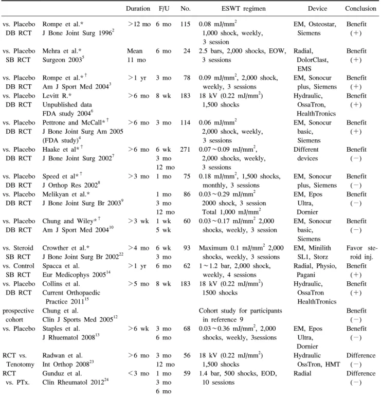
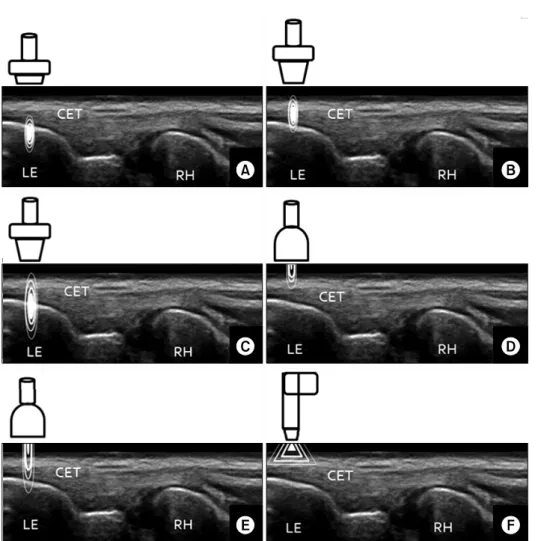
외측상과염의 체외충격파 치료: 근거와 임상적용
성균관대학교 의과대학 강북삼성병원 재활의학교실
이 용 택
전체 글
이 용 택
수치
관련 문서
Autho: Effectiveness of surgery for lumbar stenosis and degenerative spondylolisthesis in the octogenarian population: analysis of the Spine Patient Outcomes Research
The aim of this study was to provide a optimal drug therapy which secures effectiveness and safeness in elderly patients by analyzing polypharmacy and the
In order to investigate the effectiveness of the horticultural therapy program, the researcher carried out both the pre- and post-tests using the Korean Form
Bismuth-containing quadruple therapy as second-line treatment for Helico- bacter pylori infection: effect of treatment duration and antibiotic resistance on the eradication
Effects of fluid resuscitation with colloids vs crystalloids on mortality in critically ill patients presenting with hypovolemic shock: the CRISTAL randomized trial..
The effect of group exercise on physical functioning and falls in frail older people living in retirement villages: a randomized, controlled trial... Wyman JF, Croghan
1 John Owen, Justification by Faith Alone, in The Works of John Owen, ed. John Bolt, trans. Scott Clark, "Do This and Live: Christ's Active Obedience as the
(Background) The standard triple therapy used as the first-line treatment for Helicobacter pylori infection are a combination of proton pump inhibitor(PPI),
