Letter to the Editor
Vol. 27, No. 5, 2015 629
Received September 19, 2014, Revised November 5, 2014, Accepted for publication December 4, 2014
Corresponding author: Mi Woo Lee, Department of Dermatology, Asan Medical Center, 88 Olympic-ro 43-gil, Songpa-gu, Seoul 05505, Korea.
Tel: 82-2-3010-3460, Fax: 82-2-486-7831, E-mail: [email protected] This is an Open Access article distributed under the terms of the Creative Commons Attribution Non-Commercial License (http://
creativecommons.org/licenses/by-nc/4.0) which permits unrestricted non-commercial use, distribution, and reproduction in any medium,
provided the original work is properly cited. Fig. 1. A solitary well demarcated erythematous to skin colored ovoid subcutaneous nodule on the right palm.
nocytes: a characteristic feature of melasma and how it may occur. Br J Dermatol 2012;166:684-686.
10. Torres-Álvarez B, Mesa-Garza IG, Castanedo-Cázares JP, Fuentes-Ahumada C, Oros-Ovalle C, Navarrete-Solis J, et al.
Histochemical and immunohistochemical study in melasma:
evidence of damage in the basal membrane. Am J Derma- topathol 2011;33:291-295.
http://dx.doi.org/10.5021/ad.2015.27.5.629
A Case of Focal Eosinophilic Myositis Associated
with Hypereosinophilic Syndrome: A Rare Case Report
Joon Min Jung, Mi Hye Lee, Chong Hyun Won, Sung Eun Chang, Mi Woo Lee, Jee Ho Choi, Kee Chan Moon
Department of Dermatology, Asan Medical Center, University of Ulsan College of Medicine, Seoul, Korea
Dear Editor:
Hypereosinophilic syndrome (HES) is classically defined as (i) persistent eosinophilia of >1,500 eosinophils/mm3 for >6 months; (ii) the absence of any other evident cause of eosinophilia, including allergic diseases and parasitic infections; and (iii) signs or symptoms of organ involve- ment by eosinophilic infiltration. Skin involvement and cutaneous findings are frequently seen in these patients.
Although many other organs other than the skin can also be affected by HES, myopathies associated with HES have rarely been reported1. Here, we report a rare case of focal eosinophilic myositis associated with HES. A 49-year-old woman visited our clinic with a solitary ovoid subcuta- neous tender nodule on her right palm that appeared 2 weeks before her visit (Fig. 1). She denied any history of an insect bite or trauma at the site. Routine laboratory tests showed marked elevations in the eosinophil counts (6,730/mm3; reference range, 50∼500/mm3), platelet counts
(562×103/mm3; reference range, 150∼350×103/mm3), and C-reactive protein levels (1.97 mg/dl; reference range, 0∼
0.6 mg/dl); the other test results were normal. Chest ra- diography showed mild bilateral pleural effusion. Skin bi- opsy was then performed, and the patient was referred to the department of allergy to check for the cause of blood eosinophilia. Thorough medical history taking, laboratory examinations, and imaging studies excluded any known causes of hypereosinophilia such as allergic diseases, al- lergic drug reactions, parasitic infections, human immuno- deficiency virus infections, and solid tumors. The skin bi- opsy showed marked infiltration of eosinophils and lym-
Letter to the Editor
630 Ann Dermatol
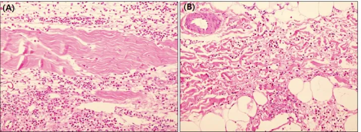
Fig. 2. (A) The marked infiltrates composed of numerous eosinophis with a slight admixture of lymphocytes in the perimysium (H&E,
×200). (B) The moderate infiltrates of eosinophils and lymphocytes in the subcutaneous fat layer (H&E, ×200).
phocytes in the muscle layer, as well as in the dermis and subcutis (Fig. 2A). The patient later developed a localized erythematous patch on her left calf. The skin biopsy at that site also showed moderate infiltration of eosinophils and lymphocytes in the dermis and subcutis (Fig. 2B). The bi- opsy specimen was insufficient for the evaluation of the muscle layer. The diagnosis of HES was made. Considering the absence of typical histologic findings of a dermal in- filtrate of eosinophils, histiocytes, and eosinophil debris between collagen bundles that form flame figures, a diag- nosis of eosinophilic cellulitis was less likely. After treat- ment with 1 mg/kg methylprednisolone for 2 weeks, the blood eosinophil counts decreased to within the reference range and the skin lesion subsided. The lungs could be an- other organ involved in HES, taking into account the sud- den disappearance of pleural effusion after the treatment.
Recently, to overcome problems with the above-men- tioned old definition, Simon et al.2 proposed a new defi- nition for HES: (i) blood eosinophilia (>1,500 eosino- phils/mm3) on at least two occasions, or evidence of prom- inent tissue eosinophilia associated with symptoms and marked blood eosinophilia; (ii) absence of secondary causes of eosinophilia, such as parasitic or viral infections,
allergic diseases, drug-induced or chemical-induced eosi- nophilia, hypoadrenalism, and neoplasm. Our case is con- sistent with this new proposed definition of HES.
To our knowledge, this is the first case of focal eosino- philic myositis associated with HES in the Korean derma- tological literature3. Furthermore, the palm is a very rare site of involvement, as muscle involvement in focal eosi- nophilic myositis is usually restricted to the lower legs4.
REFERENCES
1. Plötz SG, Hüttig B, Aigner B, Merkel C, Brockow K, Akdis C, et al. Clinical overview of cutaneous features in hypereo- sinophilic syndrome. Curr Allergy Asthma Rep 2012;12:85-98.
2. Simon HU, Rothenberg ME, Bochner BS, Weller PF, Wardlaw AJ, Wechsler ME, et al. Refining the definition of hypereo- sinophilic syndrome. J Allergy Clin Immunol 2010;126:45-49.
3. Lee MW, Suh HS, Suh DH, Choi JH, Sung KJ, Koh JK. Focal eosinophilic myositis. Ann Dermatol 1994;6:102-104.
4. Selva-O'Callaghan A, Trallero-Araguás E, Grau JM. Eosino- philic myositis: an updated review. Autoimmun Rev 2014;
13:375-378.