29
Copyrights © 2015 The Korean Society of Radiology
INTRODUCTION
Pseudoaneurysm, also known as false aneurysm, is a hemato- ma resulting from a defect in the vascular wall that freely com- municates with the parent vessel. The most common causes of massive lower gastrointestinal (GI) bleeding are diverticular dis- ease, angiodysplasia, and colitis, whereas pseudoaneurysmal lower GI bleeding is not common. We report a rare case of trau- matic superior rectal artery pseudoaneurysm presenting with recurrent lower GI and pelvic extraperitoneal bleeding resulting from a penetrating perineal injury.
CASE REPORT
A 43-year-old male patient who was a chronic alcoholic, pre- sented in a drunken state with a penetrating perineal wound,
caused by a wooden stick and resulting in massive lower GI bleeding. Computed tomography (CT) at a local hospital on the day of trauma showed an abnormal enhancing saccular lesion at the anterior wall of the rectum, 6 cm proximal from the anal verge and extending upward to the level of the bladder neck.
The lesion was thought to be supplied by the superior rectal ar- tery from the inferior mesenteric artery and was suggestive of a pseudoaneurysm (Fig. 1A). The pseudoaneurysm was saccular in shape and surrounded by a small hematoma in the posterior perivesicular space. The patient was managed at a local hospital, where he underwent an emergent laparoscopic Hartmann pro- cedure consisting of diversion sigmoid end colostomy with a su- tured rectal stump left behind without resection of the pseudoa- neurysm.
The patient had secondary lower GI bleeding after surgery on the same day. The perineal wound and rectum were packed
Case Report
pISSN 1738-2637 / eISSN 2288-2928 J Korean Soc Radiol 2015;72(1):29-32 http://dx.doi.org/10.3348/jksr.2015.72.1.29
Received July 3, 2014; Accepted October 22, 2014 Corresponding author: Jung Wook Seo, MD Department of Radiology, Ilsan Paik Hospital, Inje University College of Medicine, 170 Juhwa-ro, Ilsanseo-gu, Goyang 411-706, Korea.
Tel. 82-31-910-7389 Fax. 82-31-910-7369 E-mail: [email protected]
This is an Open Access article distributed under the terms of the Creative Commons Attribution Non-Commercial License (http://creativecommons.org/licenses/by-nc/3.0) which permits unrestricted non-commercial use, distri- bution, and reproduction in any medium, provided the original work is properly cited.
Traumatic pseudoaneurysm of the superior rectal artery is a rare cause of massive lower gastrointestinal bleeding. We reported a case of a 43-year-old male patient with pseudoaneurysm following a penetrating perineal wound. The patient had re- peat massive lower gastrointestinal and pelvic extraperitoneal bleeding and was di- agnosed as traumatic pseudoaneurysm of the superior rectal artery. To our knowl- edge, there are three case reports of traumatic pseudoaneurysm of the superior rectal artery treated by embolization. However, spontaneous regression occurred in the study subject after surgical hematoma removal, without any further pseudoan- eurysm resection.
Index terms
Traumatic Pseudoaneurysm Superior Rectal Artery
Massive Lower Gastrointestinal Bleeding
Traumatic Pseudoaneurysm of the Superior Rectal Artery with Recurrent Lower Gastrointestinal and Pelvic Extraperitoneal Bleeding: Importance of Pretreatment Recognition
재발성 하부위장관 및 골반복막외 출혈을 동반한 상직장동맥에서 발생한 외상성 가성동맥류: 치료 전 인식의 중요성
Kyung Joon Kim, MD, Jung Wook Seo, MD, You Sung Kim, MD
Department of Radiology, Ilsan Paik Hospital, Inje University College of Medicine, Goyang, Korea
Traumatic Pseudoaneurysm of the Superior Rectal Artery
30
J Korean Soc Radiol 2015;72(1):29-32 jksronline.orghematoma due to rupture of the pseudoaneurysm and active contrast leakage from the pseudoaneurysm (Fig. 1B, C). On day 3 of trauma, the patient became hemodynamically unstable and had emergent revision Hartmann procedure and hematoma evacuation because of unknown pseudoaneurysm. On day 4 of trauma, follow-up CT showed almost complete clearing of the pelvic hematoma but a small focal enhancing lesion was noted with gauze and the patient was referred to our hospital on day 2
of trauma. The patient arrived with a blood pressure of 100/40 mm Hg and a tachycardia of 141 beats per minute. He had ab- dominal distention and his abdomen was vaguely guarded. The hemoglobin level was 8.2 g/dL on arrival. The patient was pre- sumptively diagnosed as rupture of the Hartmann pouch (rectal stump). On day 2 of trauma, follow-up CT demonstrated a large
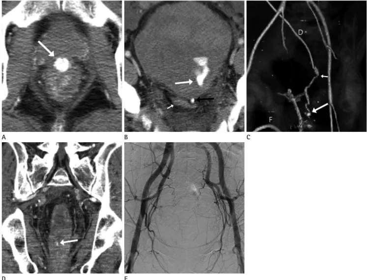
Fig. 1. Traumatic pseudoaneurysm of superior rectal artery in a 43-year-old male patient.
A. Axial contrast enhanced CT scan on the day of trauma at a local hospital shows the pseudoaneurysm (arrow) as a saccular enhancing lesion located at the anterior wall of the rectum, measuring 1.5 cm.
B. Follow-up CT scans on day 2 of trauma (next day after first surgery) show contrast leakage (long white arrow) from the pseudoaneurysm (not resected) surrounded by a hematoma in the posterior perivesical space. Drainage catheter tip (black arrow) is located in the rectovesical pouch and residual rectum stump (short white arrow) is collapsed.
C. Volume rendered three-dimensional reformatted CT on day 2 of trauma shows a saccular pseudoaneurysm (long white arrow) resulting bleed upward (short white arrow). Foley catheter (F) is inserted into urinary bladder and a drainage catheter (D) is located in intraperitoneal and pelvic cavity.
D. Follow-up coronal CT scans on day 4 of trauma (next day after second surgery) show a residual pseudoaneurysm (white arrow) as a small fo- cal enhancing lesion in the anterior wall of the rectum, measuring 0.3 × 0.6 cm.
E. Conventional angiography performed on day 17 of trauma shows no residual pseudoaneurysm on bilateral internal iliac and inferior mesen- teric arteriograms.
E B
D
A C
Kyung Joon Kim, et al
31
jksronline.org J Korean Soc Radiol 2015;72(1):29-32
garding massive lower GI bleeding resulting from a superior rectal artery pseudoaneurysm. Janmohamed et al. (3) reported a case of lower GI bleeding caused by non-traumatic pseudoan- eurysm secondary to acute diverticulitis, which was diagnosed by CT angiography. Our radiologic finding was saccular pseu- doaneurysm, similar to the previously reported case. The other two cases (4, 5) were traumatic pseudoaneurysms resulting from penetrating trauma or iatrogenic cause (colonoscopic procedure).
All three cases were treated by angiographic embolization. We performed surgical hematoma removal alone without resection of pseudoaneurysm that ruptured thereafter.
Some authors (6-8) reported spontaneous occlusion of trau- matic pseudoaneurysms resulting from thrombosis within 2 weeks. The pseudoaneurysm in our patient was apparent on CT, but not on angiography after the third bleeding following the re- vision Hartmann procedure. This was because of the 11 cm seg- mental resection of the rectosigmoid colon with ligation of the superior and middle rectal arteries. Furthermore, partial throm- bosis of the pseudoaneurysm seemed to hinder the opacifica- tion of residual pseudoaneurysm and caused spontaneous heal- ing. Yi et al. (9) reported a case of prophylactic embolization of traumatic hepatic artery pseudoaneurysm. Prophylactic emboli- zation is required to prevent severe hemorrhage from pseudoa- neurysm rupture. We likewise subsequently performed prophy- lactic embolization of the inferior mesenteric artery and the anterior branch of bilateral internal iliac arteries to treat the re- sidual pseudoaneurysm.
The patient in the present case had recurrent massive lower GI and pelvic extraperitoneal bleeding that could not be con- trolled by repeated surgical attempts. It was likely that a surgeon at the local hospital performed an emergent end colostomy on the day of trauma without recognition of the pseudoaneurysm depicted on CT. On day 3 of trauma, the surgeon at our hospital performed an emergent hematoma evacuation and revision end colostomy, again with failure to recognize the pseudoaneurysm on CT performed at our hospital. Additionally, the surgical ap- proach and visualization of the pseudoaneurysm might have been difficult due to its deep position. Hence, there was a resid- ual partially thrombosed pseudoaneurysm, whose rupture re- sulted in the third bleeding. Vascular lesions such as pseudoan- eurysms should be considered in the differential diagnosis of patients with post-traumatic lower GI bleeding, and more espe- in the anterior wall of the rectum. We diagnosed this as a par-
tially thrombosed residual pseudoaneurysm at the anterior wall of the rectum (Fig. 1D).
On day 14 of trauma, the patient’s blood pressure had dropped to 100/50 mm Hg and hemoglobin level was decreased to 7.9 g/
dL. Internal bleeding from ruptured pseudoaneurysm was sus- pected; this was the third bleeding episode. A percutaneous drainage catheter was temporarily inserted into the abdominal cavity. On day 17 of trauma, the patient became hemodynami- cally stabilized. CT showed a pelvic extraperitoneal hematoma, probably representing bleeding from the residual pseudoaneu- rysm, but active contrast leakage was not evident (not shown).
On day 19 of trauma, angiography was performed to treat the residual pseudoaneurysm. A conventional angiography via the right common femoral artery displayed no pseudoaneurysm of the superior rectal artery or extravasation from either bilateral internal iliac arteries or inferior mesenteric artery (Fig. 1E).
Therefore, we performed prophylactic Gelfoam embolization of the inferior mesenteric artery and the anterior branch of bilater- al internal iliac arteries. We concluded that spontaneous healing of the pseudoaneurysm had occurred. On day 31 of trauma, fol- low-up CT showed a decreasing pelvic hematoma and no evi- dence of residual pseudoaneurysm. The patient recovered well and no further episodes of lower GI bleeding have been report- ed. After 8 weeks, the sigmoid colostomy was reversed.
DISCUSSION
Pseudoaneurysm is a vascular out-pouching from the parent artery and lacks a complete arterial wall. Factors such as trauma, inflammation, or infection are known causes of pseudoaneu- rysm. Complications of pseudoaneurysm include early rupture, compression of adjacent structures, and combined infection (1).
Early diagnosis of pseudoaneurysm is important because at- tempts at post-rupture surgical repair lead to a high mortality rate of 50% (2). Ultrasonography, CT, and magnetic resonance imaging enable detection of visceral vascular lesions, but con- ventional angiography is important for further diagnosis and treatment. Endovascular treatment is often the first-line therapy.
Endovascular intervention or open surgical repair is necessary for all pseudoaneurysms (1).
To our knowledge, there are 3 case reports in the literature re-
Traumatic Pseudoaneurysm of the Superior Rectal Artery
32
J Korean Soc Radiol 2015;72(1):29-32 jksronline.orgcause of lower gastrointestinal haemorrhage. BMJ Case Rep 2011;2011. http://dx.doi.org/10.1136/bcr.11.2011.5102 4. Iqbal J, Kaman L, Parkash M. Traumatic pseudoaneurysm
of superior rectal artery-an unusual cause of massive low- er gastrointestinal bleed: a case report. Gastroenterol Res 2011;4:36-38
5. Zakeri N, Cheah SO. A case of massive lower gastrointesti- nal bleeding: superior rectal artery pseudoaneurysm. Ann Acad Med Singapore 2012;41:529-531
6. Gow KW, Murphy JJ 3rd, Blair GK, Stringer DA, Culham JA, Fraser GC. Splanchnic artery pseudo-aneurysms secondary to blunt abdominal trauma in children. J Pediatr Surg 1996;31:812-815
7. Dror S, Dani BZ, Ur M, Yoram K. Spontaneous thrombosis of a splenic pseudoaneurysm after blunt abdominal trau- ma. J Trauma 2002;53:383-385
8. Raghavan A, Wong CK, Lam A, Stockton V. Spontaneous occlusion of post-traumatic splenic pseudoaneurysm: re- port of two cases in children. Pediatr Radiol 2004;34:355- 357
9. Yi IK, Miao FL, Wong J, Narasimhan KL, Lo RH, Yee L, et al.
Prophylactic embolization of hepatic artery pseudoaneu- rysm after blunt abdominal trauma in a child. J Pediatr Surg 2010;45:837-839
cially in cases of penetrating injury, for effective treatments such as complete surgical removal or endovascular exclusion of flow.
In conclusion, we reported a rare case of traumatic superior rectal artery pseudoaneurysm with recurrent lower GI and pel- vic extraperitoneal bleeding that was caused by a lack of pre- treatment recognition of the pseudoaneurysm. Third bleeding on day 14 of trauma, in particular, could have been prevented if the surgeon had been informed about the pseudoaneurysm in a timely manner. Therefore, the radiologist should be aware of CT imaging findings of pseudoaneurysms and the surgeon should recognize the details regarding the parent vessel, location, and size of the pseudoaneurysm before surgery in select clinical can- didates.
REFERENCES
1. Jesinger RA, Thoreson AA, Lamba R. Abdominal and pelvic aneurysms and pseudoaneurysms: imaging review with clinical, radiologic, and treatment correlation. Radiograph- ics 2013;33:E71-E96
2. Sakamoto I, Sueyoshi E, Hazama S, Makino K, Nishida A, Yamaguchi T, et al. Endovascular treatment of iliac artery aneurysms. Radiographics 2005;25 Suppl 1:S213-S227 3. Janmohamed A, Noronha L, Saini A, Elton C. An unusual
재발성 하부위장관 및 골반복막외 출혈을 동반한 상직장동맥에서 발생한 외상성 가성동맥류: 치료 전 인식의 중요성
김경준 · 서정욱 · 김유성
외상성 가성동맥류는 대량 하부위장관 출혈의 드문 원인이다. 저자들은 회음부 관통상을 당하여 발생한 43세 남성의 가 성동맥류 증례를 보고하고자 한다. 환자는 재발성의 하부위장관 및 골반복막외 대량출혈이 발생하였고 CT 검사를 시행 하여 상직장동맥에서 발생한 외상성 가성동맥류로 진단되었다. 저자들이 조사한 바로는 상직장동맥에서 발생한 외상성 가성동맥류는 3건이 보고되었고 색전술로 치료하였다. 반면에 우리 증례는 외과적 혈종제거술이 시행되었으나 가성동맥 류는 절제하지 못하였고, 그 후에 저절로 치유되었다.
인제대학교 의과대학 일산백병원 영상의학과