Copyright © 2019. Anatomy & Cell Biology
Introduction
Venous drainage of the cerebrum is expertly modulated by highly complex superficial and the deep cerebral veins. These veins anastomose with each other and drain into the major dural venous sinuses such as superior sagittal (SSS), inferior sagittal, transverse (TS), cavernous (CS), and straight sinuses.
All these sinuses ultimately drain into the internal jugular vein (IJV) [1, 2].
The superficial middle cerebral vein (SMCV) is one of the superficial veins of the cerebrum that drains the venous
blood from most of the superolateral surface of the brain. The SMCV lies in the posterior horn of the lateral sulcus, which further course along the lateral sulcus, pterion, lesser wing of sphenoid to drains into the CS [3, 4]. Interestingly, with the help of many angiographic studies, several deviations from the normal course have been observed. The knowledge of these variant courses is vital so that different surgical ap- proaches can be applied to prevent bleeding. The termination of the SMCV and its association with the skull base is impera- tive in the preoperative planning of the skull base surgery [3].
Studies in the past have focused on the variant pattern of the SMCV and attempted to classify the same. However, most of them were computed tomography (CT) angiography based studies [3, 5]. Very few cadaveric studies on the variant pattern of the SMCV is available in the existing literature in adults and fetuses alike. Here in this study, we have made a sincere attempt to classify the various anatomical variations in the termination of SMCV in the termed fetus.
Corresponding author:
Sushma R. Kotian
Department of Anatomy, Kasturba Medical College Manipal, Manipal Academy of Higher Education, Manipal 576104, Karnataka, India Tel: +91-820-2922327, Fax: +91-820-2570061, E-mail: sushma.rk@manipal.
edu
Persistent fetal superficial middle cerebral vein: an anatomical study
Suhani Sumalatha, Sushma R. Kotian, Ashwija Shetty
Department of Anatomy, Kasturba Medical College Manipal, Manipal Academy of Higher Education, Manipal, India
Abstract: The superficial middle cerebral vein (SMCV) drains the venous blood from most of the superolateral surface of the brain and drains typically into the cavernous sinus as mentioned in standard textbooks. But the drainage of the SMCV is variable as indicated by various radiological studies. Although variations in the drainage of the SMCV exist, there is a shortage in the literature providing cadaveric evidence for the same. The present study was designed to identify the variations in the drainage pattern of the SMCV in fetal cadavers. During the dissection of formalin-fixed full-term fetuses, deviation in the drainage of the SMCV was observed in five out of 30 cases. In three out of 30 specimens (10%), SMCV was observed draining into superior petrosal sinus; and in two specimens (6.6%) into the transverse sinus. In the remaining specimens, the SMCV drained directly into the cavernous sinus. Knowledge of the variations noted in the present study is essential, not only for diagnosing several diseases involving the cavernous sinus or paracavernous sinuses but also in surgeries of paracavernous sinus lesions and endovascular treatment of arteriovenous fistulas. The SMCV and superior petrosal sinus can be a venous refluxing route in patients with arteriovenous fistulas.
Key words: Superficial middle cerebral vein, Tentorial sinus, Cavernous sinus, Superior petrosal sinus, Transverse sinus Received January 14, 2019; Revised March 26, 2019; Accepted April 12, 2019
and the dural venous sinuses were observed. The variation in the drainage pattern of the SMCV was noted and photo- graphed. The SMCV was identified and classified based on its variability using a standard reference [3].
Results
In the present study five specimens presented with varia- tion in the pattern of the drainage of SMCV. Rest of the speci-
Case 1 (superior petrosal type)
The SMCV was coursing along the lesser wing of the sphe- noid. It further passed downward and backward along the middle cranial fossa almost parallel to the CS to drain into the anterior end of the superior petrosal sinus (SPS) (Fig. 2).
Case 2 (basal type)
The SMCV was passing along the Sylvian fissure and the lesser wing of the sphenoid (LWS). It then turned abruptly
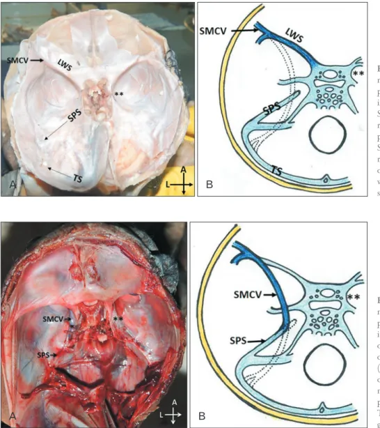
Fig. 1. Drainage pattern of superficial middle cerebral vein (SMCV)sphe no
parietal type. (A) The SMCV draining into the cavernous sinus on the left side.
Symbol ** represent the location of the right cavernous sinus. (B) Schematic re
presentation of the drainage pattern of SMCVsphenoparietal type. The Inter
rupted lines represent the re gres sion of the tentorial sinus (TS). LWS, lesser wing of sphenoid; SPS, superior petrosal sinus.
Fig. 2. Drainage pattern of superficial middle cerebral vein (SMCV)superior petrosal type. (A) The SMCV draining into the superior petrosal sinus (SPS) on the left side. This pattern of drainage of SMCV was observed in three cases (10%). Symbol ** represent the location of the right cavernous sinus. (B) Sche
matic representation of the drainage pat tern of SMCVsuperior petrosal type.
The Interrupted lines represent the re
gres sion of the tentorial sinus.
downwards and posteriorly along the middle cranial fossa, lateral to the CS. It later crossed the junction between TS and the Sigmoid Sinus and finally drained into the TS (Fig. 3). The communicating channels observed in these specimens were wider and more prominent than the other venous sinuses.
Discussion
A definitive venous pattern of the head and neck region develops in the first three months of prenatal life [6, 7]. Two embryological sinuses, i.e., tentorial sinus and the prootic si- nus take part in the formation of SMCV. Subsequent growth of cerebral hemisphere leads to elongation of the tentorial sinus. The SMCV is formed due to the medial shift of the an- terior portion of the tentorial sinus into the CS. The segment of the tentorial sinus distal to it regress leading to the adult/
definitive pattern of the SMCV. Fail in the medial shift/regres- sion of the tentorial sinus results in the anomalous course and termination of SMCV [8].
The SMCV runs typically along the LWS bone and enters into the proximal SPS. However, San Millan Ruiz et al. [9]
have raised a question regarding the drainage of the SMCV into the SPS in an anatomic study using an autopsied cadaver.
Suzuki and Matsumoto in 2000 [3] classified the drainage of SMCV into seven types based on embryological develop- ment or the remnant of the TS. The classification was done using a three-dimensional CT angiography and, 500 SMCVs were evaluated in 250 adult patients. The seven types included sphenoparietal (54%), cavernous (7%), emissary (12%), su- perior petrosal (2%), basal (2%), squamosal (2%), and under-
developed (9%). It was not possible to confirm the drainage pattern in 8 % of the cases. 3% of the cases showed multiple drainage pathways as observed in their study [3]. In the pres- ent study in fetuses, only three types of the drainage pattern of the SMCV was observed, i.e., superior petrosal type in 10% and basal in 6.6% of the cases respectively. The remain- ing 83.4% of the cases were of the sphenoparietal type. The frequency of atypical SMCV’s, i.e., superior petrosal and basal type was more in the fetuses as was observed in the present study unlike in the adults. The incidence of both types was rare/ less in adults. It could be because the fetuses born with the types above may not survive postnatal.
The basal type of SMCV as observed in the present study results due to the persistence of the entire venous route of ten- torial sinus. Thus the SMCVs routed via the sphenoparietal sinus and turned laterally along the floor of the middle cranial fossa, running along the petrous pyramid to terminate in the TS. The caudal regression of the primitive tentorial sinus is the result of the communication of the tentorial sinus to SPS leading to in superior petrosal type of the sinus [7].
A large rare squamousal type of vein running along the in- ner surface of temporal squama, connecting SMCV to into TS is reported by Kumar et al. in 2012 [8]. An unusual oblique sinus, which connected the right and the left TS was also re- ported previously [2].
In the dural arteriovenous fistulae (DAVF), the venous collaterals and venous rerouting help in the drainage of CS to the SPS, which then drains away into the cortical veins. These findings suggest that postnatal secondary anastomosis be- tween the SPS or the TS and any possible remnant variations
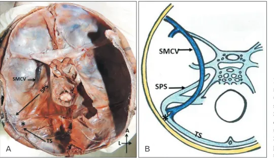
Fig. 3. Drainage pattern of superficial middle cerebral vein (SMCV)basal type.
(A) The SMCV draining into trans verse sinus (TS). This pattern of drain age of SMCV was present in 2 (6.6%) of the cases. Symbol * re pre sents the junction of the drainage of the SMCV into TS. (B) Schematic re pre sen ta tion of the drainage pattern of SMCVbasal type. SPS, superior pet ro sal sinus.
sidered to reduce the likelihood of possible venous complica- tions [12].
Gailloud et al. [13] performed a study on the termination pattern of SMCV. In their study, the SMCV was found to be absent in 19% of the cases. When present, they found the SMCV terminating directly into the CS in 20% of cases, the paracavernous sinus in 39% of cases, and the laterocavernous sinus in 22% of cases. Further, they also found that the para- cavernous and laterocavernous sinuses drain into SPS in 33%
and 18% of the cases respectively, which is the second most frequent type. Normally the paracavernous and laterocavern- ous sinuses drain into the pterygoid plexus [13].
The variation in the venous pattern as is observed in the present study in case of SMCV may also indicate the defective development of the IJV and the related venous sinuses. Iden- tification of these variant veins is therefore essential in surger- ies to avoid the dangerous catastrophic hemorrhage [14].
A variant of the SMCV mimicking an extra-axial hema- toma has been reported. Herein the patient presented with an underdeveloped type of SMCV that was drained by a large venous channel into the SSS [1]. Variation in the course of SMCV should be considered and screened by angiogram be- fore the surgery to reduce the risk during surgical procedures and the possibilities of post-operative edema [15].
Knowledge of the variations in the drainage of the SMCV as observed in the present study in fetal cadavers is essential for diagnosing several diseases involving the CS or paracav- ernous sinuses. It is also important in the correction of the le- sions of the para-cavernous sinus and endovascular treatment of arteriovenous fistulas. The SMCV and SPS can serve as a venous refluxing route in patients with arteriovenous fistulas.
ORCID
Suhani Sumalatha: https://orcid.org/0000-0002-2134-2179 Sushma R. Kotian: https://orcid.org/0000-0003-0271-3568 Ashwija Shetty: https://orcid.org/0000-0003-3031-6009
Conflicts of Interest
No potential conflict of interest relevant to this article was reported.
References
1. Saigal G, Villalobos E. A variant of the superficial middle cere- bral vein mimicking an extraaxial hematoma. AJNR Am J Neu- roradiol 2003;24:968-70.
2. Das S, Paul S. Unusual venous sinuses. Bratisl Med J 2007;108:
104-6.
3. Suzuki Y, Matsumoto K. Variations of the superficial middle cerebral vein: classification using three-dimensional CT angiog- raphy. AJNR Am J Neuroradiol 2000;21:932-8.
4. Chung JI, Weon YC. Anatomic variations of the deep cerebral veins,tributaries of Basal vein of rosenthal: embryologic aspects of the regressed embryonic tentorial sinus. Interv Neuroradiol 2005;11:123-30.
5. Fukuda M, Saito A, Takao T, Hiraishi T, Yajima N, Fujii Y. Drain- age patterns of the superficial middle cerebral vein: effects on perioperative managements of petroclival meningioma. Surg Neurol Int 2015;6:130.
6. Knosp E, Müller G, Perneczky A. Anatomical remarks on the fe- tal cavernous sinus and on the veins of the middle cranial fossa.
In: Dolenc VV, editor. The Cavernous Sinus. Vienna: Springer;
1987. p.104-16.
7. Padget DH. The cranial venous system in man in reference to development, adult configuration, and relation to the arteries.
Am J Anat 1956;98:307-55.
8. Kumar A, Chandra PS, Mankotia DS, Tripathi M, Garg A, Ma- hapatra AK. Squamosal type superficial middle cerebral vein: a rare venous drainage pattern. Neurol India 2012;60:546-7.
9. San Millan Ruiz D, Fasel JH, Rufenacht DA, Gailloud P. The sphenoparietal sinus of breschet: does it exist? An anatomic study. AJNR Am J Neuroradiol 2004;25:112-20.
10. Willinsky R, Goyal M, terBrugge K, Montanera W. Tortuous, engorged pial veins in intracranial dural arteriovenous fistulas:
correlations with presentation, location, and MR findings in 122 patients. AJNR Am J Neuroradiol 1999;20:1031-6.
11. Stiebel-Kalish H, Setton A, Nimii Y, Kalish Y, Hartman J, Huna Bar-On R, Berenstein A, Kupersmith MJ. Cavernous sinus dural arteriovenous malformations: patterns of venous drainage are re-
lated to clinical signs and symptoms. Ophthalmology 2002;109:
1685-91.
12. Sakata K, Al-Mefty O, Yamamoto I. Venous consideration in pe- trosal approach: microsurgical anatomy of the temporal bridging vein. Neurosurgery 2000;47:153-60.
13. Gailloud P, San Millán Ruíz D, Muster M, Murphy KJ, Fasel JH, Rüfenacht DA. Angiographic anatomy of the laterocavernous sinus. AJNR Am J Neuroradiol 2000;21:1923-9.
14. Murlimanju BV, Chettiar GK, Prameela MD, Tonse M, Kumar N,
Saralaya VV, Prabhu LV. Mastoid emissary foramina: an anatom- ical morphological study with discussion on their evolutionary and clinical implications. Anat Cell Biol 2014;47:202-6.
15. Dean BL, Wallace RC, Zabramski JM, Pitt AM, Bird CR, Spetzler RF. Incidence of superficial sylvian vein compromise and post- operative effects on CT imaging after surgical clipping of middle cerebral artery aneurysms. AJNR Am J Neuroradiol 2005;26:
2019-26.