Copyrights © 2017 The Korean Society of Radiology
57
Case Report
pISSN 1738-2637 / eISSN 2288-2928 J Korean Soc Radiol 2017;77(1):57-60 https://doi.org/10.3348/jksr.2017.77.1.57
INTRODUCTION
Hepatocellular carcinoma (HCC) is one of the most common cancers (1), and its incidence worldwide appears to be rising due to the increasing prevalence of hepatitis C virus infection (2). Spontaneous rupture of HCC is rare, and it has been found to be relatively more common in Asian countries than in West- ern countries (2). Because of the intraabdominal location of HCC, hemothorax resulting from ruptured HCC is extremely uncommon.
Hemothorax associated with ruptured HCC has been report- ed in only 18 cases in the English and Japanese literature (3, 4).
In one of these cases, the incidence occurred in Korea. In this case, hemothorax was caused by a ruptured metastatic mediasti- nal lymph node (3). In our study, we report a case of hemotho- rax caused by spontaneous rupture of primary HCC into the pleural cavity.
CASE REPORT
A 51-year-old male hepatitis B carrier was diagnosed with liver cirrhosis and HCC involving the right hepatic lobe in 2013.
The patient was readmitted to the hospital due to abdominal pain in December 2015.
At the time of hospitalization, no abnormalities were identified through the physical examination, and the patient,s vital signs were normal. A chest radiograph showed a moderate amount of pleural effusion in the right hemithorax. In patients with liver cirrhosis, pleural effusion can occasionally be found, especially in the right side (5). Therefore, the patient was treated to relieve the abdominal pain, and nutritional support was provided.
On the 14th day post admission, the patient experienced an abrupt onset of shortness of breath. His SpO2 level decreased to 89%, and his blood pressure fell to 80/50 mm Hg. In addition, his pulse rate (116/min) and respiration rate (40 breaths/min)
Hemothorax Caused by Spontaneous Rupture of Hepatocellular Carcinoma in the Pleural Cavity: A Case Report
간세포암의 흉강 내 자발파열로 인한 혈흉: 증례 보고
Jin Hee Seo, MD
1, Joon Young Ohm, MD
1, Song Soo Kim, MD
2, Jin Hwan Kim, MD
2*
1Department of Radiology, Chungnam National University Hospital, Daejeon, Korea
2Department of Radiology, Chungnam National University School of Medicine, Daejeon, Korea
Hemothorax resulting from ruptured hepatocellular carcinoma (HCC) is extremely rare and is generally caused by ruptured intrathoracic metastatic lesions. However, we report a rare case of hemothorax resulting from intrathoracic rupture of primary HCC.
Index terms Hemothorax
Carcinoma, Hepatocellular Rupture, Spontaneous
Multidetector Computed Tomography Embolization, Therapeutic
Received July 4, 2016 Revised September 12, 2016 Accepted October 21, 2016
*Corresponding author: Jin Hwan Kim, MD Department of Radiology,
Chungnam National University School of Medicine, 282 Munhwa-ro, Jung-gu, Daejeon 35015, Korea.
Tel. 82-42-280-7333 Fax. 82-42-253-0061 E-mail: michelan@cnu.ac.kr
This is an Open Access article distributed under the terms of the Creative Commons Attribution Non-Commercial License (http://creativecommons.org/licenses/by-nc/4.0) which permits unrestricted non-commercial use, distri- bution, and reproduction in any medium, provided the original work is properly cited.
58
Hemothorax by Rupture of HCC
jksronline.org
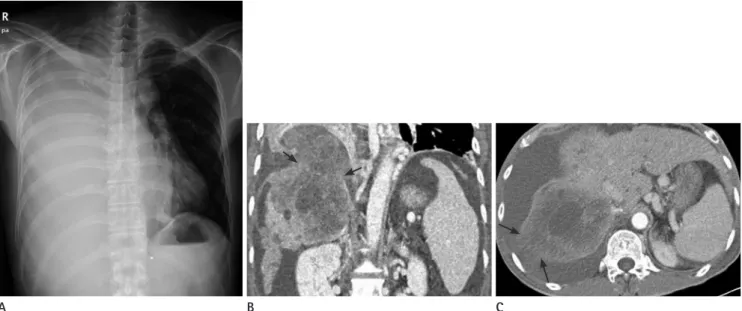
J Korean Soc Radiol 2017;77(1):57-60 were high. A chest radiograph showed complete white out of
the right hemithorax with mediastinal shifting to the left (Fig.
1A). Bloody fluid was aspirated by thoracentesis. The red blood cell count in the aspirated fluid was 1650000/mcL. Chest tube thoracostomy was performed, and the bloody fluid was drained.
The patient’s hemoglobin (Hb) level decreased from 11.2 g/dL to 6.2 g/dL.
Contrast enhanced chest and abdominal computed tomogra- phy (CT) scans were obtained at this timepoint. On comparing these CT scans to the previous CT scans, obtained 2 months earlier, it was found that HCC had rapidly increased in size from 6 cm to 10 cm. Moreover, the tumor had invaded the right diaphragm, and it had also invaded the right pleural cavity (Fig.
1B). The tumor showed extensive necrosis, and the surface of the intrapleural portion of the tumor was irregular and rup- tured (Fig. 1C). Metastatic nodules in the right lower lobe were surrounded by a collapsed lung, and intrathoracic lesions were excluded as the possible cause of hemothorax. These findings suggested that intrathoracic rupture of HCC resulted in hemo- thorax.
Transcatheter arterial embolization (TAE) was carried out immediately. Even though CT scans and angiography indicated no contrast extravasation, embolization of both the right ante- rior and middle hepatic arteries was undertaken using lipiodol
and gelatin sponges.
Subsequently, the patient,s blood pressure rose to 105/60 mm Hg, and his Hb level increased to 9.3 g/dL. The amount of bloody fluid drained through a chest catheter gradually decreased, and the catheter was removed from the patient’s chest on the 36th day post admission. Paracentesis was undertaken multiple times during admission, and a large amount of yellowish ascitic fluid was drained. Although the patient received intensive supportive care, the patient died of multiple organ failure on the 58th day post admission.
DISCUSSION
Spontaneous rupture of HCC is a rare but potentially life- threatening complication. The incidence of HCC rupture has been reported to be within the range of 2.3–26%. Since HCC is a hypervascular tumor and the pleural cavity pressure is nega- tive, hemothorax caused by spontaneous rupture of HCC leads to high possibility of mortality due to uncontrollable bleeding.
Ruptured HCC usually presents with hemoperitoneum due to its intraabdominal location. Thus, hemothorax after HCC rupture is an extremely uncommon condition, with only 18 cas- es being reported in the English and Japanese literature (3, 4). In four of the 18 cases, direct invasion of HCC into the right pleu-
A B C
Fig. 1. Hemothorax caused by spontaneous rupture of hepatocellular carcinoma in the pleural cavity in a 51-year-old male.
A. Frontal chest radiograph shows complete white out of the right hemithorax with mediastinal shifting to the left side.
B. C. Contrast-enhanced chest CT. Coronal image (B) obtained at the onset of shortness of breath demonstrates that a large hepatocellular carci- noma has invaded the right diaphragm (arrows) and the right pleural cavity. Axial CT image (C) shows that the surface of the intrapleural portion of the tumor is irregular and ruptured (arrows).
59
Jin Hee Seo, et al
jksronline.org J Korean Soc Radiol 2017;77(1):57-60
ral cavity was the cause of hemothorax, as in our case. In 13 out of the 18 cases, instead of ruptured primary HCC, ruptured in- trathoracic metastatic lesions resulted in hemothorax. In the re- maining one case, it was reported that hemothorax was caused by blood flow from hemoperitoneum into the pleural cavity (4).
Previous reports have indicated that tumors with larger sizes and greater extrahepatic protrusion rupture more frequently (6).
Besides the tumor size and the degree of extrahepatic protru- sion, vascular dysfunction in the peritumoral region, where the blood supply to the tumor is the richest, may be related to spon- taneous rupture of HCC (7). Portal vein thrombosis has also been reported to be a predictor of impending HCC rupture (8).
Multiplanar reformation images using multi-detector row CT are useful for detecting and defining the extent of extrahe- patic protrusion of HCC, which is an indicator of the possibility of HCC rupture. To detect active bleeding from ruptured HCC, contrast enhanced CT is helpful in identifying extravasation of contrast material. If extravasation of contrast material is identi- fied, this finding can be used to assist in the selection of appro- priate vessels for TAE.
Hemostasis is the first step in the management of a ruptured HCC using TAE or surgery. TAE followed by liver resection seems to achieve the best treatment results (9, 10). The progno- sis of ruptured HCC is poor compared to that of non-ruptured HCC. Prognosis is worse in advanced stage HCC rupture than in early stage HCC rupture.
In conclusion, if pleural fluid accumulates or pleural effusion abruptly increases in amount in patients with a large-sized HCC and extrahepatic protrusion, the possibility of hemotho- rax due to intrathoracic rupture should be given due consider- ation, and in the actual event of intrathoracic rupture, immedi- ate diagnosis and treatment will result in a better prognosis.
REFERENCES
1. Yoon SK, Chun HG. Status of hepatocellular carcinoma in
South Korea. Chin Clin Oncol 2013;2:39
2. Yoshida H, Mamada Y, Taniai N, Uchida E. Spontaneous ruptured hepatocellular carcinoma. Hepatol Res 2016;46:
13-21
3. Oh SY, Seo KW, Jegal Y, Ahn JJ, Min YJ, Park CR, et al. He- mothorax caused by spontaneous rupture of a metastatic mediastinal lymph node in hepatocellular carcinoma: a case report. Korean J Intern Med 2013;28:622-625
4. Ono F, Hiraga M, Omura N, Sato M, Yamamura A, Obara M, et al. Hemothorax caused by spontaneous rupture of he- patocellular carcinoma: a case report and review of the literature. World J Surg Oncol 2012;10:215
5. Strauss RM, Boyer TD. Hepatic hydrothorax. Semin Liver Dis 1997;17:227-232
6. Kanematsu M, Imaeda T, Yamawaki Y, Seki M, Goto H, Sone Y, et al. Rupture of hepatocellular carcinoma: predic- tive value of CT findings. AJR Am J Roentgenol 1992;158:
1247-1250
7. Zhu LX, Geng XP, Fan ST. Spontaneous rupture of hepato- cellular carcinoma and vascular injury. Arch Surg 2001;
136:682-687
8. Kim HC, Yang DM, Jin W, Park SJ. The various manifesta- tions of ruptured hepatocellular carcinoma: CT imaging findings. Abdom Imaging 2008;33:633-642
9. Lai EC, Lau WY. Spontaneous rupture of hepatocellular carcinoma: a systematic review. Arch Surg 2006;141:191- 198
10. Aoki T, Kokudo N, Matsuyama Y, Izumi N, Ichida T, Kudo M, et al. Prognostic impact of spontaneous tumor rupture in patients with hepatocellular carcinoma: an analysis of 1160 cases from a nationwide survey. Ann Surg 2014;259:
532-542
60
Hemothorax by Rupture of HCC
jksronline.org
J Korean Soc Radiol 2017;77(1):57-60
간세포암의 흉강 내 자발파열로 인한 혈흉: 증례 보고
서진희
1· 엄준영
1· 김성수
2· 김진환
2*
간세포암의 파열로 인한 혈흉은 매우 드물게 발생하며 그 원인으로는 흉곽 내로 전이된 병변이 파열되어 생기는 경우가 대 부분이다. 저자들은 흉곽 내 전이 병변이 아닌 원발성 간세포암의 흉강 내 자발파열로 발생한 혈흉의 드문 예를 경험하였기 에 이를 보고하고자 한다.
1충남대학교병원 영상의학과, 2충남대학교 의과대학 영상의학교실