Korean J Gastroenterol Vol. 73 No. 5, 299-302 https://doi.org/10.4166/kjg.2019.73.5.299 pISSN 1598-9992 eISSN 2233-6869
CASE REPORT
Korean J Gastroenterol, Vol. 73 No. 5, May 2019 www.kjg.or.kr
허혈성 대장염 없이 전신성 홍반성 루푸스에서 발현한 직장 궤양
손기창, 허원각, 주민수, 김의중, 정종혁, 최석채, 윤기중
1, 서검석
원광대학교 의과대학 내과학교실, 병리학교실1
Rectal Ulcer Developed in Systemic Lupus Erythematosus without Ischemic Colitis
Ki Chang Sohn, Won Gak Heo, Min Su Chu, Eui Joong Kim, Jong Hyeok Chung, Suck Chei Choi, Ki Jung Yun1 and Geom Seog Seo Departments of Internal Medicine and Pathology1, Wonkwang University School of Medicine, Iksan, Korea
Rectal involvement by systemic lupus erythematosus (SLE) is quite rare. Approximately 14 cases have been reported worldwide, but only one with ischemic colitis has been reported in Korea. A 17-year-old female patient was hospitalized with abdominal pain and hematochezia. Sigmoidoscopy revealed only a simple rectal ulcer without ischemic colitis. cytomegalovirus and bacterial infections were excluded. A sigmoidoscopic rectal biopsy indicated a rectal invasion by SLE, but the patient showed an acute worsening conditions that did not respond to treatment. This paper reports a case of rectal ulcer that developed in SLE without ischemic colitis with a review of the relevant literature. (Korean J Gastroenterol 2019;73:299-302)
Key Words: Lupus erythematosus, systemic; Rectal ulcer; Colitis, ischemic
Received November 27, 2018. Revised January 9, 2019. Accepted January 27, 2019.
CC This is an open access article distributed under the terms of the Creative Commons Attribution Non-Commercial License (http://creativecommons.org/licenses/
by-nc/4.0) which permits unrestricted non-commercial use, distribution, and reproduction in any medium, provided the original work is properly cited.
Copyright © 2019. Korean Society of Gastroenterology.
교신저자: 서검석, 54538, 익산시 익산대로 460, 원광대학교 의과대학 내과학교실
Correspondence to: Geom Seog Seo, Department of Internal Medicine, Wonkwang University School of Medicine, 460 Iksan-daero, Iksan 54538, Korea. Tel:
+82-63-859-2565, Fax: +82-63-855-2025, E-mail: [email protected], ORCID: https://orcid.org/0000-0001-8789-7989 Financial support: None. Conflict of interest: None.
서 론
루푸스는 주로 젊은 연령군에 호발하는 자가면역성 질환으 로서 신장, 중추신경계, 심장, 폐 등 전신을 침범하여 치명적 인 합병증을 일으킬 수 있고,1,2 위장관계 증상은 절반 정도의 환자에서 관찰될 정도로 비교적 흔하게 나타나지만 비특이적 이다. 그러나 루푸스에 의한 직장 궤양의 동반은 매우 드문데, 국내에서는 루푸스 환자에서 허혈성 장염에 직장 궤양이 동반 된 경우는 보고된 바 있으나3 허혈성 장염 없이 직장 궤양만 보고된 예는 없다. 이에 저자들은 내시경 육안 소견에서 허혈 성 대장염 없이 직장 궤양의 형태로 발현된 전신성 홍반성 루푸스 장염 1예를 경험하였기에 문헌고찰과 함께 보고하는 바이다.
증 례
루프스로 치료 중인 17세 여자가 내원 3일 전부터 하복부 통증, 설사 및 발열 소견을 보여 추가 검사 및 증상 조절을 위하여 입원하였다. 과거력에서 내원 3년 전 탈모, 발진, 관절 통 등의 증상으로 루푸스를 진단받았다.
내원 당시 이학적 소견에서 혈압 120/90 mmHg, 맥박 60회/분, 호흡수 20회/분, 체온 38.2℃였다. 양측 뺨 위에 고정된 융기성 홍반, 탈모, 구강 및 성기 궤양 소견은 있었지만 관절염이나 손가락, 발가락 끝의 색깔 변화 및 홍반성 결절은 관찰되지 않았다. 복부 팽만 및 복부 전반에 압통이 있었지만 간 종대나 비장 종대는 없었다. 경부나 액와 등 촉지되는 림프절 종대 소견은 없었다. 일반 혈액 검사는 백혈구 1,000/mm3, 혈색소 10.2 g/dL, 혈소판 130,000/mm3였다. 혈액화학 검사에서 나트 륨 143 mEq/L, 칼륨 3.6 mEq/L, 총 단백 4.9 g/dL, 알부민
300
손기창 등. 전신성 홍반성 루푸스에서 발현한 직장 궤양The Korean Journal of Gastroenterology Fig. 1. Abdomen and pelvic computed tomography (coronal view)
shows diffuse, severe edematous wall thickening of the entire colon (arrows), and a large amount of ascites (arrowhead).
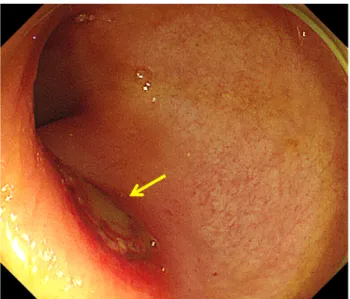
Fig. 2. Sigmoidoscopic examination showing a fairly well-marginated ulcer lesion (arrow) without an ischemic background.
Fig. 3. Sigmoidoscopic rectal biopsy shows much nuclear debris (arrows) (H&E, ×200).
3.1 g/dL, AST 19 IU/L, ALT 6 IU/L, BUN 25.0 mg/dL, 크레아티닌 2.12 mg/dL, 칼슘 8.2 mg/dL, 인 4.2 mg/dL였다.
면역학적 검사에서는 적혈구 침강속도 12 mm/hr, C-반응 단백 61 mg/L로 증가되어 있었다. Anti-ds-DNA 14 IU/L, 혈청 C3 105 mg/dL 및 C4 21 mg/dL는 정상 범위였다. 대변 검사에 서는 소수의 적혈구 및 백혈구 양성 소견이었고,
Escherichia coli, Shigella, Salmonella, Campylobactor, Clostridioides difficile
toxin PCR 음성이었다. 복부 전산화단층촬영에서 전대 장벽은 두꺼워져 있었고 다량의 복수가 관찰되었다(Fig. 1).원인 평가를 위한 구불창자 내시경 검사에서 항문연에서 10 cm 상방 직장 주위에 허혈성 손상이 동반되지 않은 경계 가 명확한 타원형의 궤양 소견이 관찰되었고(Fig. 2), 조직 검 사 소견에서 만성 염증 및 핵 잔해(nuclear debris)가 확인되 었으며(Fig. 3), 움 농양(crypt abscess) 및 육아종 소견은 관 찰되지 않았다. 조직에서 시행한 거대세포바이러스에 대한 면 역화학조직 염색도 음성이었다. 루푸스의 장 침범으로 인한 궤양 발생으로 판단하고 스테로이드를 정맥 투여하면서 경과 관찰하였지만 환자는 폐렴, 급성 호흡부전, 폐 출혈 등이 발생 하여 치료 15일만에 사망하였다.
고 찰
루푸스에 의한 대장 침범은 허혈성 손상이 발생하면서 다발 성 직장 궤양, 천공, 누공의 형태로 나타나기도 한다. 발병 기전 은 아직 명확하게 밝혀지지는 않았지만 장간막 혈관염, 거대세 포바이러스 감염에 의한 내피세포 확장, 스테로이드 사용에 따른 혈관 경화 및 항인지질 증후군(antiphospholipid syn- drome)에 의한 혈전증 등이 관여할 것으로 추정되고 있다.
저자들은 본 증례를 비롯하여 국내외에 발표된 15예의 증례 보고를 토대로 루푸스에 의한 직장 궤양의 특징을 Table 1에 정리하였다.3-16 15예 중에 12예는 병변이 직장에서 관찰되었 으며, 가장 흔한 병변 형태는 궤양성이었고 천공은 5예에서
Sohn KC, et al. Rectal Ulcer Developed in Systemic Lupus Erythematosus without Ischemic Colitis
301
Vol. 73 No. 5, May 2019 Table 1. Reported Cases of SLE Complicated with Rectal Involvementa
No References Age/sex Location Rectal involvement type Activity of SLE Therapy Prognosis
1 Tsugu et al. (1978)4 28/F R Ulcer, perforation (+) Steroid Dead
2 Yagita et al. (1981)5 18/F R-S Ulcer (multiple) (+) Steroid Alive
3 Palvio and Christensen (1987)6 43/F R Tumor (-) OP Dead
4 Iida et al. (1991)7 39/F R-S Ulcer (multiple), perforation (+) Steroid Dead
5 Igarashi et al. (1991)8 31/F R Perforation (-) OP Dead
6 Reissman et al. (1994)9 54/F R Necrosis (-) OP Alive
7 Teramoto et al. (1999)10 41/M R Ulcer, perforation (-) OP Alive
8 Amit et al. (1999)11 34/F R Ulcer (-) Steroid+OP Alive
9 Yuasa et al. (2002)12 27/M R Ulcer (multiple) (+) Steroid Alive
10 Ohara et al. (2007)13 52/F R Ulcer, perforation (+) OP Alive
11 Chattopadhyay et al. (2011)14 38/F R Ulcer (+) Steroid Alive
12 Yoo et al. (2012)3 22/M R-S Ulcer (+) Steroid Alive
13 Yau et al. (2014)15 28/F R Ulcer (multiple) (+) Steroid+CP Alive
14 Kaieda et al. (2014)16 51/M R Ulcer (+) Steroid+TL Alive
15 Present case 17/F R Ulcer (-) Steroid Dead
SLE, systemic lupus erythematosus; F, female; R, rectum; S, sigmoid colon; OP, operation; M, male; CP, cyclophosphamide; TL, tacrolimus.
관찰되었다.
임상증상은 하복부 통증, 혈변, 설사, 후중감 등의 다양한 증상이 동반될 수 있다. 내시경 소견에서는 단순 궤양, 허혈성 병변과 동반된 궤양, 궤양에 의한 천공 등이 관찰 가능하고,17 조직 검사에서 항핵 항체 등이 증가하면서 세포가 파괴되고, 이로 인하여 세포 핵의 잔해인 핵 잔해가 나타난다.18핵 잔해 는 세포가 파괴되는 상황에서 관찰될 수 있기에 루푸스를 진 단하게 하는 특징적인 소견이라고는 할 수 없지만, 루푸스처 럼 자가면역 질환에서 더 많은 다수의 핵 잔해가 발생 가능하 다. 본 환자의 경우에도 직장 궤양 조직 검사에서 다수의 핵 잔해를 확인할 수 있었다.
환자에서 관찰된 직장 궤양의 원인으로는 루푸스 장염 이외 에도 염증성 장질환, 세균성 장염, 거대세포바이러스, 기타 감 염 질환, 비스테로이드 항염증제나 면역억제제 같은 약제 등을 생각해볼 수 있다.19 본 증례에서는 내시경 소견에서 경계가 명확한 직장 궤양이 발견되어 염증성 장질환은 배제할 수 있었 고 조직 검사, 대변 PCR 및 세균 배양 검사 등을 통하여 각각 거대세포바이러스와 세균 감염 등을 배제할 수 있었다. 약물로 인한 궤양 발생 가능성에 대하여 평가하기 위하여 당시 환자가 복용 중이었던 약제에 대하여 조사하였는데, 진통 소염제의 복용력은 없었지만 루프스의 치료를 위하여 항말라리아제 (hydroxychloroquine)와 면역억제제(mycophenolate mofe- til)를 복용 중이었다. 이 중에서 hydroxychloroquine과 궤양 의 상관성은 불분명하였고, mycophenolate mofetil에 의한 장염에 의하여 궤양이 발생할 수는 있으나, 이런 경우 궤양의 발생 부위나 조직학적 소견 등20으로 배제가 가능하였다.
치료는 루프스의 질병 활성도 증가에 준하여 정맥 스테로 이드, 사이클로포스파마이드, 타크로리무스 등을 사용해볼 수 있지만16 치료에 반응하지 않을 수 있다. 루푸스에 의한 대장 침범은 루프스 진단 전에 발견되기도 하지만 대부분은 루푸스 의 활동기 때 발현되며 치료에 반응을 하지 않는 중증 경과를 보일 수 있다. 루푸스 환자에서 반복되는 복통이나 혈변의 증 상이 있는 경우 루푸스의 장 침범을 우선 고려해야 하고, 대장 내시경 검사를 시행하였을 때 루푸스 장염의 일반적인 소견이 보이지 않는다 하더라도 루푸스 장염을 배제하지 말고, 조직 검사 등의 추가 검사를 통하여 루푸스 장염을 조기 진단하여 적극적인 치료를 시행하는 것이 중요하다고 할 수 있다.
REFERENCES
1. Juvonen T, Niemelä O, Reinilä A, Nissinen J, Kairaluoma MI.
Spontaneous intraabdominal haemorrhage caused by segmen- tal mediolytic arteritis in a patient with systemic lupus eryth- ematosus--an underestimated entity of autoimmune origin? Eur J Vasc Surg 1994;8:96-100.
2. Ahn JK. Clinical manifestations and diagnosis of systemic lupus erythematosus. Korean J Med 2010;78:409-415.
3. Yoo IK, Yoo SH, Choi S, et al. A case of systemic lupus eryth- ematosus presenting as a large rectosigmoid ulcer. Korean J Med 2012;82:252-256.
4. Tsugu T, Sasho T, Abe M, Kuramochi S, Torikata C, Tadenuma T.
A case of colonic perforation caused by lupus arteritis. Naika 1978;42:515-518.
5. Yagita A, Saito H, Yuasa Y. Multiple and so-called “punched out”
ulcers at the left colon, associated with systemic lupus erythematosus. I-To-Cho (Stomach and Intestine) 1981;16:
302
손기창 등. 전신성 홍반성 루푸스에서 발현한 직장 궤양The Korean Journal of Gastroenterology 889-895.
6. Palvio DH, Christensen KS. Systemic lupus erythematosus with rectal stenosis simulating tumour or diverticulosis. Case report.
Acta Chir Scand 1987;153:63-65.
7. Iida M, Suekane K, Mochizuki Y, et al. Intestinal lesions in pa- tients with systemic lupus erythematosus. I-To-Cho (Stomach and Intestine) 1991;26:1235-1245.
8. Igarashi M, Katsumata T, Kobayashi K, et al. Systemic lupus er- ythematosus with rectal perforation. I-To-Cho (Stomach and Intestine) 1991;26:1285-1290.
9. Reissman P, Weiss EG, Teoh TA, Lucas FV, Wexner SD.
Gangrenous ischemic colitis of the rectum: a rare complication of systemic lupus erythematosus. Am J Gastroenterol 1994;89:
2234-2236.
10. Teramoto J, Takahashi Y, Katsuki S, et al. Systemic lupus eryth- ematosus with a giant rectal ulcer and perforation. Intern Med 1999;38:643-649.
11. Amit G, Stalnikowicz R, Ostrovsky Y, Prus D, Eliakim R. Rectal ul- cers: a rare gastrointestinal manifestation of systemic lupus erythematosus. J Clin Gastroenterol 1999;29:200-202.
12. Yuasa S, Suwa A, Hirakata M, et al. A case of systemic lupus eryth- ematosus presenting with rectal ulcers as the initial clinical man- ifestation of disease. Clin Exp Rheumatol 2002;20:407-410.
13. Ohara M, Takahashi H, Suzuki C, et al. A case of lupus eryth- ematosus profundus followed by systemic lupus erythematosus presenting with severe intestinal involvement. Nihon Rinsho
Meneki Gakkai Kaishi 2007;30:48-54.
14. Chattopadhyay P, Abby Philips C, Dhua D, Saha S. Systemic lupus erythematosus presenting as ischaemic proctitis. Lupus 2011;
20:653-655.
15. Yau AH, Chu K, Yang HM, Ko HH. Rectal ulcers induced by sys- temic lupus erythematosus. BMJ Case Rep 2014;2014:
bcr2014205776.
16. Kaieda S, Kobayashi T, Moroki M, et al. Successful treatment of rectal ulcers in a patient with systemic lupus erythematosus us- ing corticosteroids and tacrolimus. Mod Rheumatol 2014;24:
357-360.
17. Hiraishi H, Konishi T, Ota S, Shimada T, Terano A, Sugimoto T.
Massive gastrointestinal hemorrhage in systemic lupus eryth- ematosus: successful treatment with corticosteroid pulse therapy. Am J Gastroenterol 1999;94:3349-3353.
18. Zharkova O, Celhar T, Cravens PD, Satterthwaite AB, Fairhurst AM, Davis LS. Pathways leading to an immunological disease:
systemic lupus erythematosus. Rheumatology (Oxford) 2017;
56(suppl_1):i55-i66.
19. Lee KA, Bae SC, Bang SY, et al. A case of systemic lupus eryth- ematosus patient with ulcerative colitis. J Korean Rheum Assoc 2008;15:328-331.
20. Calmet FH, Yarur AJ, Pukazhendhi G, Ahmad J, Bhamidimarri KR.
Endoscopic and histological features of mycophenolate mofetil colitis in patients after solid organ transplantation. Ann Gastroenterol 2015;28:366-373.