pISSN: 0378-6471 eISSN: 2092-9374 DOI : 10.3341/jkos.2010.51.5.677
= 증례보고 =
새로운 인공수정체를 낭내 삽입한 미세각막절개 백내장 수술의 임상평가
신철진⋅이주은⋅김재형⋅김재용⋅차흥원 울산대학교 의과대학 서울아산병원 안과학교실
목적: 미세절개창을 통해 삽입 가능한 Akreos MI-60 인공수정체를 삽입한 군의 임상성적을 일반 인공수정체를 삽입한 군과 비교하였다.
대상과 방법: 동일한 술자가 수정체유화술을 시행한 후 Akreos MI-60 인공수정체를 삽입한 25안과 Akreos Adapt-AO 인공수정체를 삽입한 28안을 대상으로 술 후 1주, 1개월, 4개월에 최대교정시력, 고위수차, 대비감도, 수술유발난시 등을 측정하였다.
결과: 최대교정시력은 술 후 1주째 0.09±0.11 (logMAR), 0.11±0.08 (MI-60, Adapt-AO 순), 술 후 1개월째 0.06±0.07, 0.06±0.06, 술 후 4개월째 0.05±0.06, 0.06±0.05로 유의한 차이가 없었다(Mann-Whitney U test, p>0.05). 굴절오차는 술 후 1주, 1개월, 4개월 째 모두 유의한 차이를 보였다. 두 군간 고위수차, 대비감도에서 유의한 차이는 없었으나, 술 후 1개월, 4개월째 수술유발난시는 MI-60 군에서 유의하게 작았다.
결론: Akreos MI-60 인공수정체는 기존의 임상성적이 검증된 비구면 인공수정체와 대등한 구면수차, 대비감도를 보이고, 여기에 미세 절개수술의 장점으로 작은 술 후 난시값을 보였다.
<대한안과학회지 2010;51(5):677-683>
■ 접 수 일: 2009년 8월 5일 ■ 심사통과일: 2010년 2월 9일
■ 책 임 저 자: 차 흥 원
서울특별시 송파구 풍납2동 388-1 서울아산병원 안과
Tel: 02-3010-3680, Fax: 02-470-6440 E-mail: [email protected]
* 본 논문의 요지는 2008년 대한안과학회 제100회 추계학술대회에서 구연으로 발표되었음.
최근 초음파 수정체유화술 및 인공수정체 삽입술은 백내 장 수술의 표준으로 자리잡았다. 작은 각막절개창, 점안마 취, 미세수술기법, 향상된 인공수정체 재질 및 디자인은 수 많은 사람들에게 보다 나은 나안시력을 제공해 주었다. 이 제 단순히 시력표 상의 20/20의 시력이 목표가 아니라 환 자의 삶의 질을 고려한 기능적 시력(functional vision)이 중요하게 되었으며, 여러 연구들은 대비감도(contrast sensitivity)가 기능적 시력의 중요한 표지자임을 보여주었 다.1,2 이에 따라 백내장 수술과정에서 근시나 원시의 조절 뿐만 아니라 난시나 고위수차(high order aberration)를 줄 이는 것이 대비감도나 시력의 질에 있어 중요하다.3-5
현재 백내장 수술의 발달과정에 있어 보다 더 작은 절개창을 통해 초음파 수정체유화술을 시행하는 미세각막절개 백내장 수술(microincision cataract surgery, MICS)이 추세이다.
미세각막절개 백내장 수술은 phakonit6,7, bimanual phacoe- mulsification8-11, microphaco12,13등 다양하게 불렸는데 이
것을 Alio et al14이 microincision cataract surgery라고 명 명하고, 그 정의를 2 mm 투명각막절개로 인공수정체 삽입이 가능한 백내장 수술이라 하였다. 이에 술 후 안정적인 좋은 시 력 예후를 보장하면서, 보다 작은 절개창을 통해 삽입이 가능 한 새로운 미세절개 인공수정체들이 개발되었다. Akreos MI-60 인공수정체(Bausch & Lomb, Rochester, NY, USA)는 1.8 mm 각막절개창을 통해 삽입가능한 친수성 아크 릴 인공수정체(hydrophilic acrylic IOL)이다. MI-60 인공수 정체는 구면수차(spherical aberration) 값을 0에 맞추어 기 존의 구면 인공수정체보다 기능적 시력을 좀더 향상시킬 수 있 을 것으로 기대된다. 이에 더해 haptic tip이 10도 꺾여 (angulation) 있는 독특한 디자인은, 수정체 낭(capsular bag)에서 완벽한 안정성을 보장하고, 술 후 후발백내장 발생 을 줄여 줄 수 있다.
본 연구에서는 1.8 mm의 미세절개창을 통해 삽입 가능한, 얇은 광학부를 가지는 비구면 인공수정체인 Akreos MI-60 를 삽입한 군의 임상성적을 일반 비구면 인공수정체를 삽입한 군과 비교하였다.
대상과 방법
2007년 12월부터 2008년 9월까지 서울아산병원에서 노년성(senile cataract) 혹은 노년기 전 백내장(presenile cataract)으로 합병증이 없는 백내장 수술을 시행받은 51
Table 1. Comparison of sex, age, preoperative BCVA, axial length, IOL power between akreos MI-60 IOL-im- planted group and akreos Adapt-AO IOL-implanted group (Mean±SD†)
Akreos MI-60 Akreos Adapt-AO Pvalue*
Number of eyes 25 28
Age (years) 67.88±8.97 69.96±6.34 P>0.05
BCVA‡(logMAR) 0.39±0.15 0.38±0.21 P>0.05
Axial length (mm) 23.51±0.72 23.19±0.51 P>0.05
IOL Power§ (diopter) 21.00±1.46 20.93±1.04 P>0.05
Sex (M:F) 1:1 3:4
*Mann-Whitney U test; †SD=standard deviation; ‡BCVA=best corrected visual acuity; §IOL power=intraocular lens power.
명, 53안을 대상으로 하였다. 수술 전 각막질환이나 안구 내 염증, 녹내장, 그리고 당뇨병성 망막병증의 병력이 없는 환자들을 대상으로 하였고 LOCS (Lens Opacities Classi- fication System) Ⅲ 분류에 의해 핵 경화(nuclear opa- lescence) 정도가 5 이상인 환자나 전층각막이식술이나 굴 절교정수술, 녹내장 수술, 유리체-망막 수술 등을 시행받 은 환자들은 본 연구에서 제외하였다.
환자들에게 MI-60나 Adapt-AO 인공수정체 중 무작위 로 배정하여 25안은 MI-60를 삽입하였고, 28안은 Adapt- AO를 삽입하였다. 인공수정체의 도수 결정은 IOL master (Carl Zeiss Meditec, Dublin, CA, USA)에 내장된 3세대 공식 Haigis와 특화된 인공수정체 A constant를 이용하여 계산하였다.
동일한 술자가 각막절개도(keratome)를 이용하여 각막 곡률이 높은 면(steep meridian)을 따라 투명윤부절개창 (clear corneal incision)을 만들었다. MI-60와 Adapt-AO 각각 1.9 mm와 2.75 mm의 크기의 절개창을 만들고, 캘리 퍼(calipers)를 이용하여 절개창의 크기를 확인하였다. 이후 점탄물질(viscoelastic)을 전방 내에 채워 넣고, 인공수정체 의 광학부보다 약간 작은 크기의 원형전낭절개(continuous curvilinear capsulorrhexis)를 cystotome을 이용하여 시행 하였다. 평형생리식염수(balanced salt solution, BSS, Alcon, USA)를 사용하여 수력분리술과 수력윤곽술을 시행한 후, 초음파 유화기(Infinity, Alcon, USA)로 핵의 수정체유화 술과 피질 흡입을 시행하였다. 각 인공수정체를 낭내에 삽 입 후 전방 내에 남아있는 점탄물질은 관류 및 흡입장치 (Irrigation/Aspiration device)로 제거하였다.
수술 전 모든 환자들에게 최대교정시력, 구면대응치 (spherical equivalent), 세극등현미경, 안압, 안저 검사 등 의 기본 안과검사를 시행하였다. 그리고 술 후 1주, 1개월, 4개 월째에 기본검사를 포함하여 수차검사(Zywave®Aberrometer Bausch & Lomb Inc.)를 시행하였다. 수차검사는 조절을 막기 위해 암실에서, 동공 중심부 6 mm 영역에서 산동 후 검사하였다. 측정된 수차는 Zernike 상수로 표시하였으며, 구면수차(spherical aberration), 수직 및 수평 코마수차
(vertical and horizontal coma aberration), 전체고위수차 의 RMS (root mean square of total high order aberra- tions)를 측정하였다. 각막지형도 및 전방깊이는 Orbscan (version 3.12, Bausch & Lomb Inc.)을 이용하여 측정하 였다. 대비감도검사는 vision contrast test system 검사표 (Vistech Consultants Inc.)를 사용하여 중등도조명시 (mesopic condition)와 명소시(photopic condition)로 나누 어 측정하였다. 수술유발난시값(surgically induced astig- matism)은 Hill 등이 제안한 수술 시의 각막 절개방향과 수 술 전후의 각막곡률 및 방향을 이용한 벡터분석법(vector method)을 이용하여 계산하였다.15
수술 후 두 군 간의 측정치 비교는 Mann-Whitney U test 를이용하여 비교하였고, 모든 통계분석은 SPSS (version15.0, software for Windows; SPSS Inc., Chicago, IL, USA)를 이용하였다.
결 과
MI-60 인공수정체 군과 Adapt-AO 인공수정체 군 간의 평균 나이, 최대교정시력, 안축장 길이 및 삽입한 인공수정 체굴절력에는 통계적으로 유의한 차이가 없었다(Table 1).
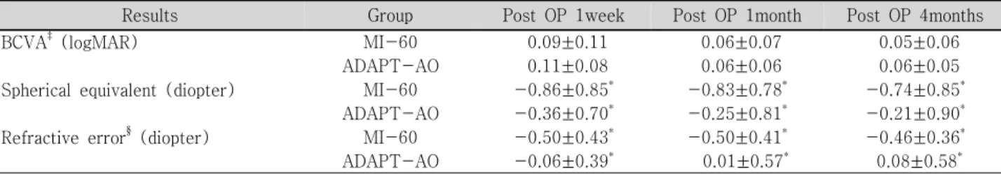
술 후 1주, 1달, 그리고 4달째 측정한 최대교정시력은 두 군 사이에 유의한 차이가 없었고, 두 군 모두 향상된 술 후 시력을 보였다. 굴절 오차(refraction error)는 술 후 1주, 1달, 그리고 4달째 모두 MI-60 IOL을 삽입한 군에서 유의 하게 컸다(Table 2).
MI-60 인공수정체 군과 Adapt-AO 인공수정체의 전체 고위수차(Total HOA)는 각각 0.753, 그리고 0.643 mi- crometer로 유의한 차이는 없었다. 구면수차(spherical aberration)와 coma 수차도 두 군 사이에 유의한 차이는 없 었다(Table 3).
두 군 간의 술 후 대비감도 검사 결과 중등도조명시와 명 소시의 모든 공간주파수 영역에서 MI-60 인공수정체 군이 Adapt-AO 인공수정체 군보다 더 나은 검사결과를 나타내 는 경향을 보였고, 이중 술 후 1개월째 중등도조명시 검사
Table 2. Comparison of postoperative BCVA, spherical equivalent, refractive error between akreos MI-60 IOL- implanted group and akreos Adapt-AO IOL-implanted group (Mean±SD†)
Results Group Post OP 1week Post OP 1month Post OP 4months
BCVA‡ (logMAR) MI-60 0.09±0.11 0.06±0.07 0.05±0.06
ADAPT-AO 0.11±0.08 0.06±0.06 0.06±0.05
Spherical equivalent (diopter) MI-60 -0.86±0.85* -0.83±0.78* -0.74±0.85*
ADAPT-AO -0.36±0.70* -0.25±0.81* -0.21±0.90*
Refractive error§ (diopter) MI-60 -0.50±0.43* -0.50±0.41* -0.46±0.36*
ADAPT-AO -0.06±0.39* 0.01±0.57* 0.08±0.58*
*Mann-Whitney U test (P<0.05); †SD=standard deviation; ‡BCVA=best corrected visual acuity; §Refractive error=postoperative spherical equivalent-target refraction.
Table 3. Comparison of postoperative total high order aberration, spherical aberration, coma aberration between akreos MI-60 IOL-implanted group and akreos Adapt-AO IOL-implanted group (Mean±SD†)
Results Group Post OP 1week Post OP 1month Post OP 4months
RMS HoA‡ MI-60 0.657±0.153* 0.649±0.188* 0.753±0.251*
ADAPT-AO 0.755±0.102* 0.826±0.262* 0.643±0.163*
Spherical aberration MI-60 0.394±0.110* 0.399±0.147* 0.424±0.110*
ADAPT-AO 0.412±0.141* 0.413±0.134* 0.408±0.103*
Coma aberration MI-60 0.233±0.140* 0.286±0.126* 0.338±0.243*
ADAPT-AO 0.328±0.150* 0.370±0.326* 0.311±0.225*
*Mann-Whitney U test (P>0.05); †SD=standard deviation; ‡RMS HoA=root mean square of total high order aberrations.
Table 4. Comparison of postoperative surgically induced astigmatism between Akreos MI-60 IOL-implanted group and akreos Adapt-AO IOL-implanted group (Mean±SD†)
Results Group Post OP 1week Post OP 1month Post OP 4months
SIA‡ MI-60 0.662±0.685 0.417±0.503* 0.394±0.235*
ADAPT-AO 0.936±0.863 1.040±0.857* 0.594±0.299*
*Mann-Whitney U test (P<0.05); †SD=standard deviation; ‡SIA=surgically induced astigmatism.
A B
Figure 1. (A) Contrast sensitivity test results of Akreos MI-60 and Akreos Adapt-AO IOL-implanted groups in photopic condition at postoperative 1 month (Mann-Whitney U test, P>0.05). (B) Contrast sensitivity test result of Akreos MI-60 and Akreos Adapt-AO IOL-implanted groups in mesopic condition at postoperative 1 month (Mann-Whitney U test, *P=0.03).
의 3.0 공간주파수 영역(MI-60 인공수정체 군:45.9±
13.94/Adapt-AO 인공수정체 군:36.07±16.77)에서 통계 적으로 의미 있는 차이를 보였다(Mann-Whitney U test, P=0.03)(Fig. 1, 2).
수술유발난시값은 술 후 1개월 및 4개월째 MI-60 인공 수정체 군에서 통계적으로 유의하게 작았다(Mann-Whitney Utest,p<0.05) (Table 4). 한편 수술 전 난시값은 Adapt- AO인공수정체 군에서 유의하게 컸지만, 그 차이는 0.5 이
A B
Figure 2. (A) Contrast sensitivity test result of Akreos MI-60 and Akreos Adapt-AO IOL-implanted groups in photopic condition at postoperative 4 months (Mann-Whitney U test, P>0.05). (B) Contrast sensitivity test result of Akreos MI-60 and Akreos Adapt-AO IOL-implanted groups in mesopic condition at postoperative 4 months (Mann-Whitney U test, P>0.05).
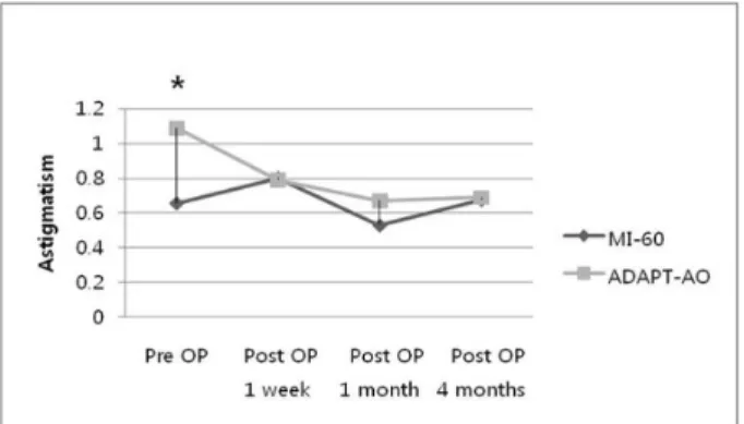
Table 5. Comparison of preoperative and postoperative astigmatism between akreos MI-60 IOL-implanted group and akreos adapt-AO IOL-implanted group (Mean±SD†)
Results Group Pre OP Post OP 1week Post OP 1month Post OP 4months
Astigmatism MI-60 0.656±0.659* 0.800±0.655 0.530±0.565 0.676±0.524
ADAPT-AO 1.090±0.767* 0.790±0.612 0.670±0.681 0.691±0.524
*Mann-Whitney U test (P<0.05); †SD=standard deviation.
Table 6. Comparison of postoperative anterior chamber depth between akreos MI-60 IOL-implanted group and akreos adapt-AO IOL-implanted group (Mean±SD†)
Results Group Post OP 1week Post OP 1month Post OP 4months
ACD‡ MI-60 3.948±0.288* 3.858±0.496 3.682±0.664
ADAPT-AO 3.721±0.376* 3.762±0.459 3.784±0.488
*Mann-Whitney U test (P<0.05); †SD=standard deviation; ‡ACD=anterior chamber depth.
Figure 3. Comparison of preoperative and postoperative astigmatism between Akreos MI-60 IOL-implanted group and Akreos Adapt-AO IOL-implanted group.
(*Mann-Whitney U test, P<0.05)
내로 수술유발난시에 의해 상쇄되었다. 그래서 수술 후 난 시는 두 군 사이에 유의한 차이가 없었다(Table 5) (Fig. 3).
전방 깊이(anterior chamber depth)는 술 후 1주째 MI- 60 인공수정체 군에서 유의하게 컸으나, 술 후 1달 및 4달 째에는 두 군 사이에 유의한 차이가 없었다. 4달 동안 전방
깊이가 유의하게 변하지는 않았지만 MI-60 인공수정체 군 이 전방이 더 얕아지는(narrowing) 경향을 보였고, 이는 MI-60 인공수정체가 앞쪽으로 이동을 하여 근시 변화(myopic shift) 를 일으키는 것을 말해 준다(Table 6).
고 찰
초음파 수정체유화술(phacoemulsification) 및 인공수정 체 후낭삽입술은 노인 환자들에서 시력을 향상시키기 위해 가장 흔히 시행되는 수술 중의 하나이다. 지난 수십 년간 백내장 수술은 수술의 안정성을 향상시키고, 술 후 빠른 시 력회복을 위해 눈부시게 발전해왔다. 그 노력의 일환으로 투명각막절개창의 크기를 줄이려는 시도가 있었고, 현재는 3.0 mm 부근의 절개 및 접힘 인공수정체(foldable IOL)의 사용이 일반화되었다. 최근에는 2.0 mm 이하의 각막절개 창을 이용한 미세각막절개 백내장 수술(microincision cat- aractsurgery, MICS)이 도입되었고, 이를 위해 더 세련되 고 정교한 백내장 수술기구와 인공수정체들이 임상에 쓰이
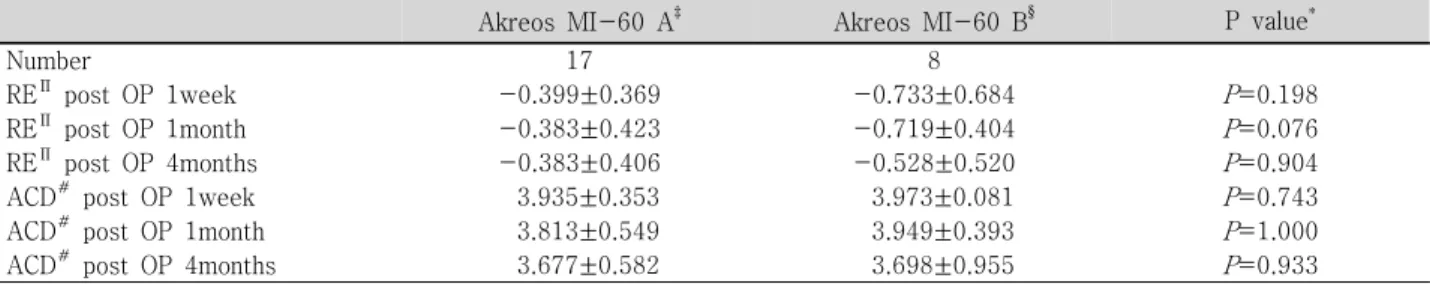
Table 7. Comparison of postoperative refractive error and anterior chamber depth between akreos MI-60 IOL- implanted group A and B (Mean±SD†)
Akreos MI-60 A‡ Akreos MI-60 B§ P value*
Number 17 8
RE∏post OP 1week -0.399±0.369 -0.733±0.684 P=0.198
RE∏post OP 1month -0.383±0.423 -0.719±0.404 P=0.076
RE∏post OP 4months -0.383±0.406 -0.528±0.520 P=0.904
ACD#post OP 1week 3.935±0.353 3.973±0.081 P=0.743
ACD#post OP 1month 3.813±0.549 3.949±0.393 P=1.000
ACD#post OP 4months 3.677±0.582 3.698±0.955 P=0.933
*Mann-Whitney U test; †SD=standard deviation; ‡group A=IOL power ≤22.0 diopter; §group B=IOL power ≥22.5 diopter; ∏ RE=refractive error; #ACD=anterior chamber depth.
고 있다. MICS의 장점으로 Donnenfeld et al은 기존의 백 내장 수술(conventional cataract surgery)에 비해 수정체 유화술 시의 초음파에너지 양을 감소시켜 술 후 각막의 손 상 및 부종을 줄일 수 있다는 보고를 하였고16, Steinert et al은 각막절개를 최소화하여 술 후 수술유발난시(surgi- cally induced astigmatism)를 줄여 줌으로써 빠른 시력회 복을 가져올 수 있다고 보고하였다.17
지금까지 미세절개 백내장수술에 이용되는 몇 가지 인공 수정체의 임상성적에 대한 여러 연구가 있었지만, Akreos MI-60 인공수정체의 임상성적에 대한 연구는 별로 없었으 며, Akreos MI-60 인공수정체와 타 인공수정체를 비교한 연구는 우리가 아는 한 없었다. 이에 본 연구에서는 미세각 막절개창을 통해 삽입이 가능한 새로운 비구면 인공수정체 를 기존의 임상성적이 검증된 비구면 인공수정체와 비교하 여 보았다. 과거의 연구들을 살펴보면 Akreos MI-60 인공 수정체의 장점으로 시력과 고위수차(high-order aberration) 에 관해서 기술하고 있다. Jorge et al은 Akreos MI-60 인 공수정체가 비구면수차 인공수정체(aberration-free IOL) 로서 안전하고 효과적이며, 탁월한 시력 및 대비감도를 보 인다고 보고하였다.18Tong et al은 백내장 수술로 인한 각 막 웨이브프론트 수차(wavefront aberration)는 절개창의 크기에 따라 좌우되고, MICS technique은 절개창의 크기를 최소화함으로써 각막의 광학적 성질(optical quality)을 유 지할 수 있는 장점이 있다고 보고하였다.19Denoyer et al의 연구도 비슷한 결과를 보였다.20본 연구에서도 술 후 4개월 까지의 결과를 살펴보면 MI-60 인공수정체와 ADAPT- AO 인공수정체 모두 술 후 작은 수차(aberration)를 나타 내었고, 두 군 모두 훌륭한 술 후 최대교정시력 및 대비감 도를 보였다.
한편 Wilczynski et al은 58명의 환자들을 대상으로 Akreos MI-60 인공수정체를 삽입한 전향적 연구에서 bi- manual MICS technique과 coaxial MICS technique 두 방 법 모두 아주 작은 양의 수술유발난시(surgically induced
astigmatism, SIA)를 보인다고 보고하였다.21 본 연구에서 도 술 후 1개월 및 4개월째 MI-60 인공수정체 군에서 더 낮은 수술유발난시를 나타내었다.
그러나 MI-60 인공수정체 군에서 수술 후 목표로 한 구 면대응치(target refraction) 값보다 약간 근시성 구면대응 치를 나타내었다. MI-60 인공 수정체의 경우 인공수정체 의 굴절력(IOL power)에 따라 각기 다른 수정체 크기 (optic body size)를 가지는데, 10.0에서 15.0 디옵터까지 는 6.2 mm, 15.5 에서 22.0 디옵터까지는 6.0 mm, 그리고 22.5 에서 30.0 디옵터까지는 5.6 mm의 수정체 크기를 가 진다. 25안의 MI-60 인공수정체 군을 다시 수정체 크기에 따라 두 군으로 나누어 굴절오차 및 전방깊이를 비교해 보 았으나, 이에 따른 굴절오차 및 인공수정체 전방이동의 차 이는 관찰되지 않았다(Table 7). 이에 대해서는 수술 후 정 확한 인공수정체 도수 계산을 위하여, 향후 MI-60 인공수 정체의 A constant에 관한 추가적인 연구 및 조정이 필요할 것으로 사료된다.
본 연구는 두 인공수정체 군 간의 수술유발난시 비교에 있어 절개창의 크기가 다르므로 두 렌즈의 직접적인 특성 비교에 있어서 한계가 있다. 절개창의 크기 차이로 인한 수 술유발난시값을 보정하기 위해서는 MI-60 인공수정체 삽 입 시 절개창의 크기를 1.8 mm와 2.75 mm로 나누어 비교 해 보거나, MI-60 인공수정체와 Adapt-AO 인공수정체 두 군 모두 2.75 mm의 절개창을 만들어 비교해 보는 것이 도움이 될 것이다. 그리고 50명의 인원으로 비교적 짧은 4 개월 동안 경과관찰하였다는 점에서 제약이 있다.
결론적으로, Akreos MI-60 인공수정체는 기존의 임상 성적이 검증된 인공수정체와 비교하여 시력과 대비감도 면 에서 동등한 임상결과를 나타내고, 여기에 더하여 작은 절 개창으로 인하여 수술유발난시를 유의하게 줄여 줄 수 있 다는 장점이 있다. 하지만 미세절개 백내장수술의 단점으로 Milla et al은 미세절개 백내장수술이 고전적 백내장 수술 에 비해 긴 습득 과정(long learning curve)이 필요하며 미
세절개 백내장수술에 익숙해지기 전까지는 기존의 수술보 다 수술 시간이 길어져서 오히려 각막내피세포의 손상을 더 유발할 수 있다고 하였다.22 그러므로 수술 전 환자의 기 저 난시를 고려하여 미세절개백내장 수술로 도움을 받을 수 있는 환자의 선택에 신중을 기하는 한편, 술자의 미세절 개 백내장 수술실력 등을 고려하여 단단한 수정체 핵 소견 을 보이는 등 수술이 힘들거나 길어질 것 같은 경우 미세절 개백내장수술을 피하여야 할 것이다. 향후 보다 많은 사람 들을 대상으로 한 장기간의 임상연구가 추가적으로 필요할 것이다.
참고문헌
1) Ginsberg AP, Evans DW, Sekuler R, et al. Contrast sensitivity predicts pilots’ performance in aircraft simulation. Am J Optom Physiol Opt 1982;59:105-9.
2) Pesudovs K, Hazel CA, Doran RM, Elliot DB. The usefulness of Vistech and FACT contrast sensitivity charts for cataract and re- fractive surgery outcomes research. Br J Ophthalmol 2004;88:11-6.
3) Applegate RA, Hilmantel G, Howland HC, et al. Corneal surface opti- cal aberrations and visual performance. J Refract Surg 2000;16:507-14.
4) Mierdel P, Kaemmerer M, Mrochen M, et al. Ocular optical aberr- ometer for clinical use. J Biomed Opt 2001; 6:200-4.
5) Bhattacharjee H, Bhattacharjee K, Medhi J. Visual performance: com- parison of foldable intraocular lenses. J Cataract Refract Surg 2006;
32:451-5.
6) Agarwal A, Agarwal A, Agarwal S, et al. Phakonit: phacoemulsification through a 0.9 mm corneal incision. J Cataract Refract Surg 2001;27:
1548-52.
7) Agarwal A, Agarwal S. Phakonit with an AcriTec IOL. J Cataract Refract Surg 2003;29:854-5.
8) Fine IH, Hoffman RS, Packer M. Optimizing refractive lens exchange with bimanual microincision phacoemulsification. J Cataract Refract Surg 2004;30:550-4.
9) Braga-Mele R, Liu E. Feasibility of sleeveless bimanual phacoemulsifi- cation with the Millenium microsurgical system. J Cataract Refract Surg 2003;29:2199-203.
10) Mackool RJ. Temperature during bimanual phacoemulsification. J
Cataract Refract Surg 2004;30:732.
11) Soscia W, Howard J, Olson R. Bimanual phacoemulsification through 2 stab incision a wound-temperature study. J Cataract Refract Surg 2002;28:1039-43.
12) Soscia W, Howard JG, Olson RJ. Microphacoemulsification with WhiteStar. A wound-temperature study. J Cataract Refract Surg 2002;
28:1044-6.
13) Olson RJ. Clinical experience with 21-gauge manual microphacoe- mulsification using Sovereign Whitestar technology in eyes with dense cataract. J Cataract Refract Surg 2004;30:168-72.
14) Alio JL, Rodriguez Prats JL, Galal A. Advances in microincision cata- ract surgery intraocular lenses. Curr Opin Ophthalmol 2006; 17:80-93.
15) Hill W. Expected effects of surgically induced astigmatism on AcrySof toric intraocular lens results. J Cataract Refract Surg 2008;34:364-7.
16) Donnenfeld ED, Olson RJ, Solomon R, et al. Efficacy and wound- temperature gradient of whitestar phacoemulsification through a 1.2 mm incision. J Cataract Refract Surg 2003;29:1097-100.
17) Steinert RF, Brint SF, White SM, et al. Astigmatism after small incision cataract surgery. A prospective, randomized, multicenter comparison of 4- and 6.5 mm incisions. Ophthalmology 1991; 98:417-23.
18) Alió JL, Piñero DP, Ortiz D, Montalbán R. Clinical outcomes and postoperative intraocular optical quality with a microincision aberra- tion-free aspheric intraocular lens. J Cataract Refract Surg 2009;35:
1548-54.
19) Tong N, He JC, Lu F, et al. Changes in corneal wavefront aberrations in microincision andsmall-incision cataract surgery. J Cataract Refract Surg 2008;34:2085-90.
20) Denoyer A, Denoyer L, Marotte D, et al. Intraindividual comparative study of corneal and ocular wavefront aberrations after biaxial micro- incision versus coaxial small-incision cataract surgery. Br J Ophthal- mol 2008;92:1679-84.
21) Wilczynski M, Supady E, Piotr L, et al. Comparison of surgically in- duced astigmatism after coaxial phacoemulsification through 1.8 mm microincision and bimanual phacoemulsification through 1.7 mm microincision. J Cataract Refract Surg 2009;35:1563-9.
22) Milla E, Verges C, Cipres M. Corneal endothelium evaluation after phaocoemulsification with continuous anterior chamber infusion.
Cornea 2005;24:278-82.
23) Choi JA, Kim CY, Na KS, et al. Clinical results after implantation of a spherical aberration-free intraocular lens: Effect of contrast sensitivity and wavefront aberration-a clinical comparative study. Ophthalmo- logica 2009;223:320-5.
=ABSTRACT=
Clinical Outcomes After Microincision Cataract Surgery and In-the-bag Implantation of a New Intraocular Lens
Chul Jin Shin, MD, Joo Eun Lee, MD, Jae Hyung Lee, MD, Jae Yong Kim, MD, PhD, Hungwon Tchah, MD, PhD
Department of Ophthalmology, Asan Medical Center, University of Ulsan College of Medicine, Seoul, Korea
Purpose: To compare visual performance after microincision cataract surgery (MICS) with the implantation of the Akreos MI-60 (MI-60) intraocular lens (IOL) through a 1.8-mm microincision with that after conventional cataract surgery with implantation of the Akreos Adapt-AO IOL (Adapt-AO).
Methods: All MICS procedures were performed by the same surgeon. The MI-60 was implanted into 25 eyes, and the Adapt-AO was place in 28 eyes. Best corrected visual acuity (BCVA), total high-order-aberration (HOA), contrast sensitivity, and surgi- cally-induced astigmatism (SIA) were recorded one-week, one-month, and four-months postoperatively.
Results: There were no statistically significant differences in BCVA between eyes implanted with the MI-60 or those with the Adapt-AO (MI-60 vs. Adapt-AO, 0.09±0.11 at baseline (logMAR), 0.11±0.08 at one-week, 0.06±0.07, 0.06±0.06 at one-month, 0.05±0.06, 0.06±0.05 at four-months according to the Mann-Whitney U test, p>0.05). Refractive errors were significantly less with the Adapt-AO than with the MI-60 (MI-60 vs. Adapt-AO, -0.50±0.43 at baseline (diopter), -0.06±0.39 at one-week, -0.50±0.41, 0.01±0.57 at one-month, -0.46±0.36, 0.08±0.58 at four-months according to the Mann-Whitney U test, p<0.05).
There were no statistically significant differences in total HOA or contrast sensitivity between eyes implanted with the MI-60 and those implanted with the Adapt-AO. SIAs were significantly reduced in eyes implanted with the MI-60 than in those with the Adapt-AO at one-month and four-months postoperatively (Mann-Whitney U test, p<0.05).
Conclusions: Implantation with either the MI-60 or the Adapt-AO produced clinically acceptable outcomes, including good spherical aberration and contrast sensitivity. Furthermore, implantation with the MI-60 caused less SIA at one- and four-months postoperation, as compared to that with the Adapt-AO.
J Korean Ophthalmol Soc 2010;51(5):677-683
Key Words: Contrast sensitivity, Microincision cataract surgery, Phacoemulsification, Spherical aberration, Surgically-induced astigmatism
Address reprint requests to Hungwon Tchah MD, PhD Department of Ophthalmology, Asan Medical Center
#388-1 Pungnap2-dong, Songpa-gu, Seoul 138-736, Korea
Tel: 82-2-3010-3680, Fax: 82-2-470-6440, E-mail: [email protected]