Introduction
With the development of techniques, to raise the success rate of implants even in cases with the poor bone volume and bone quality, diverse surface treatment methods have been investigated and introduced.
The methods to treat implant surface could be classified broadly into two methods. One is the addition of the implant surface by diverse methods [1-3]. Another method is the abrasion of the implant surface for the increasing in surface
area of the implant [4].
The purpose of such surface treatments could be considered to promote the osseointegration between the bone and implants in cases with a poor bone volume and bone quality.
First, considering from the physical aspects, the addition and abrasion of the implant surface performed to increase and roughen the surface area of the implant. This is based on the fact that osteoblasts attach better to a rough surface [5], which increases bone deposition [6]. In addition, several investigators explained the induction of faster osseointegration by the rough surfaced implants in comparison with the machined-surface implants by presenting a higher bone to implant contact (BIC) rate and the removal torque value [7-9].
All materials used as additives by diverse methods on the implant surface are materials with an osseointegration potential.
Reviewing studies performed on implant materials, diverse materials have been reported to have the osseointegration
Histomorphometric evaluation for osseointegration after particulated- dentin coated implant placement
Jae-Seek You
1, Su-Gwan Kim
1,*, Ji-Su Oh
1, Sung-Chul Lim
2, Jae-Sung Kim
31
Department of Oral and Maxillofacial Surgery, School of Dentistry, Chosun University,
2Department of Pathology, School of Medicine, Chosun University,
3Pre-Dentistry, College of Dentistry, Chosun University, Gwangju, Korea
ABSTRACT
Purpose: The purpose of this study was to evaluate osseointegration after placement of a machined surface implant and particulated-dentin-coated implant in a canine model.
Materials and Methods: Four dogs were randomly assigned to two groups, and each group was further divided into two subgroups 4 and 8 weeks after implantation. The implant was placed under different conditions. Group 1 included the machined surface implant and group 2 included the particulated-dentin-coated implant. Histologic sections and histomorphometric analysis were obtained 4 and 8 weeks after surgery.
Results: Group 2 showed a higher bone-implant contact rate and bone formation rate than group 1 in the 8 week group. In group 2, the 8 week group revealed significant elevation of both bone-implant contact rate and new bone formation rate versus the 4 week group.
Conclusion: According to these results, particulate-dentin-coated implants can provide satisfactory stability and increase the quantity and maturity of new bones in the later stages of implant placement.
Key Words: Bone to implant contact, Dental implant, New bone formation, Surface
Received Mar 3, 2015; Revised version received Mar 12, 2015 Accepted Mar 13, 2015
Corresponding author: Su-Gwan Kim
Department of Oral and Maxillofacial Surgery, School of Dentistry, Chosun University, 309 Pilmun-daero, Dong-gu, Gwangju 501- 759, Korea
Tel: 82-62-220-3815, Fax: 82-62-228-7316
E-mail: [email protected]
potential [10-14]. And this showed that materials from metal (such as vitallium, tantalum and titanium) to hydroxyapatite (HA) had the ability to integrate into bone tissues directly.
In addition, recent studies on particulate dentin have been conducted actively with successful results in cases that used particulate dentin as bone graft materials. Such particulate dentin is obtained easily from human or porcine teeth and it could be considered economical in comparison with other surface treatment methods. In addition, through diverse experiments, it has been evaluated to be a successful bone substitution material.
In this study, implants coated with particulate dentin and implants with the machined-surface were placed to the mongrels, the level of osseointegration on the implant interface was analyzed histomorphometrically, and the differences reported.
Materials and Methods
Materials
The subjects were 4 adult mongrels, 8 to 9 months, and weighing approximately 12 kg. They were used regardless of their sex, and their health condition was good.
The implants used were 10 mm in length, 3.9 mm in diameter and either the implant coated with particulate dentin (Jeil Medical, Seoul, Korea) for the experimental group or the machined-surface implant (Jeil Medical) for the control group. The number of implants was 4 in each control group and experimental group per animal, for a total of 32 implants placed.
Experiment methods
Anesthesia
Systemic anesthesia was induced by the injection of 3 mL Zylaxine Hydrochloride (Rompum
®; Bayer Korea Ltd., Seoul, Korea), Tiletamin, and Zolazepam (Zoletil
®50; Virbac Lab., Carros, France) to each femoral area, and for the prevention of hemorrhage in the implant area and to suppress pain, infiltration anesthesia was performed by the use of 2%
Hydrochloride Lidocaine (Yuhan Co. Ltd., Seoul, Korea).
Tooth extraction
A total of 4 teeth (2nd, 3rd, and 4th premolar and the 1st
molar) were extracted from each adult dog using a pair of forceps. In all adult dogs, at the time of extraction, the tooth was extracted completely without leaving aresidual root rest or granulation tissues. After extraction, sutures were placed, and in all groups, to prevent infection, 2 mL Gentamicin Sulfate (Deasung Gentamicin Inj.; Deasung Microbiological Labs. Co.
Ltd., Uiwang, Korea) was injected for 3 days. Subsequently, 1 mL was injected for 2 days.
Implant placement
Two months after tooth extraction, Anesthesia was performed, incision was made, and the periosteum was elevated using a periosteal elevator. In each group, 4 implants were placed at distance intervals of approximately 5 mm.
After implant placement, suturing was performed. Gentamicin Sulfate (Deasung Gentamicin Inj., Deasung Microbiological Labs. Co. Ltd) was injected intramuscularly, using the method similar to the time of extraction.
Classification of experimental groups
The groups were divided according to the type of implant placed. They were divided into group 1 (control group), which placed machined-surface implants (n=16) and group 2 (experimental group), which placed implants coated with particulate dentin (n=16). They were divided again into the 4-week groups and the 8-week groups according to their sacrifice time.
Evaluation of experiments
Histomorphometric evaluation
After the sacrifice of animals, implants were removed as en bloc which included adjacent bones. The removed samples were immediately fixated in 70% alcohol for 6 days, dehydrated by alcohol washing, and embedded in glycol- methacrylate resin (Spurr Low-viscosity Embedding Media;
Polysciences, Warrington, PA, USA). The polymerized samples were sectioned as 200 μm thickness using a high-precision diamond disc (Low speed diamond Wheel Saw 650; South Bay Technology Inc., San Clemenate, CA, USA), and finally, using a lapping and polishing machine (OMNILAP 2000; South Bay Technology Inc.), samples 30 μm in thickness were prepared.
One slide per implant was prepared, and after Villanueva
osteochrome bone staining, they were examined under a light
microscope (Olympus BX 50; Olympus, Tokyo, Japan), and histomorphometric analysis was performed.
BIC and new bone formation rate (NBFR) within the threads (from the lowest point of the implant head to the last apical thread) were calculated using the following formulas:
×
×
Statistical analysis
Mann-Whitney test, which applied the SPSS for window version 16.0 (SPSS Inc., Chicago, IL, USA), was performed for statistical analysis of the evaluation results of each group. In addition, in each statistical analysis, p-value lower than 0.05 was considered to be statistically significant.
Results
Histological results
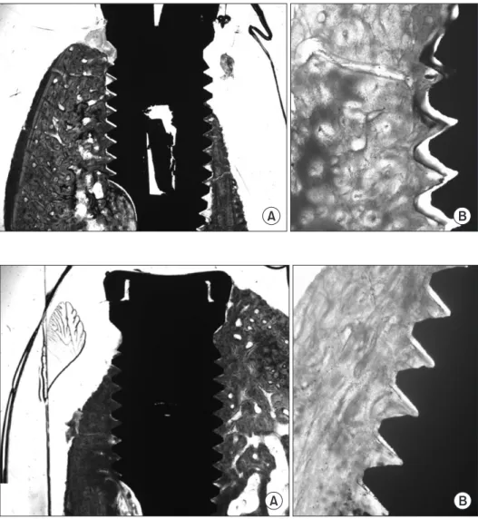
Machined-surface implants (control group, group 1) In the 4-week group, the implants were placed well in the mandibular bone and new bone formation between implant threads was observed. The presence of initial trabecular bone growth was noted, and but complete osseointegration on the implant interface was not shown (Fig. 1). In the 8-week group, it was placed well in the mandibular bone. New bone formation between the threads of implant was observed. The pattern of compact bone together with trabecular bone was detected and the process of osseointegration on the implant interface was detected in some areas (Fig. 2).
Fig. 1. Machined-surface implants at 4 weeks. Partial trabecular bone growth was seen between the threadsin im- plant (Villanueva osteochrome bone stain; A: ×12.5, B: ×40).
Fig. 2. Machined-surface implants at 8 weeks. Not only the trabecular bone but also the compact bone shape was seen between the threads in implant (Villanueva osteochrome bone stain;
A: ×12.5, B: ×40).
A B
A B
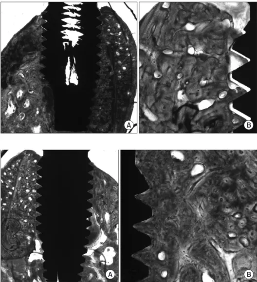
Particulated-dentin coated implant (experimental group, group 2)
In the 4-week group, the implant was placed well in the mandibular bone. The growth of new bone between the threads of the implant was detected. It showed the pattern of trabecular bone, and partly showed the pattern of cortical bone (Fig.
3). In the 8-week group, the implant was placed well in the mandibular bone. New bone formation between the implant threads was well observed. It showed the pattern of compact bone without the growth of trabecular bone, and the complete osseointegration in the implant interface was observed (Fig. 4).
Histomorphometric result
In the control group (group 1) and the experimental group (group 2), animals were sacrificed at 4 weeks and 8 weeks each, and the BIC rate, as well as the NBFR was evaluated. The results
showed that the experimental group showed a higher BIC rate and the NBFR than the control group, and a significant difference was shown (p<0.05). In addition, in the experimental group, the 8-week group observed a significant increase of the BIC rate as well as a NBFR in comparison with the 4-week
Table 1. Mean Percentages of New Bone Formation in Group 1 and 2 at 4 and 8 Weeks after Placement
Time period (wk) Group 1 Group 2
4 8
51.48±18.08 65.93±24.33
62.45±10.71 92.98±4.15
*,+Values are presented as mean±standard deviation.
Group 1: the machined surface implant, group 2: the particulated- dentin coated implant.
*Statistically significant difference compared to group 1, p<0.05.
+
Statistically significant difference compared to group 2 at 4 weeks, p<0.05.
Fig. 3. Particulated-dentin coated implants at 4 weeks. Trabecular bone growth was seen between the threads in implant and also compact bone was partially seen at the same area (Vil- lanueva osteochrome bone stain; A:
×12.5, B: ×40).
Fig. 4. Particulated-dentin coated im- plants at 8 weeks. Only compact bone shape without any trabecular bone was revealed between the threads in implant (Villanueva osteochrome bone stain; A: ×12.5, B: ×40).
A B
A B
group (p<0.05) (Tables 1, 2).
Discussion
As materials with the osseointegration potential, titanium and HA have been known to be most useful. In particular, HA is a major inorganic component of bone tissues in vivo. Its affinity to the bone surface is excellent. The bonding property to bone tissues is known to be far superior to other materials and, thus it has been used for the treatment of the titanium surface [15-18]. The HA coated implant showed good bone growth in animal experiments, and thus it was used from the mid-1980s in clinics. A method of placing a thin coating of HA on titanium surface was suggested [19]. Because of its superior biological characteristic observed in such HA coating implant cases, numerous other successful cases have been reported [20].
However, such HA coated implants also showed the separation of the coated HA, and such detachment of HA induced many problems [21]. Darimont et al. [22] observed the decrease of the thickness of HA coating in the area where the contact with the bone was absent.
Diverse surface treatment methods have been introduced, and each surface treatment method have advantages as well as shortcomings, nonetheless, for good osseointegration, diverse surface treatment methods have been applied. Therefore, in this study, to improve osseointegration, on implant surface, particulate dentin was prepared as a target, and the surface treatment was performed by attaching particulate dentin onto the implant surface through the sputtering method.
This particulate dentin is prepared by washing extracted teeth, particulating at 900
oC to 1,200
oC high temperature for 90 to 120 minutes, grinding, and removing contaminants.
The development was initiated in 1992, and through diverse experimental processes, its safety and efficacy were proven, having good results obtained in clinical studies [23,24].
Particulate dentin has no noticeable foreign body reaction or inflammation, preservation of the osteoinduction potential, in vivo absorbability, and low costcould be pointed out as advantages. Kim et al. [25] reported in an animal experiment examined the cytotoxicity and hypersensitivity of particulate dentin and found it lacks cytotoxicity and specific allergic reaction, and therefore could be used safely in vivo. A retrospective study was conducted on 10 patients whose bone defect area was restored with the mixture of particulate dentin and plaster, and it was mentioned that particulate dentin is a useful bone substitute that could be manipulated readily [26].
In addition, Kim et al. [26,27] also showed in a study that it was used for the restoration of the bone defect area in the vicinity of an implant. Good contact of the bone and the implant was formed, which showed the possibility of the wide use of particulate dentin in clinics. Choe and Ko [28] conducted a study on the attachment of such particulate dentin to titanium, and they reported that titanium attached particulate dentin was superior to titanium without the attachment in an anti-erosion property.
In this study, the implant surface was coated with particulate dentin, and the bone-implant contact rate and NBFR was compared with the machined-surface implant group.
The result showed that at 8 weeks, the group of implants coated with particulate dentin, the bone-implant contact rate as well as the NBFR showed superior results. In addition, in the group of implants coated with particulate dentin, both bone- implant rate and NBFR showed better results than at 4 weeks.
Our experiment performed a short term analysis that ranged from 4 to 8 weeks, and it showed the achievement of early stability after transplant by the particulate dentin coating. This shows that particulate dentin coating implants are useful to obtain the early stability after implant placement.
The results of this experiment found that particulated-dentin coated implant showed satisfactory results in osseointegration through the results of BIC (%) and NBFR (%) as well as in the stability of the implant. But this is the study of the short term of 4 to 8 weeks.
We need additional research on whether the separation of dentin particle doesn’t occur on the implant surface and if it Table 2. Mean Percentages of Bone to Implant Contact Ratio in
Groups 1 and 2 at 4 and 8 Weeks after Placement
Time period (wk) Group 1 Group 2
4 8
45.05±8.22 66.08±16.15
53.85±14.61 93.58±2.10
*,+Values are presented as mean±standard deviation.
Group 1: the machined surface implant, group 2: the particulated- dentin coated implant.
*Statistically significant difference compared to Group 1, p<0.05.
+