대한치주과학회지 : Vol. 35, No. 3, 2005
Histological response of anodized titanium implant
Svetlana Lim1ㆍSeong-Joo Heo2ㆍChong-Hyun Han3ㆍTae-II Kim1ㆍ Yang-Jo Seol1ㆍYoung Ku1ㆍKyoung-Uk Chung1ㆍChong-Pyoung Chung1ㆍ
Soo-Boo Han1ㆍIn-Chul Rhyu1
1Department of Periodontology, College of Dentistry, Seoul National University
2Department of Prosthodontics, College of Dentistry, Seoul National University
3Department of Prosthodontics, Yonsei University, Yongdong Severance Hospital
Ⅰ. Introduction
1)Osseointegration is the essential biological basis of current dental implants1). Osseoin- tegration was initially defined on the light microscopic level as “a direct structural and functional connection between ordered, liv- ing bone and the surface of a load-carrying implant”2).
Today, by definition, osseointegtation re- quires the absence of a fibrous layer and im- plies that the biological response of the bone is not one of inertness towards a foreign ma- terial but rather one of active integration of the material with the bone as part of the body3).
According to Giavaresi. et al.4) osseointe- gration is defined not only as the absence of a fibrous layer around the implant with an
active response in terms of integration to host bone, but also as a chemical(bonding osteogenesis) or physico-chemical(connective tissue osteogenesis) bond between implant and bone.
Endosseous integration can be deconvo- luted into three distinct bony healing phases. The first, osteoconduction, relies on the migration of differentiating osteogenic cells to the implant surface. The second, de novo bone formation, results in a minera- lized interfacial matrix equivalent to that seen in cement lines in natural bone tissue.
Implant surface design will have a profound effect on osteoconduction, while the surface topography of the implant will play a critical role in bone bonding concomitant with de novo bone formation. The third healing phase, that of bone remodeling, will also, at Corresponding author : In-Chul Rhyu, Department of Periodontology, College of Dentistry, Seoul National University, 28 Yongon-Dong, Chongno-Ku, 110-749, Seoul, Korea, E-mail:[email protected]
discrete sites, create a bone-implant inter- face comprising de novo bone formation.
Treatment outcomes in dental implantology will be critically dependent on implant sur- face designs that optimize the biologic re- sponse during each of these three distinct integration mechanisms5).
A successful long-term implant requires biocompatibility, toughness, strength, corro- sion resistance, wear resistance, and frac- ture resistance6).
The surface of implantable biomaterials is in direct contact with the host bone and soft tissue and plays a critical role in deter- mining the biocompatibility, functional com- patibility, osteoinduction of bone ald osseo- integration of implants. Placing of implants into bone tissue should in principle lead to bone-implant osseointegration. The bone for- mation that occurs during osseointegration may be an osteoblast activity which is af- fected by the implant surface3).
Very few studies have been carried out to systematically investigate the role of indi- vidual surface(oxide) properties of titanium in the biological response, although recent in vitro and in vivo studies provide strong indications that biological responses to tita- nium are influenced both by surface struc- ture(roughness) and chemical composition.
In most studies, however, the type of sur- face preparation and/or characterization methods used prevent any firm conclusions as to which surface properties were the de- termining factor for the observed differences in biological response7).
Differences in shape and surface charac- teristics of different implant systems could
also influence handling, time and other re- sources used, and clinical success8).
The quality of the implant surface is one of the 6 factors described by Albrektsson that influence wound healing at the implan- tation site and subsequently affect osseo- integration.
Plasma spray-coation is one of the most common methods for surface modification.
Plasma spraying is used for the application of both Ti or HA on metallic cores with a coating thickness of 10 to 40 μm for Ti and 50 to 70 μm for HA.
Blasting with particles of various diame- ters is another frequently used method of surface alteration. In this approach, the im- plant surface is bombarded with particles of aluminum oxide(Al2O3) or titanium oxide (Ti- O2), and by abrasion, a rough surface is pro- duced with irregular pits and depressions.
Chemical etching is another process by which surface roughness can be increased.
The metallic implant is immersed into an acidic solution. which erodes its surface.
creating pits of specific dimensions and shape, Concentration of the acidic solution.
time. and temperature are factors determi- ning the result of chemical attack and mi- crostructure of the surface2).
Sandblasting, plasma spraying and acid etching have become the three most common approaches used to alter the surface top- ography and increase the surface area of im- plants4).
Greater surface roughness increases the implant surface area and increases the po- tential for biomechanical interlocking of bone into the implant surface. These character-
istics are thought to enhance the gone im- plant interface and improve stabilization9).
A certain degree of surface roughness may have a positive effect on adsorption of mole- cules, local factor production, and pro- liferation and differentiation of cells at the implant surface. Differences in topography and roughness can be obtained by treating the titanium surface with additive and re- ductive techniques10).
Titanium plasma spray(TPS) surface has since become one of the benchmarks for rough implant surfaces, with over twenty years of clinical experience and a large number of published articles. In spite of the success of the TPS surface, numerous re- searchers and clinicians have been working on new and improved micro rough surfaces.
In 1998 and 1999, some major changes were introduced to the ITI. After more than 10 years of intensive research and clinical testing, a major breakthrough-the SLA (Sand-blasted, Large grit, Acid-etched) sur- face-was launched in mid-June 1998 at the ITI World Symposium in Boston. The SLA surface was first tested in cell cultures and animals in 1990.
The SLA surface of the ITI implant is prepared by sandblasting the implant sur- face with large grid(250-500 μm) corundum, washing in ultrasonic deionized water bath, and drying. The implant surface is then acid-etched in a hot hydrochloric acid/
sulthuric acid(HCL/H2SO4) mixture, followed by thorough rinsing in deionized water befor drying in hot air. This procedure leads to a surface roughness between 20 and 40 μm11).
The intensive testing and successfully on-
going clinical and field trials have shown that the SLA surface clearly has the poten- tial to replace the TPS surface. Due to the macro/micro double roughness of the SLA surface, osseointegration of ITI implants has been improved and time to loading has been reduced to a maximum of 50%.
In recent years, attempts have been made to improve bone anchorage of dental im- plants. Thomas and Cook(1985) examined the variables that influenced the apposition of bone to an implant surface. Of 12 para- meters studied, only surface characteristics had a significant effect on the integration of the implant. This observation has been con- firmed in a histometric study by Buser et al.
(1991) that showed a positive correlation be- tween the percentage of bone-to-implant con- tacts and the roughness values of five differ- ent titanium surface tested. Implant surfaces that were sandblasted and acid etched ach- ieved a greater bone-implant contact than theroughertitanium plasma sprayed(TPS) surfaces. However, this study was carried out in long bones of miniature pigs and eval- uated only short-term healing periods of un- loaded experimental implants12).
A well-documented sandblasted and acid- etched surface(SLA), as a new development in this area, consistently showed better re- sults both in histometric and biomechanical testing in comparison with alternative surfa- ces, such as the well-documented TPS(tita- nium plasma-sprayed) surface; and fur- thermore proved advantageous in clinical application13).
Cochran DL et al.12) compared TPS and acid-etched implants placed in canine man-
dible. They found the acid-etched surface resulted in a significantly higher degree of bone-implant contact after 3 months of heal- ing, but 3months of loading(6 months of healing) no significant difference was found between the SLA and TPS surfaces im- plants. After 12 months of loading the SLA implants had a significantly greater percent- age of BIC than did the TPS implants.
These results are consistent with earlier studies on SLA implants and suggest that this surface promotes greater osseous con- tact at earlier time points compared to TPS-coated implants.
Experimental studies in animals have found stronger bone fixation for TiO₂ blast- ed implants than for turned implants14).
Most metals form oxide layers when ex- posed to the atmosphere. The nature of this oxide depends on the metal and the con- ditions under which it was oxidized. Any- thing that comes in contact with the implant surface has the potential to change it.
Assuming that the physiologic conditions of the body remain fairly constant, the behav- ior of a metal in the body depends on the character of the oxide layer. TiO₂ is the most stable and therefore the most com- monly used under physiologic conditions.
When an implant is introduced into the body, complex reactions begin to take place at the oxide / bioenvironment interface. The oxide film grows as ions diffuse outward from the metal and inward from the environment. The oxide that forms in the body may, therefore, be somewhat different than that which forms in air. The rate of formation and composition of this film is
important. Titanium, both as a pure metal and as on alloy, is easily passivated, form- ing a stable TiO₂ surface oxide will repair itself instantaneously on damage such as might occur during insertion of an implant.
Values of 100 to 300 ppm are frequently ob- served in soft tissues surrounding Ti implants. At these levels, tissue discolora- tion with Ti pigments can be seen. this rate of dissolution is one of the lowest of all pas- sivated implant metals and seems to be well tolerated by the body6).
Most of the studies referred to have used machined screw implants, and it is possible that surface modification can facilitate im- plant healing and improve the clinical outcome. Recently, wide application in im- plantlogy was received with technique of anodic oxidation.
Anodic oxidation is one technique of sur- face modification that results in growth of the implant surface oxide to a thickness of about 1to 10 μm and a porous surface struc- ture; an increase in oxide thickness, from 1 to 2 μm at the coronal part to7 to10 μm at the apical part of the implant. The surface exhibited nimerous pores of varying size, pre- dominantly 1 to 2 μm, as measured at the ap- ical part of the implant. Surface roughness increased continuously from the conical upper part to the apical end, with an average Ra value of 1.2 μm. Experimental studies have demonstrated higher bone-to- implant contact values for oxidized implants compared with those for machined implants, indicating a rapid development of a strong bone-implant fixation for the oxidized implants15).
Titanium dioxide blasting represents a re-
ductive technique, in which a plastic de- formation of the surface is obtained by blasting particles of TiO₂ toward the sur- face. The technique was introduced to in- crease the roughness without adding foreign elements to the surface and to enable the surface configuration to be changed by using different particle sizes for the blasting pro- cedure10).
Gotfredsen et al.(1992) and Ericsson et al.(1994) demonstrated that titanium im- plants blasted with titanium-dioxide par- ticles(TiO2) provided better anchorage to bone than implants with conventional, ma- chine-prepared surfaces17).
Improved understanding of this inter- action between cells and the implant surfa- ces would be of importance in the selection of an optimal surface texture17).
The rough surface obtained not only im- proves the positive effects of coated surfaces on osseointegration but also solves the prob- lems created by coating techniques. It was found that this method can facilitate initial bone healing at the dental implant-bone in- terface1).
The purpose of the present study was to compare SLA and anodized surfaces tita- nium implants histologically.
Ⅱ. Materials and Methods
6 rabbits were used in the study. The rabbits were anaesthetized and cleaned.
All animals were supplied with the latter an antibiotic the following 3 days after
surgery.
Twenty four screw-shaped titanium im- plants*(12 - SLA surfaced implants and 12 - anodized surfaced implants with a length of 8 mm and a diameter of 4.3mm) were installed in every left and right femur of animals.2)
After 12 weeks the animals were killed with overdose of a thiopental sodium.
The implants with surrounding bone were prepared and fixed for 2 weeks in 10% neu- tral formalin. Subsequently implants and surrounding bone with average thickness 10mm were put in 70%, 80%, 90%, 95%, 100% alcohol for 2 days and they were em- bedded in light-curing resin(Technovit 7200 VLC, Kultzer & Co, Germany). After poly- merization, the cutting and grinding were performed in an Exakt cutting and grinding system machine. The implants were polished with 800, 1200 and 4000 grit paper until 20-50 μm of thickness, and stained with Multiple staining solution(Ethylene alcohol [64-17-5], methyl alcohol[67-56-1], Tolui- dine BlueO[92-31-9], Basic Fuchsin [569- 61-9], water[7732-18-5]. Polysciences, Inc.).
The sections were seen by the computer connected with the digital camera(OLYM- PUS B×50) and optical microscope(Uplan Apo, 2×/0,08, 4×/0,16, 10×/0.40) and the bone and the bone-to-implant contact, % were measured by TDI Scope Eye software (Techsan Co, Korea).
Statistical analysis:
Statistical analysis involved comparisons of BIC, (%) between SLA and anodized surfaces
*:WARANTEC.Co.,Ltd.,Seoul, Korea
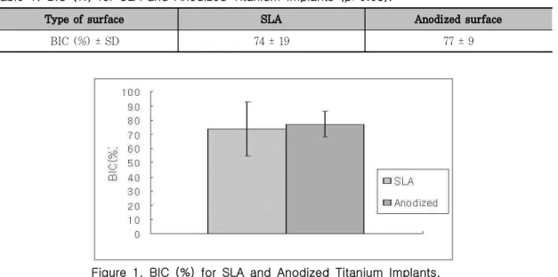
Table 1. BIC (%) for SLA and Anodized Titanium Implants (p>0.05).
Type of surface SLA Anodized surface
BIC (%) ± SD 74 ± 19 77 ± 9
Figure 1. BIC (%) for SLA and Anodized Titanium Implants.
of titanium implants. BIC data were com- pared by paired student t-test (p<0.05).
Ⅲ. Results
The results of histomorphometric analysis (Table 1) showed a higher percentage of bone-to-implant contact for implants with anodized surface(77±9) compared with SLA implant(74.±19).However this difference did not show statistical significance(p>0.05).
Ⅳ. Discussion
In present study between SLA and ano- dized surfaces implants there are not stat- istically significat differences in bone im- plant contact % representing bone healing response. The exact mechanism as to why osteoblasts produce more bone in the pres-
ence of a microrough surface is not yet well-understood18).
The degree of surface roughness may not be the only aspect of surface topography that effects osseointegration. The intimacy of bone contact with the implant surface may be important as may the ionic charge, surface energy and surface tension or other still undefined properties of the surface9).
Anodic oxidation of the electropolished surfaces, which produced areas of increased roughness and a thicker surface oxide, had an enhancing effect on the rate of bone formation. Increasing the oxide thickness of rough machined implants had no significant effect on the bone response7). But Ivanoff CJ et al.19)supposed, that with increasing oxide thickness there is a change in oxide crystal- linity, in that the amorphous oxide changes to anatase and rutile forms, which may cre-
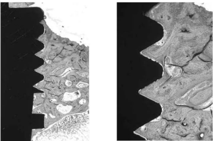
Figure 2. Histological section in mesiodistal direction of anodized implant; Multiple stain, magnification ☓40 (left), ×100 (right)
Figure 3. Histological ection in mesiodistal direction of SLA implant; Multiple stain, magnification ☓40 (left),
×100 (right)
ate a stronger bone reaction.
Several surface treatments(particle blast- ing, plasma-spray coatings, acid etching) have been proposed to improve implant sur- face characteristics and to increase the quantity and quality of bone at the inter- face, with increased interlocking20).
Within a defined immediate loading treat- ment protocol, the use of oxidized surfaced implants help to reduce the risk of a stabil-
ity loss in the posterior maxilla in the early healing period21), and use of TiUnite im- plants was successful in treating regions of bone quality 4 and no implant was lost re- sulting in a cumulative success rate of 100%22).
The preliminary results of prospective mul- ticenter clinical study by Friberg B et al.23) on mainly TiUnite implants showed an early cumulative survival rate(CSR) of 99.7%.
In the study by Rocci A. et al.,24) the Ti- Unite implant showed bone-to-implant con- tact along the full length of the implant and to a level coronal to the first thread. It was concluded that successful osseointegration of a TiUnite implant placed in soft bone was also possible when the implant was immedi- ately loaded.
Salata L. and coworkers25) have suggested that faster development of implant stability for oxidized implants than turned compo- nents when placed in bone defects. Bone formation towards the rough surfaced im- plant is facilitated by a more stable con- nection between bone matrix and the im- plant surface, which can be explained by a higher degree of protein adsorption on the anodic-oxidised implants.
Glauser R et al.26) concluded that the ap- plied immediate loading protocol in combina- tion with a slightly tapered implant and a modified implant surface texture was shown to be a successful treatment alternative even in regions exhibiting soft bone.
The results from study by Olsson M et al.27)indicated that early loading can be ap- plied to cross-arch dental bridges supported by six to eight oxidized implants in the maxilla. Authors suggested that the use of surface-modified implants may have played an important role in the favorable outcome of study.
According to Li DH et al.,1) the rough surface of titanium dental implants created by the modified sandblasting treatment can greatly enhance the shear strength at the dental implant-bone interface and that, with this enhancement, the secondary micropores
play a much more important role in im- plant-bone bonding.
The tissue response may not depend on only one specific surface property but rather on a number of different alterations. Bone tissue responses may greatly depend on the surface chemistry of implants, the porous oxide structures,28) the crystallinity of tita- nium oxide and mechanical interdigita- tion29-31). However, it is not fully understood whether these oxide properties influence the bone tissue response separately or syner- gisticcally.
However, the lack of improvement of bone-bonding ability in the later stages of implantation may be attributed to the low porosity and to the superficial ingrowths of apatite-like deposits into the pores of the anodic oxidation Ti layer. In addition to the anatase surface crystal structure, other ox- ide properties, such as micropore size and configuration, may also play important roles in the bone-bonding ability of anodically oxi- dized titanium in the body32).
However, results from our animal studies may not always be extrapolated to the clin- ical situation. this may be due to differences in bone anatomy, physiology and loading conditions. Therefore, it may be more rele- vant to study the bone tissue response to implants in human bone.
Ⅴ. Conclusions
In present animal histological study new bone formation occurred along anodized tita- nium implant surface in apical direction and was filled within fixture threads. Anodized
titanium implant showed high score of bone implant contact %. But anodized titanium implants did not show statistically sig- nificant difference with SLA titanium im- plant in bone response.
Ⅵ. References
1. Li DH, Liu BL, Zou JC, Xu KW.
Improvement of osseointegration of tita- nium dental implants by a modified sandblasting surface treatment: An in vivo interfacial biomechanics study.
Implant Dent 1999;8: 289-294.
2. Sykaras N, Lacopino AM, Marker VA, Triplett RG, Woody RD. Implant materi- als, designs, and surface topographies:
their effect on osseointegration. A liter- ature review. Int J Maxillofac Implants 2000;15: 675-690.
3. Letic-Gavrilovic A, Scandurra R, Abe K.
Genetic potential of interfacial guided osteogenesis in implant devices. Dental Materials Journal. 2000; 19(2): 99-132.
4. Giavaresi G, Fini M, Cigada A, Chiesa R, Rondelli G, Rimondini L, Torricelli P, Aldini NN, Giardino R. Mechanical and histomorphometric evaluations of tita- nium implants with different surface treatments inserted in sheep cortical bone. Biomaterials 2003; 24: 1583-1594.
5. Davies JE. Machanisms of endosseous integtation. Int J Prosthodont 1998; 11:
391-401.
6. Parr GR, Gardner LK, Toth RW.
Titanium: The mystery metal of implant dentistry. Dental materials aspects. The journal of prosthetic dentistry.1985;
54(3): 410-414.
7. Larsson C, Thomsen P, Aronsson BO, Rodahl M, Lausmaa J, Kasemo B, Ericson LE. Bone response to sur- face-modified titanium response to ma- chined and electropolished implants with different oxide thicknesses. Biomaterials 1996; 17(6): 605-615.
8. Moberg LE, Kondell PA, Sagulin GB, Bolin A, Heimdahl A, Gynther GW.
Branemark System and ITI Dental Implant System for treatment of man- dibular edentulism. Clin Oral Impl Res.
2001; 12: 450-461.
9. Klokkevold PR, Nishimura RD, Adachi M, Caputo A. Osseointegration enhanced by chemical etching of the titanium surface. Clin Oral Impl Res 1997; 8:
442-447.
10. Gotfredsen K, Berglundh T, Linghe J.
Anchorage of titanium implants with dif- ferent surface characteristics: An ex- perimental study in rabbits. Clin Impl Dent and Relat Res 2000; 2(3): 120-128.
11. Onur MA, Cehreli MC, Tas Z, Sahin S.
Effect of machined/turned, TiO₂-blasted and sandblasted/acid-etched titanium or- al implant surfaces on nerve conduction:
A study on isolated rat sciatic nerves. J Biomed Mater Res 2003; Part B: Appl Biomater 67B: 772-778.
12. Cochran DL, Schenk RK, Lussi A, Higginbottom FL, Buser D. Bone re- sponse to unloaded and loaded titanium implants with a sandblasted and acid-etched surface: A histometric study in the canine mandible. J Biomed Mater Res 1998; 40: 1-11.
13. Li D, Ferguson SJ, Beutler T, Cochran DL, Sittig C, Hirt HP, Buser D.
Biomechanical comparison of the sand- blasted and acid-etched and the ma- chined and acid-etched titanium surface for dental implants. J Biomed Mater Res. 2002; 60: 325-332.
14. Ivanoff CJ, Hallgren C, Widmark G, Sennerby L, Wennerberg A. Histologic evaluation of the bone integration of TiO
₂ blasted and turned titanium microim- plants in humans. Clin Oral Impl Res.
2001; 12: 128-134.
15. Rocci A, Martignoni M, Burgos PM, Gottlow J, Sennerby L. Histology of re- trieved immediately and early loaded oxidized implants: light microscopic ob- servations after 5 to 9 months of loading in the posterior mandible. Clinical Implant Dentistry and Related Research 2003; 5(Suppl 1): 88-98.
16. Ericsson I, Johansson CB, Bystedt H, Norton MR. A histomorphometric evalua- tion of bone-to-implant contact on ma- chine-prepared and roughened titanium dentalimplants. Clin Oral Impl Res 1994;5:202-206.
17. Mustafa K, Wroblewski J, Hultenby K, Silva Lopez B, Arvidson K. Effects of ti- tanium surfaces blasted with TiO₂ par- ticles on the initial attachment of cells derived from human mandibular bone.
Clin Oral Impl Res 2000; 11: 116-128.
18.Buser D, Broggini N, Wieland M, Schenk RK, Denzer AJ, Cochran DL, Hoffmann B, Lussi A, Steinemann SG. Enhanced bone apposition to a chemically modified SLA titanium surface. J Dent Res 2004;
87(3):529-533.
19. Ivanoff CJ, Widmark G, Johansson C, Wennerberg A. Histologic evaluation of bone response to oxidized and turned ti- tanium micro-implants in human jawbone. Int J Oral Maxillofac Implants 2003; 18: 341-348.
20. Degidi M, Petrone G, Iezzi G, Piattelli A. Histologic evaluation of a human im- mediately loaded titanium implant with a porous anodized surface. Clin Impl Dent Relat Res 2002; 4(2): 110-114.
21. Glauser R, Portmann M, Ruhstaller P, Lundgren AK, Hammerle C, Gottlow J.
Stability measurements of immediately loaded machined and oxidized implants in the posterior maxilla(A comparative clinical study using resonance frequency analysis). Applied Osseointegration Research. 2001; 2(1): 27-29.
22. Glauser R, Gottlow J, Lundgren AK, Sennerby L, Portmann M, Ruhstaller P, Hammerle C. Immediate occlusal loading of Branemark Mk Ⅳ TiUnite implant placed in bone quality type 4. Applied Osseointegration Research 2002; 3(1):
22-24.
23. Friberg B, Billstrom C. Preliminary re- sults of a prospective multicenter clinical study on TiUnite implants. Applied Osseointegration Research 2002; 3(1):
29-31.
24. Rocci A, Martignoni M, Sennerby L, Gottlow J. Immediate loading of a Branemark System implant with the TiUnite surface. Histological evaluation after 9 months. Applied Osseointegration Research 2002; 3(1): 25-28.
25. Salata L, Rasmusson L, Novaes A, Papalexiou V, Sennerby L. The influence of anodic oxidation on implant in- tegration and stability in bone defects.
An RFA study in the dog mandible.
Applied Osseointegration Research 2002;
3(1): 32-34.
26. Glauser R, Lundgren AK, Gottlow J, Sennerby L, Portmann M, Ruhstaller P, Hammerle C. Immediate occlusal loading of Branemark TiUnite implants placed predominantly in soft bone: 1-year re- sults of a prospective clinical study. Clin Implant Dent Relat Res. 2003; 5 (1) Suppl: 47-56.
27. Olsson M, Urde G, Andersen JB, Sennerby L. Early loading of maxillary fixed cross-arch dental prostheses sup- ported by six or eight oxidized titanium implants: Results after 1 year of load- ing, case series. Clinical Implant Dentistry and Related Research 2003;
5(1) Suppl: 81-87.
28. Son WW, Zhu XL, Shin HI, Ong JL, Kim KH. Invivohistological response to ano- dized and anodized /hydrothermally treated titanium implants. J Biomed
Mater Res Part B; 2003; Appl Biomater 66B: 520-525.
29. Sul YT, Johansson CB, Jeong Y, Roser K, Wennerberg A, Albrektsson T.
Oxidized implants and their influence on the bone response. Journal of Materials Science: Materials in Medicine. 2001;
12: 1025-1031.
30. Sul YT, Johansson CB, Kang Y, Jeon DG, Albrektsson T. Bone reactions to oxidized titanium implants with electro- chemical anion sulphuric acid and phos- phoric acid incorporation. Clin Impl Dent Relat Res 2002; 4(2): 78-87.
31. Sul YT. The significance of the surface properties of oxidized titanium to the bone response: special emphasis on po- tential biochemical bonding of oxidized titanium implant. Biomaterials 2003; 24:
3893-3907.
32. Liang B, Fujibayashi S, N대 M, Taumra J, Kim HM, Uchida M, Kokubo T, Nakamura T. Hisological and mechanical investigation of the bone-bonding ability of anodically oxidized titanium in rabbits. Biomaterials 2003;24:4959-4966.
-국문초록-
양극 산화한 티타늄 임프란트의 조직학적 반응
임 스베틀라나1․허성주2․한종현3․김태일1․설양조1․ 구 영1․정경욱1․정종평1․한수부1․류인철1
1서울대학교 치과대학 치주과학교실
2서울대학교 치과대학 치과보철학교실
3연세대학교 영동세브란스 병원
여러 연구들을 통해 많은 학자들이 임프란트 안정성(stability)은 표면의 특징에 달려있다고 생각하게 되었 다. 표면의 구조, 에너지, 산화물(oxide) 두께와 표면성상(topography)등 임프란트의 표면의 특징은 임프란 트와 골조직의 반응에서 중요한 역할을 하는 것이 알려짐에 따라 티타뮨 임프란트의 표면의 처리 방법에 큰 관 심을 가지게 되었다.
그 중에서 티타늄 임프란트 표면의 산화피막화(anodization)가 한 방법으로 대두되었다. 이 방법은 전기화 학적 방식으로 임프란트 표면에 거칠고(rough)두꺼우며(thick), 기공(pore)을 가지는 산화물 막을 형성하는 것으로 산화물의 두께는 coronal 부분(1-2 ㎛)으로부터 apical부분(7-10 ㎛)까지 증가하게 된다. 산화피막의 표면에는 다양한 크기의 수많은 기공이 주로 1-2 ㎛ 두께로 임프란트의 apical 부분에서 존재하며, 임프란트 표면의 거칠기는 conical 위부분에서 apical 부분까지 계속 증가한다(평균 Ra value=1.2 ㎛).
또 다른 표면 처리 방법으로는 blasting 후에 etching을 한 SLA 표면이 있다.
이 연구의 목적은 일반적으로 많이 이용되고 있는 anodized 표면과 SLA 표면의 조직학적 반응을 비교 분 석하는 것이다.
24개 임프란트를(anodized surfaced implant-12개 , SLA-12개, 8mm ×φ 4.3) 6마리 토끼의 오른쪽 과 왼쪽 femur에 식립하였다. 12주후에 동물들을 희생하여 EXACT cutting-grinding system을 이용하여 샘플을 절단하고 800, 1200 및 4000 번 연마제(abrasive) paper로 20-50 ㎛ 까지 grinding하였다. 샘플은 Multiple staining 용액으로 염색하여 SLA 임프란트 군과 비교하였다. 골과 임프란트 사이에 연결을 TDI 프로그램을 이용하여 %로 측정하였다.
SLA 임프란트 군 경우에는 골과 임프란트 사이의 연결이 74±19% 이고, 양극 산화한 임프란트 군 경우에 는 77±9%이었다. 양극 산화한 티타늄 임프란트의 골 접촉률이 SLA 표면 임프란트 경우과 통계학적으로 유의 한 차이는 보이지 않았다.3)
주요어 : 골접촉률, 양극 산화한 임프란트, 골 반응, 산화피막화