i
Table of Contents
List of Tables ··· ii
List of Figures ··· iii
Abstract ··· iv
I. Introduction ··· 1
II. Materials and Methods ··· 5
1. Animal ··· 5
2.Sample fabrication ··· 5
3. Experimental design ··· 6
4. Surgical protocol ··· 6
5. Histologic and histomorphometric analysis ··· 7
6. Statistical analysis ··· 9 III. Results ··· 10 1. Clinical observations ··· 10 2. Histologic analysis ··· 10 3. Histomorphometric analysis ··· 11 IV. Discussion ··· 13 V. Conclusion ··· 18 References ··· 19 Tables ··· 25 Figures ··· 26 Korean abstract (국문초록) ··· 30
ii
List of Tables
iii
List of Figures
Figure 1. Representative photomicrograph CPRS implant for histomorphometric
analysis ··· 26
Figure 2. Representative photomicrographs of all experimental groups ··· 27
Figure 3. Representative photomicrographs with high magnification of all groups ··· 28
Figure 4. Representative photomicrographs with high magnification of fibrous
iv
ABSTRACT
Bone regeneration and collagen fiber orientation around
machined and rough surface of CaP-coated implants
in circumferential defect
: A histomorphometric study in dogs
Yea-Won Kim, D.D.S.,M.S.D.
Department of Dental Science Graduate School, Yonsei University
(Directed by Professor Seong-Ho Choi, D.D.S., M.S.D., Ph.D.)
Objectives: The aim of this study was to compare the bone formation and tissue reactions around CaP-coated machined surface and CaP-coated rough surface implants (CPMS and CPRS) at the critical-sized circumferential ridge defects in dogs at 4 and 8 weeks of the observation period
.
Materials and methods: All mandibular premolars were extracted bilaterally in five
male mongrel dogs. After eight weeks, circumferential marginal defects (2mm gap and 5mm depth) were created unilaterally using a customized drill and CPMS and CPRS implants were inserted. Four weeks later, the contralateral side was prepared using the same protocol. The dogs were then sacrificed four weeks after the last
v
surgery. Specimens were analyzed histologically and histomorphometrically.
Results: Both CPMS and CPRS implants showed successful osseointegration at the
non-defect area, normal bone area regardless of the observation periods (4 and 8 weeks). All circumferential defects were incompletely filled and osseointegration was limited at the half lower area of the defect. In unfilled defect area, collagen fibers were arranged perpendicularly or obliquely onto the exposed implant surfaces, especially on CPRS implants.
Conclusion: Within the limits of this study, it can be suggested that CPMS and CPRS
implants show no difference in bone-to-implant contact and bone regeneration in the circumferential defects in 4 and 8 weeks healing period, and unique features of perpendicularly/obliquely arranged collagen fibers onto the exposed implant surfaces.
Key Words: Calcium phosphate, Dental implant, Bone regeneration, Titanium
1
Bone regeneration and collagen fiber orientation around
machined and rough surface of CaP-coated implants
in circumferential defect:
A histomorphometric study in dogs
Yea-Won Kim, D.D.S.,M.S.D.
Department of Dental Science Graduate School, Yonsei University
(Directed by Professor Seong-Ho Choi, D.D.S., M.S.D., Ph.D.)
I. Introduction
Clinical success of dental implant treatment can be achieved with the direct contact between bone and installed implant fixture, i.e. osseointegration (Branemark et al. 1969; Linder et al. 1983). Titanium alloy for the dental or orthopedic implant provides a biocompatible surface where osteoblasts can be differentiated and grown. Therefore, bone tissue formation on the surface of titanium implant (contact osteogenesis) has been emphasized, as well as bone formation and growth from the adjacent existing bone tissue (distance osteogenesis) (Davies 1998, 2003). Previous studies demonstrated that the surface topography influenced differentiation and maturation of osteoblasts and fibroblasts (Lossdorfer et al. 2004; Galli et al. 2012;
2
Olivares-Navarrete et al. 2012). It has been suggested that the implant surface treatment accelerates contact osteogenesis and decreases healing time for functional loading.
Surface modification of dental implant includes mechanical and chemical treatment on the titanium surface. Since the development of a rough surfaceimplant, both short and long- term survival rates have increased along with a reduce in the healing period for functional loading when compared with the use of machined surface implants (Cochran 2000; Ganeles et al. 2008; Karabuda et al. 2011). However, some dental implants have been shown to fail, particularly at sites with poor bone quality or severe bone defect. Therefore, many researchers have studied and developed a more ‘bone-friendly’ surfaced implant within the last two decades using chemical modification (Li et al. 2008; Mamalis et al. 2011; Mertens & Steveling 2011; Hamlet et al. 2012).
Surface coating techniques via bioactive ceramic compounds has been addressed among the chemical modification of the dental implant surface (Tomisa et al. 2011). A thin ceramic layer can promote cell attachment, differentiation and bone formation, and accelerate osseointegration between the implant surface and its surrounding bone tissues (Yang et al. 2003; Jimbo et al. 2011; Palaiologou et al. 2012). When the ceramic coated implants are used, a carbonated apatite layer forms on their surfaces, which has the same composition and structure to the mineral phase of bone tissue (Tomisa et al. 2011).
3
implant surface coating techniques (Li et al. 2008; Fontana et al. 2011; Jimbo et al. 2011). Plasma spraying was used as a primary method for the CaP coating technique, but could not provide adequate adherence strength of the CaP coating to the titanium alloy (7 MPa) (Tomisa et al. 2011). Therefore, current studies suggested applying various ions to increase the adherence strength of titanium to the CaP coating, where the ion-beam-assisted deposition (IBAD) method reported a high adherence strength (35-70 MPa) of CaP coating (Liu et al. 2000; Jung et al. 2001) and additional synergistic effects with SLA implant surface without loss of micro-structure (Kim et al. 2010).
Machined surface implants have been showed lower bone to implant contact (BIC) and survival rate compared with those of rough surface implants in various studies. However, the CaP-coated machined surface implant (CPMS) resulted in an enhanced BIC to the comparable level of CaP-coated rough surface implant (CPRS) in normal bone model of the previous study (Choi et al. 2012, Kim et al. 2008). In addition, An exposed rough surface implant can make a situation more complicated compared to machined surface implant, due to their lack of decontamination in the roughened micro-structure (Lang & Berglundh 2011; Renvert et al. 2011). From this point of view, machined surface implants possess more advantages in controlling the bacterial load during maintenance phase. Therefore, CPMS could be more clinically suitable than a CPRS in normal bone.
On the other hand, Bone regeneration in clinically challenged bony defects such as gap defect and large dehiscence defect can be different from the healing observed in
4
bones without defects. Several studies using the IBAD method have shown that CaP coating may improve the bone response and produce beneficial effects in resolving bony defects (Chae et al. 2008; Yoon et al. 2009). According to the previous result, There was more defect fill in CPRS group compared with CPMS group in three-wall intrabony defect (Choi et al. 2012). However, these were evaluated at 12 weeks healing period, which may be enough time to make favorable osseointegration around dental implants. Therefore further evaluation was required for the two types of CaP-coated implants in various experimental environments and healing time.
Thus, the aim of this study was to compare bone formation and tissue reactions around CPMS and CPRS implants at the critical-sized circumferential ridge defects in dogs at short- and mid-term healing period.
5
II. Materials and methods
1. Animal
Five male mongrel dogs, 18–24 months old and weighing about 30 kg, were chosen. The animals had intact dentition and healthy periodontium. Animal selection, management, preparation, and surgical protocol followed the routine procedure approved by the Animal Care and Use Committee, Yonsei Medical Center, Seoul, Korea.
2. Sample fabrication
Dental implants of commercially pure titanium (Ø3.4mm, 10mm length) were fabricated(Dentium, Seoul, Korea), and two types of surface were prepared for this study; machined and SLA. Both surfaces were treated by calcium phosphate nanocoating at a thickness of 500nm, using Ion beam-assisted deposition (IBAD). In brief, disc-form evaporants (hydroxyapatite + 37% Calcium oxide) were sintered for 2h at 1000°C. The IBAD method was used, in which an ion beam was introduced into the vacuum chamber and the resulting evaporants adhered to the surface of the implants (Lee et al. 2007).
6
3. Experimental design
Implants were allocated to four groups according to types of surface (machined and SLA) and observation period (4 and 8 weeks): 4-CPMS or 8-CPMS coated-machined surface for 4 or 8 weeks healing period), and 4-CPRS or 8-CPRS (CaP-coated-SLA surface for 4 and 8 weeks healing period). Two types of implants were installed in unilateral side of mandible, the same types of implants were inserted in contralateral side of mandible at four weeks later.
4. Surgical protocol
Teeth were extracted under general anesthesia under sterile conditions in an operating room using atropine 0.05 mg/kg SQ, xylazine (Rompun, Bayer Korea, Seoul, Korea) 2 mg/kg, ketamine hydrochloride (Ketalar, Yuhan Co., Seoul, Korea), and 10 mg/kg IV. Dogs were placed on a heating pad, intubated, administered 2% enflurane, and monitored with an electrocardiogram. After disinfecting the surgical sites, 2% lidocane HCl with epinephrine 1:100,000 (Kwangmyung Pharm., Seoul, Korea) was administered by infiltration at the surgical sites. Second, third, and fourth premolars on the both sides of mandible were extracted. After 8 weeks of healing, the prefabricated implants were placed under the same surgical conditions as those for the tooth extraction. A mid-crestal incision was made, and mucoperiosteal flaps were carefully reflected on the buccal and lingual aspects. The edentulous ridge was
7
carefully flattened with a surgical bur and sterile saline irrigation. Submerged-type, CaP nanocoated implants (3.5 mm diameter, 10 mm length) with machined or SLA surface were placed on the unilateral side of the mandible. Four weeks later, implants with the same features were inserted on the contralateral side. Implant site preparation was performed by sequential drilling, and parallel circumferential defect (2mm gap and 5mm length) was created surgically with a customized drill. Experimental implants were installed, and flaps were closed without grafting at circumferential defect using 5-0 resorbable suture materials (Vicryl 5/0, Polyglactin 910, Ethicon, Johnson & Johnson, Somerville, NJ, USA). Sutures were removed after 7–10 days and a soft diet was provided throughout the study period. Four weeks after the last surgery, animals were sacrificed with an anesthesia drug overdose. Block sections including segments of implants were preserved and fixed in 10% neutral-buffered formalin. The specimens were dehydrated in ethanol, embedded in methacrylate, and sectioned in the mesio-distal plane using a diamond saw (Exakt, Apparatebau, Norderstedt, Germany). From each implant site, the central section was taken to a final thickness of about 30 μm, and the sections were stained with hematoxylin-eosin. The most central section was included in histologic and histometric analysis.
5. Histologic and histomorphometric analysis
8
polarized light microscopy (Olympus® Research System Microscope BX51, Olympus, Tokyo, Japan) and a PC-based image analysis system (Image-Pro Plus™, Media Cybernetic, Silver Spring, MD, USA; Adobe® Photoshop® CS3, Adobe Systems Inc., San Jose, CA, USA). The following measurements were made:
• Bone regeneration height: proportion of distance from the base of the defect (5mm apical point from the top of fixture platform) to the coronal extension of newly formed bone contacting the implant surface in a defect height (5mm); • Bone regeneration area: proportion of newly formed alveolar bone within a
standardized template as a proxy for the defect site (2mm width X 5mm length). The template was aligned parallel to the implant surface interfacing the base of the defect;
• BIC: bone-to-implant contact ratio at the defect area (upper area from the base of the defect; BIC-Defect) and normal bone area (lower area from the base of the defect; BIC-Non-Defect) were measured separately;
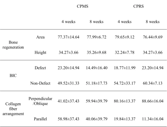
Additionally measurement was made at the unfilled defect area, for evaluating the attachment of connective tissue at the exposed surface of implant (Fig. 1):
• Collagen fiber arrangement: proportion of collagen fiber attachment or adhesion around implant surface within the unfilled defect area. Collagen fibers perpendicularly/obliquely ran and inserted into the implant surface (Attachment), or ran parallel to the implant surface (Adhesion).
9
6. Statistical analysis
The statistical analysis was performed using a commercially available software program (PASW Statistics 18, SPSS inc., Chicago, IL, USA). Kruskal-Wallis test was used to analyze the effects of time and experimental conditions. The level of significance set at 5%.
10
III. Results
1. Clinical observations
All experimental sites healed uneventfully with minimal signs of inflammation and exposure. At the time of implant insertion surgery, one animal lacked vertical length of the alveolar bone, and three implants except one (8-CPMS) could not show any primary stability. In the histologic evaluation, these sites showed a short height of the alveolar bone (distance from inferior alveolar nerve to the crest of the bone) less than 5mm (height of the crestal defect). Therefore, three sites were excluded from the analysis; 1 from 4-CPMS, 1 from 4-CPRS, and 1 from 8-CPRS.
2. Histologic analysis
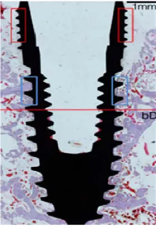
Incompletely, but considerable amount of new bone formed in all circumferential defects independent of the types of surface and observation period (Fig. 2). The dimensions of the defect significantly decreased and the apical border of the defect could not be distinguished in all sites. The defect-filled area including three macro-threads showed newly formed bone contacting the implant surface. In contrast, the coronal area of the defect in all groups showed fibrous attachment onto the implant surface despite of the considerable resolution of the defect (Fig. 1 and 2). In this
11
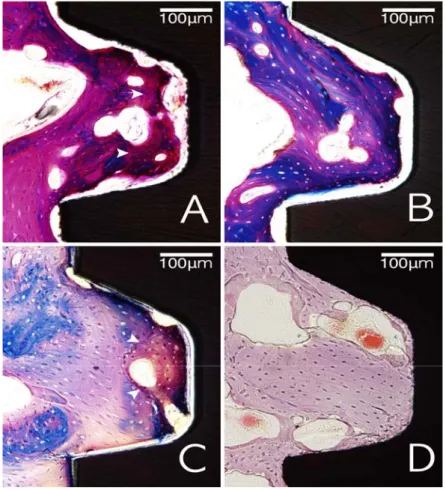
unfilled defect area, most of the collagen fibers were arranged perpendicularly or obliquely to the exposed implant surface on the CPRS implants (Fig. 4).
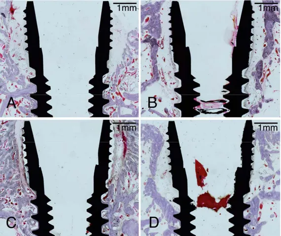
In the non-defect area, all experimental sites showed direct contact of newly formed bone to implant surface (osseointegration) for the whole length of the implant within the this area with no significant differences of bone healing between the CPMS and CPRS implants. Although newly formed bone could be seen to contact with the implant surface in the threaded region, the sites of the 4-weeks groups showed immature woven bone at some area between the two consecutive threads. In the 8-week experimental sites, mature alveolar bone with the lamellar structure and osteon was observed around the implant surface regardless of the implant surface type, and there was no distinguishable line between the existing and newly formed bone (Fig. 3).
3. Histomorphometric analysis
The results of histomorphometric analyses are presented in Table 1. All experimental groups showed similar bone regeneration of circumferential defect in both aspects of the area and height measurements, regardless of the type of coated-surface and healing time. There were no statistically significant differences for BIC measurements at both levels (natural bone and defect area), as well as bone regeneration.
12
were arranged perpendicularly/obliquely to the implant surface for both 4 and 8 weeks healing periods: 80.16 ± 13.37 and 88.66 ± 16.04% for 4-CPRS and 8-CPRS versus 58.98 ± 37.43 and 40.06 ± 39.79% for 4-CPMS and 8-CPMS, respectively. However, there were no statistically significant differences between all groups.
13
IV. Discussion
The objective of this study was to compare bone formation and tissue reactions around CPMS and CPRS implants at the critical-sized circumferential ridge defects in dogs. The thickness of the coating layer to implant surface varies from micro to nano size depending on selected coating technique. If the coating layer is too thick, the outer layer may detach, and if the coating layer is too thin, the stability of the coating layer may decrease due to poor cyrstallinity. In a previous studies on coating thickness using IBAD method, CaP was coated on anodized implants with a thickness of 200 nm and 500 nm. The results of the histological studies revealed better coronal defect fill with 500 nm of CaP coating thickness than 200 nm of thickness. However, comparable defect filling was observed with 200 nm CaP coated anodized implants (Um et al. 2011). Thus, implant surfaces were treated by IBAD calcium phosphate coating method at a thickness of 500nm in present study.
The development and use of a rough surfaced implant in clinical implant dentistry showed a decrease in early failure as well as a reduction in the healing time for osseointegration compared to machined surface implant. The previous studies described two types of bone healing patterns around dental implant; distance osteogenesis and contact osteogenesis (Davies 1998, 2003). As the rough surface of titanium can increase cell attachment, the author suggested that rough surface dental implant could enhance contact osteogeneis and reduce healing period prior to loading.
peri-14
implantitis will increase once it is exposed to the oral environment compared to machined surface implants (Lang & Berglundh 2011; Renvert et al. 2011). It may be caused by influences of surface characteristics on the amount and composition of biofilm formation (Renvert et al. 2011). Therefore, exposed rough surfaced implants can create challenges for controlling bacterial load during the maintenance period, as well as the progression of the disease. Even though rough surfaced implants have more advantages in early healing phase, machined surface implants can be more suitable during the maintenance phase.
The previous studies found that CaP-coated surface made the enhancement of bone healing around machined surface implants (Li et al. 2008; Choi et al. 2012). Choi et al. demonstrated similar bone-to-implant contact (54.9 vs. 57.4%) and bone density (76.0 vs. 75.6%, for machined versus SLA surface) in the sites of normal bone received CPMS and CPRS without defect induction. The present study used 4 and 8 weeks as the observation period, and confirmed the enhancement of BIC on machined surface implant to the similar level with the SLA implants using a CaP coating technique in short- and mid-term healing time frame. While previous studies demonstrated a significant increase in BIC and bone regeneration around the SLA implant than the machined surface implant (Cochran 2000; Ganeles et al. 2008; Karabuda et al. 2011), more recent studies demonstrated an increase in enhanced bone regeneration and BIC around both surface implants using CaP coating surface modification (Yoon et al. 2009; Kim et al. 2010). The present study demonstrated similar bone regeneration and BIC regardless of surface roughness. It is suggested
15
that higher synergistic effects of the CaP coating technique in early healing phase were shown in machined surface implants compared to SLA surface one.
This study also used experimental models which included two types of bony environments; critical-sized circumferential defect at the coronal 5mm area and normal bone at apical 5mm area (Fig. 1). However, there was an incomplete bone regeneration within the circumferential defects, which are contrary to the previous study of Botticelli et al. (2005). They illustrated a successful defect fill around SLA implants in smaller-sized circumferential defect model (1.25mm) at 4 months. This may be caused by the differences in defect size and healing period. Circumferential defect with a size smaller than 1.5mm has been known as a contained defect with a ‘jumping distance’ (Botticelli et al. 2003), in which bone tissue could regenerate naturally without a barrier membrane. At this defect, a rough surface characteristic of dental implants could make an enhancement of appositional bone growth and contact osteogenesis, which may in turn accelerate the bone regeneration around the dental implant (Botticelli et al. 2005). However, in spite of the synergistic effects of the surface modification with CaP, both types of implants did not show complete bone regeneration in the critical-sized circumferential defects of the present study. In this context, it can be suggested that CaP coating surface modification could accelerate osseointegration of dental implants installed in normal bone, but not enhance bone healing in the circumferential defect at short-term observation period.
Tooth and dental implants are surrounded by three different tissues; epithelium, connective tissue, and alveolar bone (Stern 1981; Berglundh et al. 1991).
16
Although there are some variations in amounts of all dento(implanto)-gingival tissues, there is considerable evidence supporting the fundamental roles of soft tissue layers as a barrier against the oral environment (Comut et al. 2001). The connective tissue layer around dental implant contains collagen fibers that are arranged parallel to the implant surface providing a cuff-like soft tissue sealing (Berglundh et al. 1991; Buser et al. 1992; Listgarten et al. 1992). Whereas, natural tooth has dentogingival collagen fibers inserted into the cementum which are oriented perpendicularly or obliquely to the tooth surface (Stern 1981). Therefore, ‘self-limiting’ process against inflammatory lesion exists in connective tissue around teeth by functionally oriented fibers (Berglundh et al. 2011; Lang & Berglundh 2011).
The present study showed that most of collagen fibers were arranged perpendicularly or obliquely to the implant surface within the unfilled defect area on CPRS implant group regardless of the healing period. (Fig. 4). Previous studies found that a rough surfaced characteristic may influence the attachment or proliferation of fibroblasts on titanium materials (Guy et al. 1993; Mustafa et al. 1998), and several surface modifications of chemistry, surface charge, and topography have been developed to improve soft tissue attachment as a barrier around dental implant (Schwarz et al. 2007). However, most of the surfaces including SLA showed parallel oriented collagen fiber network with limited perpendicular insertion into the surface. This study showed that eighty percent of connective tissue around the exposed implant surface demonstrated a perpendicular/oblique oriented appearance. Therefore, a surface coating with bioactive calcium phosphate may influence the arrangement of
17
collagen fibers around an implant surface. However, unlike previous studies observing supra-alveolar region, these results appeared within the unfilled bone defect. To confirm the effects of the CaP coating technique on the collagen fiber arrangement, further studies are needed to focus on the implanto-gingival attachment at CaP-coated titanium surface in the other experimental environments.
18
V. Conclusion
Within the limits of this study,
1. CPMS and CPRS implants showed incomplete healing in the critical size circumferential defect in 4 and 8 weeks healing period.
2. It can be suggested CPMS and CPRS implants show no difference in bone-to-implant contact and bone regeneration in the circumferential defects in 4 and 8 weeks healing period.
3. CPRS implants showed unique features of perpendicularly/obliquely arranged collagen fibers onto the exposed implant surfaces.
19
Reference
Berglundh T, Lindhe J, Ericsson I, Marinello CP, Liljenberg B, Thomsen P: The soft tissue barrier at implants and teeth. Clin Oral Implants Res 2: 81-90, 1991. Berglundh T, Zitzmann NU, Donati M: Are peri-implantitis lesions different from
periodontitis lesions? J Clin Periodontol 38 Suppl 11: 188-202, 2011.
Botticelli D, Berglundh T, Buser D, Lindhe J: The jumping distance revisited: An experimental study in the dog. Clin Oral Implants Res 14: 35-42, 2003. Botticelli D, Berglundh T, Persson LG, Lindhe J: Bone regeneration at implants with
turned or rough surfaces in self-contained defects. An experimental study in the dog. J Clin Periodontol 32: 448-455, 2005.
Branemark PI, Adell R, Breine U, Hansson BO, Lindstrom J, Ohlsson A: Intra-osseous anchorage of dental prostheses. I. Experimental studies. Scand J Plast Reconstr Surg 3: 81-100, 1969.
Buser D, Weber HP, Donath K, Fiorellini JP, Paquette DW, Williams RC: Soft tissue reactions to non-submerged unloaded titanium implants in beagle dogs. J Periodontol 63: 225-235, 1992.
Chae GJ, Jung UW, Jung SM, Lee IS, Cho KS, Kim CK, Choi SH. Healing of surgically created circumferential gap around Nano-coating surface dental implants in dogs. Surf Interface Anal 40:184-187, 2008
Choi JY, Jung UW, Kim CS, Jung SM, Lee IS, Choi SH: Influence of nanocoated calcium phosphate on two different types of implant surfaces in different bone environment: an animal study. Clin Oral Implants Res, 2012.
20
Cochran DL: The scientific basis for and clinical experiences with Straumann implants including the ITI Dental Implant System: a consensus report. Clin Oral Implants Res 11 Suppl 1: 33-58, 2000.
Comut AA, Weber HP, Shortkroff S, Cui FZ, Spector M: Connective tissue orientation around dental implants in a canine model. Clin Oral Implants Res 12: 433-440, 2001.
Davies JE: Mechanisms of endosseous integration. Int J Prosthodont 11: 391-401, 1998.
Davies JE: Understanding peri-implant endosseous healing. J Dent Educ 67: 932-949, 2003.
Fontana F, Rocchietta I, Addis A, Schupbach P, Zanotti G, Simion M: Effects of a calcium phosphate coating on the osseointegration of endosseous implants in a rabbit model. Clin Oral Implants Res 22: 760-766, 2011.
Galli C, Piemontese M, Lumetti S, Ravanetti F, Macaluso GM, Passeri G: Actin cytoskeleton controls activation of Wnt/beta-catenin signaling in mesenchymal cells on implant surfaces with different topographies. Acta Biomater, 2012.
Ganeles J, Zollner A, Jackowski J, ten Bruggenkate C, Beagle J, Guerra F: Immediate and early loading of Straumann implants with a chemically modified surface (SLActive) in the posterior mandible and maxilla: 1-year results from a prospective multicenter study. Clin Oral Implants Res 19: 1119-1128, 2008. Guy SC, McQuade MJ, Scheidt MJ, McPherson JC, 3rd, Rossmann JA, Van Dyke
TE: In vitro attachment of human gingival fibroblasts to endosseous implant materials. J Periodontol 64: 542-546, 1993.
21
Hamlet S, Alfarsi M, George R, Ivanovski S: The effect of hydrophilic titanium surface modification on macrophage inflammatory cytokine gene expression. Clin Oral Implants Res 23: 584-590, 2012.
Jimbo R, Xue Y, Hayashi M, Schwartz-Filho HO, Andersson M, Mustafa K, et al.: Genetic responses to nanostructured calcium-phosphate-coated implants. J Dent Res 90: 1422-1427, 2011.
Jung YC, Han CH, Lee IS, Kim HE: Effects of ion beam-assisted deposition of hydroxyapatite on the osseointegration of endosseous implants in rabbit tibiae. Int J Oral Maxillofac Implants 16: 809-818, 2001.
Karabuda ZC, Abdel-Haq J, Arisan V: Stability, marginal bone loss and survival of standard and modified sand-blasted, acid-etched implants in bilateral edentulous spaces: a prospective 15-month evaluation. Clin Oral Implants Res 22: 840-849, 2011.
Kim H, Choi SH, Chung SM, Li LH, Lee IS: Enhanced bone forming ability of SLA-treated Ti coated with a calcium phosphate thin film formed by e-beam evaporation. Biomed Mater 5: 044106, 2010.
Kim MK, Choi JY, Chae GJ, Jung UW, Kim ST, Lee IS, Cho KS, Kim CK, Choi SH. The histometric analysis of osseointegration in hydroxyapatite surface dental implants by ion beam-assisted deposition. J Korean Acad Periodontol 38:363-372, 2008
Lang NP, Berglundh T: Periimplant diseases: where are we now?--Consensus of the Seventh European Workshop on Periodontology. J Clin Periodontol 38 Suppl 11: 178-181, 2011.
22
Lee IS, Zhao B, Lee GH, Choi SH, Chung SM: Industrial application of ion beam assisted deposition on medical implants. Surface and Coatings Technology 201: 5132-5137, 2007.
Li Y, Lee IS, Cui FZ, Choi SH: The biocompatibility of nanostructured calcium phosphate coated on micro-arc oxidized titanium. Biomaterials 29: 2025-2032, 2008.
Linder L, Albrektsson T, Branemark PI, Hansson HA, Ivarsson B, Jonsson U, et al.: Electron microscopic analysis of the bone-titanium interface. Acta Orthop Scand 54: 45-52, 1983.
Listgarten MA, Buser D, Steinemann SG, Donath K, Lang NP, Weber HP: Light and transmission electron microscopy of the intact interfaces between non-submerged titanium-coated epoxy resin implants and bone or gingiva. J Dent Res 71: 364-371, 1992.
Liu JQ, Luo ZS, Cui FZ, Duan XF, Peng LM: High-resolution transmission electron microscopy investigations of a highly adhesive hydroxyapatite coating/titanium interface fabricated by ion-beam-assisted deposition. J Biomed Mater Res 52: 115-118, 2000.
Lossdorfer S, Schwartz Z, Wang L, Lohmann CH, Turner JD, Wieland M, et al.: Microrough implant surface topographies increase osteogenesis by reducing osteoclast formation and activity. J Biomed Mater Res A 70: 361-369, 2004. Mamalis AA, Markopoulou C, Vrotsos I, Koutsilirieris M: Chemical modification of
an implant surface increases osteogenesis and simultaneously reduces osteoclastogenesis: an in vitro study. Clin Oral Implants Res 22: 619-626, 2011.
23
Mertens C, Steveling HG: Early and immediate loading of titanium implants with fluoride-modified surfaces: results of 5-year prospective study. Clin Oral Implants Res 22: 1354-1360, 2011.
Mustafa K, Silva Lopez B, Hultenby K, Wennerberg A, Arvidson K: Attachment and proliferation of human oral fibroblasts to titanium surfaces blasted with TiO2 particles. A scanning electron microscopic and histomorphometric analysis. Clin Oral Implants Res 9: 195-207, 1998.
Olivares-Navarrete R, Raines AL, Hyzy SL, Park JH, Hutton DL, Cochran DL, et al.: Osteoblast maturation and new bone formation in response to titanium implant surface features are reduced with age. J Bone Miner Res, 2012. Palaiologou A, Stoute D, Fan Y, Lallier TE: Altered cell motility and attachment with
titanium surface modifications. J Periodontol 83: 90-100, 2012.
Renvert S, Polyzois I, Claffey N: How do implant surface characteristics influence peri-implant disease? J Clin Periodontol 38 Suppl 11: 214-222, 2011.
Schwarz F, Herten M, Sager M, Wieland M, Dard M, Becker J: Histological and immunohistochemical analysis of initial and early subepithelial connective tissue attachment at chemically modified and conventional SLA titanium implants. A pilot study in dogs. Clin Oral Investig 11: 245-255, 2007.
Stern IB: Current concepts of the dentogingival junction: the epithelial and connective tissue attachments to the tooth. J Periodontol 52: 465-476, 1981.
Tomisa AP, Launey ME, Lee JS, Mankani MH, Wegst UG, Saiz E: Nanotechnology approaches to improve dental implants. Int J Oral Maxillofac Implants 26 Suppl: 25-44; discussion 45-29, 2011.
Um YJ, Lee JS, Jung UI, Kim ST, Kim CS, Chung SM, Lee IS, Choi SH: The Evaluation of Calcium Phosphate Coated Implants by Ion-beamAssisted
24
Deposition (IBAD) Method in Dogs : A Preliminary Study. Biomater Res 13: 21-25, 2009
Yang Y, Bumgardner JD, Cavin R, Carnes DL, Ong JL: Osteoblast precursor cell attachment on heat-treated calcium phosphate coatings. J Dent Res 82: 449-453, 2003.
Yoon HJ, Song JE, Um YJ, Chae GJ, Chung SM, Lee IS, et al.: Effects of calcium phosphate coating to SLA surface implants by the ion-beam-assisted deposition method on self-contained coronal defect healing in dogs. Biomed Mater 4: 044107, 2009.
25
Tables
Table 1. Results of histomorphometric analysis: proportions of bone regeneration in
the defect, BIC, and collagen fiber arrangement (%).
CPMS CPRS
4 weeks 8 weeks 4 weeks 8 weeks
Bone regeneration Area 77.37±14.64 77.99±6.72 79.65±9.12 76.44±9.69 Height 34.27±3.66 35.26±9.68 32.24±7.78 34.27±3.66 BIC Defect 23.20±14.94 14.49±16.40 18.77±11.99 23.20±14.94 Non-Defect 49.52±31.33 51.18±17.73 54.72±33.17 60.34±7.13 Collagen fiber arrangement Perpendicular /Oblique 41.02±37.43 59.94±39.79 80.16±13.37 88.66±16.04 Parallel 58.98±37.43 40.06±39.79 19.84±13.37 11.34±16.04
CPMS (CaP-coated machined surface implant), CPRS (CaP-coated rough surface implants), BIC(bone-to-implant contact ratio)
26
Figures
Figure 1. Representative photomicrograph CPRS implant indicating the separated
area by the base of the defect (red horizontal line) for histomorphometric analysis; lower area for normal bone environment without any bony defects and upper area for circumferential defect area. The proportions of these collagen fiber arrangements in the unfilled defect area (red boxed) were calculated histomorphometrically. Blue box showed defect-filled area. Scale bars =1mm.
27
Figure 2. Representative photomicrographs of all experimental groups (A, 4-CPMS;
B, 8-CPMS; C, 4-CPRS; D, 8-CPRS). All circumferental defects were incompletely filled with the newly formed bone and limited osseointegration regardless of observation period and coated-implant surface. Scale bars = 1mm.
28
Figure 3. Representative photomicrographs with high magnification in defect-filled
area (blue box in Fig.1) of all groups (A, 4-CPMS; B, CPMS; C, 4-CPRS; D, 8-CPRS). Both sites at 4 weeks (A and C) showed the small parts of woven bone near the implant surface and clearly distinguishable reversal line between the woven and lamellar bone (arrowheads), whereas all 8-week sites showed lamellar mature bone within the space between the threads. Scale bars = 100 μm.
29
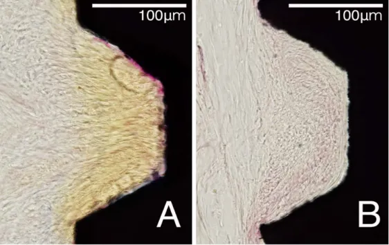
Figure 4. Representative photomicrographs with high magnification of fibrous
attachment area in unfilled defect areas (red box in Fig. 1). Most of unfilled defect area around CPRS implant (A) showed perpendicularly or obliquely arranged onto the exposed implant surface, whereas parallel-arranged collagen fibers could be observed more frequently in the unfilled defect area around CPMS implant (B). Scale bars = 100 μm.
30 국문요약
환상형 골결손 모델에서 기계절삭형 표면과 거친 표면에
칼슘 포스페이트 코팅된 임플란트 주위 골재생과 콜라겐
섬유의 주행성 : 성견에서의 조직형태계측학적 연구
<지도교수 최 성 호>
연세대학교 대학원 치의학과
김 예 원
목적: 본 연구의 목적은 성견에서 환상형 골결손을 형성한 뒤 4 주와 8 주의 치유 기간에서 칼슘 포스페이트 코팅된 기계 절삭형 표면과 거친 표면의 임플란트의 골형성과 조직 반응을 비교하는 것이다. 방법: 다섯 마리의 성견의 양측 하악 소구치를 모두 발치하였다. 8 주의 치유 기간이 지난뒤 환상형 골결손 (2mm 간극, 5mm 깊이)을 형성한 뒤 두 그룹의 임플란트를 식립하였다. 4 주가 지난 뒤 반대측에 같은 술식을 시행하였다. 4 주의 치유 기간 후 희생시켜 조직 계측학적 분석을 위한 시편을 제작하였다.31 결과: 칼슘 포스페이트 코팅된 기계절삭형 표면 임플란트와 칼슘 포스페이트 코팅된 거친 표면의 임플란트는 치유 기간에 상관없이 아래쪽 정상골에서 성공적인 골유착을 보였다. 모든 환상형 골견손에서 골유착은 아래쪽에 국한되어 있었다. 특히 칼슘 포스페이트 코팅된 거친 표면의 임플란트에서 채워지지 않은 골결손 부위에 콜라겐 섬유가 임플란트 표면에 수직적이거나 사선으로 배열되어 있었다. 결론: 본 연구에서 칼슘 포스페이트 코팅된 기계절삭형 표면 임플란트와 거친 표면의 임플란트는 환상형 골결손에서 골유착과 골재생에서 유의한 차이를 보이지 않았다. 칼슘 포스페이트 코팅된 거친 표면의 임플란트에서는 노출된 임플란트 표면으로 수직적 혹은 사선으로 배열된 콜라겐 섬유의 특징을 나타내었다. 핵심되는 말: 칼슘 포스페이트, 치과 임플란트, 골재생, 티타늄 표면, 콜라겐 섬유