대한치주과학회지 : Vol. 36, No. 4, 2006
The effect of Ca-P coated bovine bone mineral on bone regeneration around dental implant in dogs
Su-Yeon Cho
1, Hye-Ran Jeon
2, Sun-Kyoung Lee
1,2, Seoung-Ho Lee
1,2,*Jun-Young Lee
2, Keum-Ah Han
21. Department of Periodontology, Graduate School of Clinical Dentistry, Ewha Womans University
2. Department of Periodontics, Ewha Medical center, Mokdong Hospital, Ewha Womans University
I. INTRODUCTION
There are many obstacles to overcome in im- plant dentistry. The bony defect around implant can be seen in immediate installation procedures.
Following tooth extraction, however, a socket often presents dimensions that may be consid- erably greater than the diameter of a conven- tional implant.1)
The placement of implants in fresh ex- traction sockets was advocated by many authors as a means of reducing the time required for rehabilitation1)-5). Carlsson et al6). used a rabbit model and placed implants in recipient sites that provided gaps of varying size (group A = 0 mm; group B = 0.35 mm; group C = 0.85 mm) between the implant and the host bone. In bi- opsies obtained after 6 and 12weeks of healing it was observed that residual gaps (between 0.22 and 0.54 mm in width) occurred both in
group B and C.
In a recent experiment, Botticelli et al7). de- scribed a model in the dog for the study of bone reaction to implant installation and bone regeneration in marginal defects lateral to ti- tanium rods. The authors observed that self-contained, that is, four-wall, marginal defects after a 4-month period of submerged healing were more or less fully resolved and that the newly formed bone was in direct con- tact with the sand-blasted, large-grit, acid- etched(SLA) surface of the implant. The defects studied by Botticelli et al7). were about 5 mm deep and 1.25 mm wide, that is, larger than the size that would allow for proper hard tissue bridging, that is, the jumping distance27),28).
In a series of clinical studies8)-12), it was demonstrated that substantial hard-tissue fill could also occur in marginal defects around implants in fresh extraction sites if during
* Corresponding author : Seoung-Ho Lee, Department of Periodontology, Graduate School of Clinical Dentistry, Ewha Womans University, 911-1 MokDong, YangCheon-Ku, Seoul, Korea (E-mail : [email protected])
healing they were not submerged under the ridge mucosa but protected with a barrier membrane.
Deproteinated bovine bone powder(DBBP) is the graft material from calves and composed of hydroxyapatite and carbonate in which all or- ganic components are removed13)-15). It re- sembles human cancellous bone. Biocera (Oscotec, Cheonan, Korea) is DBBP coated with biocompatible calcium-phosphate(Ca-P) nano- crystal thin film. The Ca-P has negative charges and thus attracts growth factors(PDGF, TGF-β, etc.) from body fluids and differ- entiates mesenchymal cells into osteoblasts to induce new bone formation16).
The purpose of this experiment is to inves- tigate the effect of Ca-P coated bovine bone mineral on bone regeneration in circumferential bone defect around implants.
II. MATERIALS AND METHODS
1. Surgical procedures
Two adult mongrels were used for this study.
Prior to surgery, each dog was anesthetized with an intramuscular injection of 50mg/ml Ketamine(Ketarlar; Yuhan- Kimberly, Seoul, Korea) and 1.5mg/10kg Xylazine(Rompun;
Bayer-Korea, Seoul, Korea). In addition, the surgical area was locally anesthetized with 2%
lidocaine solution containing 1:80,000 epinephrine. In each dog the mandibular pre- molars and 1st molars were extracted. After 6 weeks of healing, defect preparation and im- plant installations were performed. Following a
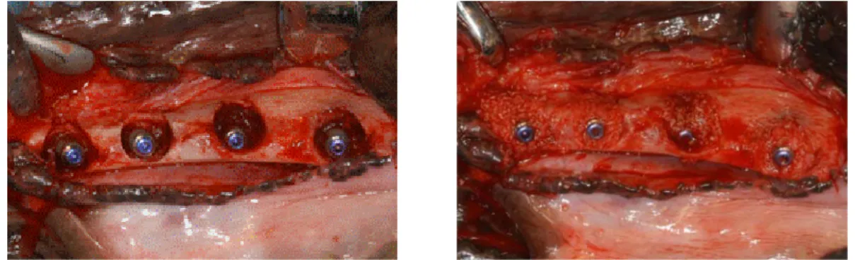
crestal incision on the each side of the man- dible, buccal and lingual full-thickness muco- periosteal flaps were elevated. Traditional im- plant site preparation was performed in four sites of each side of mandible. In order to make the experimental defect, a 7.5 mm diam- eter trephine bur was used and the depth of defect was 5.0 mm(figure 2). The harvested bone during making bone defects is used as autografts. Following the installation of the implant (Osstem, Korea, GS Ⅱ: diameter = 3.5 mm; length = 15 mm), a circumferential gap occurred between the bone and implants that was 5 mm deep and 2 mm wide. The defects were filled with Biocera and autogenous bone(figure 1).
The mucoperiosteal flaps were repositioned and sutured using a Vicryl(Ethicon; Somerville, NJ, USA) 4-0 suture material with continuous locking suture technique. The same surgical procedures were used for the other dog. From the day of surgery until the day the dogs were sacrificed, dogs fed on soft diets and plaque control was maintained by topical application on teeth and surrounding gingivae, twice a week, of 0.2% chlorhexidine digluconate solution.
Figure 1. The location of the autograft sites and xenograft sites.
Figure 2. (Left) Surface porosity of Ca-P nano crystal coated on BioceraⓇ, (Right) Defect preparation (Diameter : 7.5 mm, Depth : 5 mm)
Figure 3. (Left) Four identical 3.5mm-diameter titanium implants with 15mm length were placed into the defect sites. (Right) The each gap was filled with autogenous particulate bone or BioceraⓇ
2. Histologic examination
Two dogs were sacrified in postoperative 4 and 8 weeks. The mandibles were removed and placed in the 10% neutral buffered formalin.
The implant site was dissected into blocks. The tissue blocks were rinsed with water, dehy- drated in a graded series of increasing ethanol concentrations and embedded in super low vis- cosity embedding media(Polyscience Inc.
Warrinton, PA, USA). Each block was sectioned mesiodistally through the center of the implant using Exakt cutting-grinidng system(Exakt Appreateb, Hamburg, Germany). The sections, 50 ㎛ thick, were stained in hematoxylin and eosin(H&E). Each stained specimen was eval-
uated under a light microscope at varying magnifications. After initial evaluation, each stained section was magnified and photo- graphed using the KAPPA Image Base(KAPPA opto-electronics, Gottingen, Germany). The de- gree of bone-to-implant contact(BIC percent- age) and the bone density were measured.
∙ BIC : The length of implant in contact with the bone / Total length of implant × 100
∙ Bone density : The area of the part where the bone was formed within implant thread / The area between a thread and a thread × 100
3. Statistical Analysis
Mixed model analysis was carried out for a
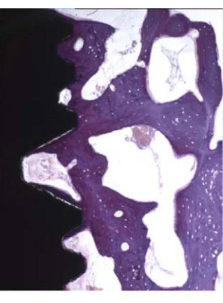
Figure 4. Ground section (mesio-distal plane) of xenograft sites after 4 weeks(left) and 8 weeks(right) of healing. Note the dense layer of mainly lamellar bone that occupies the marginal portion of the implant site. Magnification × 10
Figure 5. Magnification(×40) of the 8 week healing shown in Figure 4. A thin layer of appa- rently newly formed bone was found to be in direct contact with the implant surface. A re- versal line is observed between a newly formed bone and an older bone tissue.
test of significance in terms of bone-implant contact and bone density by materials and by time. Materials and time were considered as fixed effects, and location and repeated meas- urement of specimens according to time were considered as random effects. As a result, it was found that any factor was not significant at the 0.05 level of significance. Because nor- mality of bone-implant contact and bone den- sity, which are dependent variables, is pre- supposed in a mixed model, normality was tested using Kolmogorov- Smirnov test. As a result, it was found that both bone-implant contact and bone density were not significant at the 0.05 level of significance(p>0.05).
III. RESULTS
1. Clinical evaluation
All surgical sites showed uneventful healing and all implants were covered with newly
growing bone. There were no clinical signs of inflammation in the mucosa in all experimental sites. Clinically, it was impossible to dis- tinguish xenograft sites from autograft sites.
There were more bone covering of 8 week specimens than that of 4 week specimens.
2. Histological evaluation
1) Healing after 4 weeks
Although newly formed bone around implant was observed in the Biocera (test) sites, the appearance of the structure was more sparse than that of the autogenous bone graft(control) sites. The BIC of Biocera (test) sites was less than that of autogenous bone graft(control) sites. On the autogenous bone graft(control) sites more matured and lamellated bone was observed than that of the Biocera (test) sites.
The tissue in the zone next to the implant appeared to be undergoing a process of remodeling. This was illustrated by the large
Fig. 4. Fig. 4. Fig. 5.
Figure 6. Ground sections of xenograft sites. Magnification×100. Individual particles of Biocera® were embedded in lamellar bone.
Figure 7. Ground section (mesio-distal plane) of autograft sites after 4 weeks(left) and 8 weeks(right) of healing. The newly formed bone appeared to have properly filled the mar- ginal defect. Magnification×10
number of secondary osteons present in the tissue immediately lateral to the implant surface. Also, in more lateral areas, there were marked signs of remodeling and lamellar bone formation. The non-mineralized tissue included large numbers of adipocytes and vascular structures.
2) Healing after 8 weeks.
The bone density of 8 week specimens was increased than that of the 4 week specimens in
both groups. The bone tissue formed during the healing appeared to have properly filled the surgically prepared marginal defect. The bone tissue in the defect region was comprised of a mixture of lamellar bone and woven bone. A comparatively large portion of the implant sur- face was in direct contact with bone after 8 weeks of healing. A higher magnification view of the tissue is presented in Figure 5. The tis- sue in this area exhibited obvious signs of remodeling. A thin layer of apparently newly
material Time(weeks) BIC(%) Bone density(%)
autogenous 4 28.2 ± 19.1 39.7 ± 21.0
8 45.0 ± 9.8 41.7 ± 11.1
xenograft 4 34.6 ± 27.5 32.7 ± 25.4
8 27.6 ± 23.1 37.4 ± 17.6
* Mean values and standard deviations ± SD are shown.
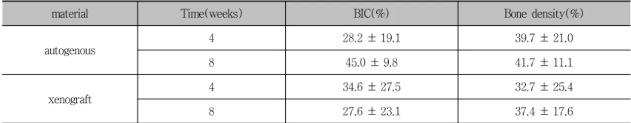
Table 1. Results of Bone-to-implant contact percentage (BIC %) and bone density per materials and times.
formed bone was found to be in direct contact with the implant surface. Lateral to this layer, large areas of woven bone were seen to be continuous with old bone tissue. In the non-mineralized tissue included large numbers of adipocytes and vascular structures.
3. Histomorphometric analysis.
On the autogenous bone graft(control) sites the average of BIC was 28.2±19 % and 44.9±9
% at 4 and 8 weeks respectively. On the Biocera (test) sites the average of BIC was 34.6±27 % at 4 weeks and 27.6±23 % at 8 weeks.
On the autogenous bone graft(control) sites the average of bone density was 39.7 ±21% and 41.7±11 % at 4 and 8 weeks respectively. On the Biocera (test) sites the average of bone density was 32.7±25 % at 4 weeks and 37.4±17
% at 8 weeks (Table 1).
There was more increased bone density of the 8 week specimens than that of the 4 week specimens in both group. There was no sig- nificant difference between autogenous bone graft(control) group and Biocera (test) group.
These results were not statistically significant (p>0.05).
IV. DISCUSSION
The findings of the present experiment re- vealed that 2 mm wide marginal defect, present at the time of implant installation, after 4 weeks and 8 weeks of healing had been filled with newly formed bone. It was also observed that the degree of bone-to-implant contact of both sites was similar. Although clinically complete bone fill was observed at 4 week, the histological examination showed that the bone fill was not complete. In this model, the gap at the time of implant placement had a negative effect on bone-to-implant contact, confirming the findings by Carlsson et al6), who indicated that histologically, as the initial gap increases, the amount of bone-to-implant contact diminishes.
Botticelli et al17). reported that the BIC was 74.1±4.2 % in 1.24 mm wide and 5.0 mm depth defect after 4 months healing period. According to his report, the BIC was 68.1±9.7 % after 4 months healing period in the marginal gap sizes of 1-2.25 mm. Botticelli et al17). showed that such hard tissue bridging is a time-dependent phenomenon. Thus, using the dog model it was demonstrated that healing periods of 1 and 2 months were not long enough to allow hard tissue to form on the surface of the implant in the defect region. In other words, the reso-
lution of defects adjacent to implants seems to be dependent both on defect size and time of healing. Hence, it is possible that the four re- maining defects in the present sample that were not filled with bone – after 4 months – may also have been resolved if the healing pe- riod had been extended.
The present study shows relatively less BIC percentage than above mentioned studies. The reason may partially be short healing period than previous studies. Other possible con- tributing factors are inadequate oral hygiene and improper animal management. In order to obtain more predictable results, careful surgical techniques and meticulous post-surgical care must be required. Also complete initial stability could not be achieved in some fixtures after installation.
From the similar result concerning bone fill in both groups, the present study suggest that Biocera can be used to overcome the bony de- fect around implant instead of autogenous par- ticulate bone graft. The finding that localized marginal bone defects after immediate implant installation may heal without the use of space maintaining barrier membranes or filler mate- rial confirms findings made in previous studies in man18),19),33).
Botticelli et al13). reported from experiments in dogs that mechanically produced defects of varying dimension(1.25~2.25 mm in width and 5 mm in depth) in the marginal portion of im- plant sites following 4 months of healing were consistently filled with newly formed bone.
The clinical protocol used in the present clinical trial called for re-entry after 4 months of healing. This decision was based on findings made in experiments7),17). It was reported that
hard-tissue formation in marginal defects that were ≥1.25 mm wide was complete after 4 months of healing. It may be argued that soft- and hard-tissue healing occurs faster in dogs than in man. The present results, however, documented that also defects of larger di- mensions could be resolved without the use of membrane.
Based on the findings made in the current experiment and in the studies referred to, it can be argued that it may not be the size of the marginal gap per se but rather the for- mation of a coagulum in the defect, its re- tention and replacement with a provisional ma- trix that determine whether defect resolution will occur. This hypothesis is supported by findings presented by Scipioni et al20). They used the so-called edentulous ridge expansion technique in a dog experiment and demon- strated that defects larger than 5mm could be entirely resolved21). Further, it was recently demonstrated that defects(sockets) of com- paratively large dimensions that occurred fol- lowing extraction of premolars in dogs within a 1-month period were filled with newly formed bone22).
Bone grafting materials have widely been utilized in bone augmentation procedures.
These materials include autogenic human bone, demineralized freeze-dried human bone, and xenogenic bone substitutes like natural and synthetic hydroxyapatite, deproteinized bovine bone mineral, and calcium phosphate compounds.
Among these materials, deproteinized bovine bone mineral(DBBM) has been shown to exhibit especially favorable properties. Animal and clinical human research have demonstrated DBBM to be biocompatible and to promote
growth of bone into its natural cavities23)-28). Recent studies have evaluated a deproteinized bovine bone mineral as a filler in a GBR proce- dure model on the rabbit skull29),30). In combi- nation with a stiff bioresorbable membrane made of polylactic acid, DBBM improved the amount of initial soft tissue formation and in- creased the rate of mineralized bone formation compared to blood-filled control sites.
Biocera (Oscotec, Cheonan, Korea) is a bone substitute coated with biocompatible cal- cium-phosphate(Ca-P) nano-crystal thin film.
The Ca-P has negative charges and thus at- tracts growth factors(PDGF, TGF-β, etc.) from body fluids and differentiates mesenchymal cells into osteoblasts to induce new bone for- mation16). There was no significant difference between autogenous bone graft group and Biocera group. Biocera has a bone-forming ability just as good as that of autogenous bone.
V. CONCLUSION
1. The marginal gap that occurred between the titanium implant fixture and the bone tissue following implant installation may predict- ably heal with new bone and defect resolution.
2. The mean values of BIC and bone density showed no significant difference between in autogenous bone graft(control) sites and Biocera (test) sites.(p>0.05)
3. There was no significant difference in BIC and bone density by materials or by time at the 0.05 level of significance.(p>0.05)
4. Histological studies showed that new bone formation occurred around implants and a similar pattern for healing was observed between the two groups.
VI. REFERENCES
1. Lazzara RJ. Immediate implant placement into extraction sites: surgical and re- storative advantages. Int J Periodontics Restorative Dent 1989;9:332-343.
2. Gher ME, Quintero G, Sandifer JB, et al.
Combined dental implant and guided tissue regeneration therapy in humans. Int J Periodontics Restorative Dent 1994;14:
332-347.
3. Becker W, Becker BE. Guided tissue re- generation for implants placed into ex- traction sockets and for implant de- hiscences: surgical techniques and case report. Int J Periodontics Restorative Dent 1990;10:376-391.
4. Attard NJ, Zarb GA. Immediate and early implant loading protocols: a literature re- view of clinical studies. J Prosthet Dent 2005;94:242-258.
5. Nemcovsky CE, Artzi Z, Moses O. Rotated split palatal flap for soft tissue primary coverage over extraction sites with imme- diate implant placement. Description of the surgical procedure and clinical results. J Periodontol 1999;70:926-934.
6. Carlsson L, Rostlund T, Albrektsson B, et al. Implant fixation improved by close fit.
Cylindrical implant-bone interface studied in rabbits. Acta Orthop Scand 1988;59:272 -275.
7. Botticelli D, Berglundh T, Buser D, et al.
The jumping distance revisited: An ex- perimental study in the dog. Clin Oral Implants Res 2003;14:35-42.
8. Cochran DL, Douglas HB. Augmentation of osseous tissue around nonsubmerged endo- sseous dental implants. Int J Periodontics
Restorative Dent 1993;13:506-519.
9. Bragger U, Hammerle CH, Lang NP.
Immediate transmucosal implants using the principle of guided tissue regeneration (II).
A cross-sectional study comparing the clinical outcome 1 year after immediate to standard implant placement. Clin Oral Implants Res 1996;7:268-276.
10. Hammerle CH, Bragger U, Schmid B, et al.
Successful bone formation at immediate transmucosal implants: a clinical report.
Int J Oral Maxillofac Implants 1998;13:
522-530.
11. Cornelini R, Cangini F, Covani U, et al.
Immediate restoration of implants placed into fresh extraction sockets for sin- gle-tooth replacement: a prospective clin- ical study. Int J Periodontics Restorative Dent 2005;25:439-447.
12. Lang NP, Bragger U, Hammerle CH, et al.
Immediate transmucosal implants using the principle of guided tissue regeneration. I.
Rationale, clinical procedures and 30- month results. Clin Oral Implants Res 1994;
5:154-163.
13. Scoop IW, Morgan FH, Dooner JJ, et al.
Bovine bone(Boplant) implants for in- trabony lesion(Clinical trials in humans).
Periodontics 1966;4:169-178.
14. Thaller SR, Hoyt J, Borjeson K, et al.
Reconstruction of calvarial defects with anorganic bovine bone mineral (Bio-oss ) in a rabbit model. J Craniofac Surg 1993;4:79-84.
15. Benke D, Olah A, Mohler H. Protein-chem- ical analysis of Bio-oss bone substitute and evidence on its carbonate content.
Biomaterials 2001;22:1005-1012
16. 성선주, 정현주, 박흥주 외. 백서 두개골 결손부
에서 Ca-P 피복된 이종골의 골재생 효과. 대한 치주과학회지 2004;34:475-487.
17. Botticelli D, Berglundh T, Buser D, et al.
Appositional bone formation in marginal defects at implants. Clin Oral Implants Res 2003;14:1-9.
18. Covani U, Cornelini R, Barone A.
Bucco-lingual bone remodeling around im- plants placed into immediate extraction sockets: a case series. J Periodontol 2003;
74:268-273.
19. Fiorellini JP, Engebretson SP, Donath K, et al. Guided bone regeneration utilizing ex- panded polytetrafluoroethylene membranes in combination with submerged and non- submerged dental implants in beagle dogs.
J Periodontol 1998;69:528-535.
20. Scipioni A, Bruschi GB, Giargia M, et al.
Healing at implants with and without pri- mary bone contact. An experimental study in dogs. Clin Oral Implants Res 1997;8:
39-47.
21. Scipioni A, Bruschi GB, Calesini G. The edentulous ridge expansion technique: a five-year study. Int J Periodontics Restorative Dent 1994;14:451-459.
22. Cardaropoli G, Araujo M, Lindhe J.
Dynamics of bone tissue formation in tooth extraction sites. An experimental study in dogs. J Clin Periodontol 2003;30: 809-818.
23. Berglundh T, Lindhe J. Healing around im- plants placed in bone defects treated with Bio-Oss. An experimental study in the dog.
Clin Oral Implants Res 1997;8:117-124.
24. Fukuta K, Har-Shai Y, Collares MV, et al.
Comparison of inorganic bovine bone min- eral particles with porous hydroxyapatite granules and cranial bone dust in the re- construction of full-thickness skull defect.
J Craniofac Surg 1992;3:25-29.
25. Klinge B, Alberius P, Isaksson S, et al.
Osseous response to implanted natural bone mineral and synthetic hydroxylapatite ce- ramic in the repair of experimental skull bone defects. J Oral Maxillofac Surg 1992;50:241-249.
26. Schlickewei W, Riede UN, Yu J, et al.
Influence of humate on calcium hydrox- yapatite implants. Arch Orthop Trauma Surg 1993;112:275-279.
27. Thaller SR, Hoyt J, Borjeson K, et al.
Reconstruction of calvarial defects with anorganic bovine bone mineral (Bio-Oss) in a rabbit model. J Craniofac Surg 1993;4:
79-84.
28. Wetzel AC, Stich H, Caffesse RG. Bone ap- position onto oral implants in the sinus area filled with different grafting materials. A histological study in beagle dogs. Clin Oral Implants Res 1995;6:
155-163.
29. Hammerle CH, Olah AJ, Schmid J, et al.
The biological effect of natural bone mineral on bone neoformation on the rabbit skull.
Clin Oral Implants Res 1997;8:198-207.
30. Schmid J, Wallkamm B, Hammerle CH, et al. The significance of angiogenesis in guided bone regeneration. A case report of a rabbit experiment. Clin Oral Implants Res 1997;8:244-248.
31. Schenk R, Willenegger H. Zur Histologie der primaren Knochenheilung. Modifikationen une Grenzen der spaltheilung in Abhangigkeit von der Defktgrosse. Unfallheilkunde.
1977;80:155-160.
32. Schenk RK. Bone regeneration: biologic basis. In: Guided Bone Regeneration in Implant Dentistry, eds. Buser D, Dahlin C, Schenk RK. pp. 49-100. Berlin:
Quintessence Book. 1994.
33. Paolantonio M, Dolci M, Scarano A, et al.
Immediate implantation in fresh extraction sockets. A controlled clinical and histo- logical study in man. J Periodontol 2001;
72:1560-1571.
개 모델에서의 임플란트 주위 골결손시 Ca-P 표면 처리된 이종골의 효과
조수연
1, 전혜란
2, 이선경
1,2, 이승호
1,2, 이준영
2, 한근아
21. 이화여자대학교 임상치의학대학원 치주과학교실 2. 이화여자대학교 부속 목동병원 치주과
목적 :
최근 발치 후 즉시 임플란트 식립은 널리 사용되는 수술 방식이다. 이 연구의 목적은 임플란트 주위 골결손 시 Ca-P으로 표면 처리된 이종골을 사용하여 골재생을 평가하기 위함이다.
재료와 방법 :
두 마리의 개 모델에서 하악 소구치와 제일 대구치를 발치하였다. 발치 6주 후 trephine bur를 이용하여 7.5 mm 지름과 5 mm 깊이를 가진 결손부를 형성하였다. 이 후 이 결손부의 중앙에 3.5 mm 지름과 15mm 길 이의 fixture(GS Ⅱ)를 식립하였다. 결과적으로 임플란트와 주변을 둘러싸고 있는 골 사이에는 2.0 mm정도의 gap이 만들어진다. 준비된 결손부 내로 자가골 또는 Biocera 를 채웠다. 각각 4주, 8주 후 조직 절편을 제작 하였다. 조직학적 평가를 위해 Block biopsy를 시행하였다.
결과 :
두 집단 모두 임상적으로 골이 완전히 채워졌다. 자가골이 이식된 부위(control)의 평균 골-임플란트 접촉 (BIC)은 각각 4주째 28.2±19%였고, 8주째 44.9±9%였다. Biocera 가 이식된 부위(test)의 평균 BIC는 각각 4주째 34.6±27%였고, 8주째 27.6±23%였다.
자가골이 이식된 부위(control)의 평균 골밀도는 각각 4주째 39.7±21%, 8주째 41.7±11%였다. Biocera 가 이식된 부위(test)의 평균 골밀도는 각각 4주째 32.7±25%, 8주째 37.4±17% 였다.
골-임플란트 접촉(BIC)과 골밀도의 평균 비율(%)은 비슷하였다.
조직학적으로 자가골과 이종골 이식 부위 모두 주변골과 잘 조화를 이루었고 유사한 치유 양상이 관찰되었 다. 자가골과 이종골 이식 부위간 유의한 차이는 없었다.(P>0.05)
결론 :
임플란트 주위 2 mm의 골 결손부위에 자가골 또는 이종골로 채운 경우 유사한 결과를 얻었다.
이 결과는 임플란트 fixture 주위의 골 결손부 해소를 위해 자가골을 대체할 수 있는 재료로 Biocera 를 사 용할 수 있음을 보여준다. 2)
주요어 : 골재생, 임플란트, Biocera