241 http://dx.doi.org/10.4196/kjpp.2014.18.3.241
ABBREVIATIONS: IP3, inositol 1,4,5-triphophate; PKC, protein kinase C; PLC, phospholipase C; PUFA, polyunsaturated fatty acid;
ROCC, receptor operated Ca2+ channels; VDCC, voltage-operated Ca2+ channels.
Received February 19, 2014, Revised March 20, 2014, Accepted April 23, 2014
Corresponding to: Sang Soo Sim, College of Pharmacy, Chung-Ang University, 84, Heukseok-ro, Dongjak-gu, Seoul 156-756, Korea. (Tel) 82-2-820-5615, (Fax) 82-2-816-7338, (E-mail) [email protected]
This is an Open Access article distributed under the terms of the Creative Commons Attribution Non-Commercial License (http://
creativecommons.org/licenses/by-nc/3.0) which permits unrestricted non-commercial use, distribution, and reproduction in any medium, provided the original work is properly cited.
Effects of C18 Fatty Acids on Intracellular Ca
2+Mobilization and Histamine Release in RBL-2H3 Cells
Myung Chul Kim, Min Gyu Kim, Young Soo Jo, Ho Sun Song, Tae In Eom, and Sang Soo Sim College of Pharmacy, Chung-Ang University, Seoul 156-756, Korea
To investigate the underlying mechanisms of C18 fatty acids (stearic acid, oleic acid, linoleic acid and α -linolenic acid) on mast cells, we measured the effect of C18 fatty acids on intracellular Ca2+
mobilization and histamine release in RBL-2H3 mast cells. Stearic acid rapidly increased initial peak of intracellular Ca2+ mobilization, whereas linoleic acid and α -linolenic acid gradually increased this mobilization. In the absence of extracellular Ca2+, stearic acid (100 μ M) did not cause any increase of intracellular Ca2+ mobilization. Both linoleic acid and α -linolenic acid increased intracellular Ca2+
mobilization, but the increase was smaller than that in the presence of extracellular Ca2+. These results suggest that C18 fatty acid-induced intracellular Ca2+ mobilization is mainly dependent on extracellular Ca2+ influx. Verapamil dose-dependently inhibited stearic acid-induced intracellular Ca2+ mobilization, but did not affect both linoleic acid- and α-linolenic acid-induced intracellular Ca2+ mobilization. These data suggest that the underlying mechanism of stearic acid, linoleic acid and α -linolenic acid on intracellular Ca2+ mobilization may differ. Linoleic acid and α -linolenic acid significantly increased histamine release. Linoleic acid (C18:2: ω -6)-induced intracellular Ca2+ mobilization and histamine release were more prominent than α -linolenic acid (C18:3: ω -3). These data support the view that the intake of more α -linolenic acid than linoleic acid is useful in preventing inflammation.
Key Words: Ca2+ mobilization, C18 fatty acids, Histamine release, PLC assay
INTRODUCTION
Mast cells play a central role in the innate immune response. They are particularly important in helping to re- cruit other cells, such as basophils, neutrophils and lym- phocytes. Mast cells play an important role in a number of allergic diseases including atopic dermatitis and asthma, and contribute to chronic inflammatory conditions like atherosclerosis, vasculitis and rheumatoid arthritis. Mast cells have a number of secretory granules including hista- mine and 5-hydroxytryptamine, and rapidly synthesize lip- id-derived mediators, such as prostaglandins and leuko- trienes from arachidonic acid (C20:4: ω-6) released by the activation of phospholipase A2 [1]. The addition of arach- idonic acid to RBL-2H3 cells distinctly alter the fatty acid composition of the cell membrane and enhance Fcε RI-mediated degranulation and tumor necrosis factor-alpha (TNF-α) release with augmented tyrosine phosphorylation of some proteins and Ca2+ influx [2]. We previously reported
that exogenous arachidonic acid significantly increases his- tamine release from RBL-2H3 cells [3].
On the other hand, ω-3 polyunsaturated fatty acids (PUFAs) reportedly decrease the production of arachidonic acid-derived eicosanoids. These fatty acids decrease the production of inflammatory cytokines, such as TNF, inter- leukin (IL)-1, and IL-6, and the expression of adhesion mol- ecules involved in inflammatory interactions between leu- kocytes and endothelial cells [4]. Babcock et al. reported that ω-3 PUFAs, such as eicosapentaenoic acid and docosa- hexaenoic acid, significantly inhibit the production of TNF- α and IL-10 in monocytes [5]. The anti-inflammation ef- fects of ω-3 eicosapentaenoic acid and the inflammation re- actions of ω-6 arachidonic acid have been studied largely, but α-linolenic acid (a precursor of eicosapentaenoic acid) and linoleic acid (a precursor of arachidonic acid) had been not. In this study, to investigate the underlying mecha- nisms of C18 fatty acids on anti-inflammatory response, we measured the effect of C18 fatty acids such as stearic acid (C18, saturated fatty acid), oleic acid (C18:1: ω-9), linoleic acid (C18:2: ω-6) and α-linolenic acid (C18:3: ω-3) on in- tracellular Ca2+ mobilization and histamine release in RBL-2H3 mast cells.
chased from BIO-RAD (Hercules, CA, USA). The materials for cell culture were purchased from Life Technologies (Grand Island, NY, USA). Unless otherwise stated, all re- agents were of the highest purity and were purchased from Sigma Chemical.
Cell culture
Rat basophilic leukemia (RBL)-2H3 cells were obtained from the American Type Culture Collection (ATCC, Manas- sas, VA, USA) and cultured as recommended by ATCC at 37°C in Dulbecco’s modified Eagle’s medium (DMEM) sup- plemented with 10% heat-inactivated fetal bovine serum (FBS) and antibiotics (100 U/mL penicillin G, 100 μg/mL streptomycin) in a 5% CO2 incubator. Cells between 5 and 20 passages were used for experiments at a density of 1
×106 cells/mL.
Measurement of cell viability
Cell viability testing was performed using a MTT-based colorimetric assay [6]. Cells were diluted and cultured in 96-well plates (1×10⁴ cells/mL), with the select C18 fatty acid added (100∼200 μM). An equal amount of ethanol was added in the control group, which resulted in a final concentration of 0.1% in the culture medium. MTT was add- ed to the medium to a final concentration of 200 μg/mL and incubated for another 4 h at 37oC. After removing the culture medium, 200 μL of dimethylsulfoxide was added to the cells to dissolve the formazan. The absorbance of each well was measured by using a FlexStation device (Molecular Devices, Sunnyvale, CA, USA) at wavelength of 520 nm [7].
Measurement of intracellular Ca2+ mobilization The intracellular Ca2+ level was measured using Fura- 2/AM by monitoring a fluorospectrometer [8]. Briefly, cul- ture medium was replaced and cells were washed three times with phosphate buffered saline (PBS). Cells were de- tached using 0.5% trypsin/EDTA buffer and suspended in 10 mL Krebs buffer [120.8 mM NaCl, 4.5 mM KCl, 1.2 mM MgCl2, 1.8 mM CaCl2, 5.6 mM glucose, 1.2 mM NaH2PO4
and 25 mM HEPES (pH 7.4)]. Fura-2/AM was added to a final concentration of 2 μM and incubated at 37oC for 1 h. The cells were washed twice with Krebs buffer and cen- trifuged at 3,000×g for 10 min. For the experiments per- formed in the absence of external calcium, Krebs-EGTA buffer [120.8 mM NaCl, 4.5 mM KCl, 1.2 mM MgCl2, 1.0 mM EGTA, 5.6 mM glucose, 1.2 mM NaH2PO4 and 25 mM HEPES (pH 7.4)] was used. Fura-2 fluorescence was moni- tored by a Quanta Master device (Photon Technology Inter-
serum albumin (1 mg/mL) and then resuspended at a den- sity of 2×10⁷ cells/mL. A portion (1 mL) of the cell suspen- sion was transferred to a microcentrifuge tube and in- cubated at 37°C for 15 min. Phosphatidylinositol 4,5-bi- sphosphate (PIP2) hydrolysis was initiated by adding 100 μM fatty acids for 30 min. Reactions were terminated by adding 200 μL of ice-cold 10% perchloric acid (HClO4).
After 30 min in an ice bath, the tubes were centrifuged and the supernatants were diluted 5-fold with distilled water and applied to Dowex AG 1-X8 anion exchange columns.
Each column was washed with 2 mL of distilled water fol- lowed by 10 mL of 60 mM ammonium formate containing 5 mM sodium tetraborate. Total inositol phosphates were eluted with a solution containing 1 M ammonium formate and 0.1 M formic acid. The radioactivity of the [3H]inositol phosphates was determined using a Tri-Carb Liquid Scin- tillation Counter (PerkinElmer, Waltham, MA, USA) [11].
Histamine release assay
The method that the determination of histamine is based on the reaction of histamine with OPT, which results in a highly fluorescent condensation product [12]. The har- vested RBL-2H3 cells were washed with Krebs buffer and suspended in Krebs buffer at a density of 106 cells/mL. The cells were treated with BIM for 10 min and histamine re- lease was induced by C18 fatty acids for 30 min in 37oC.
After centrifugation, histamine contents in both super- natant and pellet were measured with 0.1 mL of 1% OPT in methanol. After 4 min, the reaction was terminated by adding 0.2 mL of 3 N HCl. The fluorescence intensity was measured using excitation and emission wavelengths of 355 and 455 nm, respectively, with a FlexStation apparatus.
Data are expressed as % release (histamine contents in su- pernatant / histamine contents in supernatant and pellet
×100).
Statistical analyses
Results are presented as mean±S.D. and significance was determined by analysis of variance (ANOVA) and differ- ences between groups were determined with a Newman- Keul’s test. The level of significance was set at less than 5% (p<0.05).
RESULTS
Effects of C18 fatty acids on the viability of RBL 2H3 cells
To confirm cytotoxicity of C18 fatty acids, cell viability
Fig. 1. Cytotoxicity of C18 fatty acids in RBL-2H3 cells. Cells were incubated with C18 fatty acids (A: stearic acid, B: oleic acid, C: linoleic acid, D: α-linolenic acid) for 24 h in a 5% CO2 incubator at 37oC. The number of living cells was measured using MTT. Results are mean±S.D.
from four separate experiments.
was determined by the ability of the cells to metabolically reduce MTT to a formazan dye. It was performed after 6, 12 and 24 h exposure to the C18 fatty acid stearic acid, oleic acid, linoleic acid or α-linolenic acid. After 24 h ex- posure to oleic acid, linoleic acid and α-linolenic acid, which contain a double bond, no cytotoxicity was evident at a concentration of 200 μM. In contrast, stearic acid, which lacks a double bond, reduced the cell viability by 16%
at 200 μM (Fig. 1). However, stearic acid showed no cyto- toxicity at 6 h.
C18 fatty acid-induced intracellular Ca2+ mobilization To investigate the effects of C18 fatty acids on intra- cellular Ca2+ concentration, we measured Ca2+ mobilization in Fura-2/AM-loaded RBL-2H3 cells. As shown in Fig. 2A, stearic acid rapidly increased the initial peak of intra- cellular Ca2+ mobilization, whereas linoleic acid and α-lino- lenic acid gradually increased intracellular Ca2+ mobili- zation. Oleic acid (100 μM) slightly increased intracellular Ca2+ concentration, which was smaller than the effects of other C18 fatty acids (Fig. 2A). In the next experiment, we
excluded oleic acid. Stearic acid, linoleic acid and α -linolenic acid dose-dependently increased intracellular Ca2+ mobilization (Figs. 2B-2D). In the absence of ex- tracellular Ca2+, stearic acid (100 μM) did not cause any increase of intracellular Ca2+ mobilization, but both linoleic acid and α-linolenic acid increased intracellular Ca2+ mobi- lization; the increase was smaller than that in the presence of extracellular Ca2+ (Fig. 3). These data suggest that C18 fatty acid-induced intracellular Ca2+ mobilization is mainly dependent on extracellular calcium influx.
Effects of calcium antagonists and BIM on C18 fatty acid-induced intracellular Ca2+ mobilization
Verapamil (10 μM), a voltage-dependent calcium channel blocker, dose-dependently inhibited stearic acid-induced intracellular Ca2+ mobilization (Figs. 4A and 5), but did not affect linoleic acid- and α-linolenic acid- induced intracellular Ca2+ mobilization (Figs. 4B and 4C).
At 10 μM TMB-8, a intracellular calcium release blocker, did not influence intracellular Ca2+ mobilization induced by stearic acid, linoleic acid and α-linolenic acid (data not
Fig. 2. C18 fatty acid-induced intracellular Ca2+ mobilization. Intracellular Ca2+ mobilization was measured in Fura-2/AM-loaded RBL-2H3 cells. Intracellular Ca2+ concentration induced by a 100-sec exposure to the C18 fatty acids [A: C18 fatty acids, B: stearic acid (SA), C:
linoleic acid (LA), D: α-linolenic acid (LN)] was expressed as the ratio of F340/F380. Results are the representative data of four separate experiments.
Fig. 3. C18 fatty acids-induced intracellular Ca2+ mobilization in the presence or absence of extracellular Ca2+. Intracellular Ca2+ concentration was induced by a 100-sec exposure to the C18 fatty acids [A: stearic acid (SA, 100 μM), B: linoleic acid (LA, 100 μM), C: α-linolenic acid (LN, 100 μM)]. Results are the representative data of four separate experiments.
Fig. 4. Effect of verapamil on intracellular Ca2+ mobilization induced by C18 fatty acids. After a 50-sec treatment with 10 μM verapamil (VP), intracellular Ca2+ concentration was induced by a 100-sec exposure to the C18 fatty acids [A: stearic acid (SA), B: linoleic acid (LA), C: α-linolenic acid (LN)]. Results are the representative data of four separate experiments.
Fig. 5. (A) Dose-response of verapamil (VP) to intracellular Ca2+ mobilization and (B) peak ratio induced by stearic acid (SA, 100 μM).
Results are the representative data of four separate experiments. *Significantly different from control (p<0.05).
Fig. 6. Effect of bisindolylmaleimide (BIM) on intracellular Ca2+ mobilization induced by a 100-sec exposure to C18 fatty acids [A: stearic acid (SA), B: linoleic acid (LA), C: α-linolenic acid (LN)]. Results are the representative data of four separate experiments.
Concentration (μM)
[3H]Inositol phosphates (dpm/mg protein)
Control 0 278±20
Stearic acid 100 293±24
Linoleic acid 100 284±19
α-Linolenic acid Melittin
100 10
296±27 682±48*
Results are mean±S.D. from four separate experiments.
*Significantly different from control (p<0.05).
Table 1. Production of [3H]inositol phosphates by C18 fatty acids
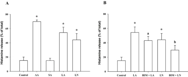
Fig. 7. C18 Fatty acid-induced histamine release in RBL-2H3 cells. (A) Histamine release induced by arachidonic acid (AA, 100 μM), stearic acid (SA, 100 μM), linoleic acid (LA, 100 μM) and α-linolenic acid (LN, 100 μM). (B) Effect of 10 μM bisindolylmaleimide (BIM) on histamine release induced by linoleic acid (LA, 100 μM) and α-linolenic acid (LN, 100 μM). *Significantly different from Control (p<0.05); aSignificantly different from LA (p<0.05); bSignificantly different from LN (p<0.05).
er, three C18 fatty acids did not increase [H]inositol phos- phate formation (Table 1), indicating that C18 fatty acids-induced intracellular Ca2+ mobilization is not asso- ciated with intracellular Ca2+ release by the PLC pathway.
Effects of C18 fatty acids on histamine release in RBL-2H3 cells
At 100 μM, linoleic acid and α-linolenic acid signifi- cantly increased histamine release from 15.2±4.8% (control) to 54.3±7.7% and 44.2±8.8%, respectively, which was small- er than the effect of 100 μM arachidonic acid (69.7±2.4%).
However, stearic acid, which lacks a double bond, did not
exposed to the cells for 1 h. This exposure was not toxic, since no cytotoxicity was apparent after 6 h exposure of the tested fatty acids. These data suggest that exocytosis of histamine may be independent on cell membrane rupture. It has been reported that exposure of stearic acid at 150 μM for 24 h induces cell death and that ω-6 PUFAs have a greater protective effect on the deleterious effect caused by stearic acid in ECV-304 endothelial cells than ω-3 PUFAs [13].
Stearic acid rapidly increased the initial peak of intra- cellular Ca2+ mobilization, whereas linoleic acid and α-lino- lenic acid gradually increased intracellular Ca2+ mobili- zation. In the absence of extracellular Ca2+, stearic acid (100 μM) did not cause any increase of intracellular Ca2+
mobilization. Both linoleic acid and α-linolenic acid in- creased intracellular Ca2+ mobilization, but the increase was smaller than that in the presence of extracellular Ca2+. These results suggest that C18 fatty acid-induced intra- cellular Ca2+ mobilization is mainly dependent on ex- tracellular calcium influx. A prior study reported that 40 μM of three ω-6 polyunsaturated fatty acids (arachidonic acid, γ-linolenic acid and linoleic acid) increased intra- cellular calcium concentration, while 40 μM ω-3 and ω-9 PUFAs did not [14]. The discrepancy between the present and previous study may be due to the concentration of C18 fatty acids because we used 100 μM of the C18 fatty acids.
At 10 μM, verapamil, a voltage-dependent calcium chan-
nel blocker [15], inhibited stearic acid-induced intracellular Ca2+ mobilization, but did not affect both linoleic acid and α-linolenic acid-induced intracellular Ca2+ mobilization.
The PKC inhibitor BIM used at 10 μM significantly in- hibited both linoleic acid and α-linolenic acid-induced in- tracellular Ca2+ mobilization, but it did not affect stearic acid-induced intracellular Ca2+ mobilization. These data suggest that the underlying mechanism of stearic acid, lino- leic acid and α-linolenic acid on intracellular Ca2+ mobi- lization may be different from each other. Also, C18 fatty acids-induced intracellular Ca2+ mobilization appeared not to be associated with intracellular Ca2+ release by the PLC pathway, since they failed to induce [3H]inositol phosphates formation in [3H]inositol-labeled cells. This finding is con- sistent with a previous report that 40 μM arachidonic acid did not result in the formation of inositol-1,4,5-triphosphates [14]. Long chain fatty acids and ω-3 PUFAs (EPA and DHA) have been demonstrated to act as ligands of several G-pro- tein-coupled receptors, such as GRP40 and GRP120, re- spectively [16,17]. Especially GRP40 activation which is strongly expressed in the pancreas results in an increase of intracellular Ca2+ mobilization via the phosphoinositide path- way. Further study will be needed to elucidate novel physio- logical roles of GRP40 and GRP120 in RBL-2H3 cells.
Clinical studies [18-20] indicated that the ingested ratio of ω-6 to ω-3 (especially linoleic vs. α-linolenic) fatty acids is important for maintaining cardiovascular health. While ω-3 PUFAs are extremely beneficial in preventing heart disease in humans, the levels of ω-6 PUFAs are insignificant [21,22]. Both ω-3 α-linolenic acid and ω-6 linoleic acid are essential fatty acids in humans. ω-3 and ω-6 C18 PUFAs com- pete for the same metabolic enzymes; thus, the ω-6: ω-3 ratio will significantly influence the ratio of the ensuing eicosanoids (hormones) including prostaglandins, leukotrienes and throm- boxanes, and will alter the body's metabolic function [23].
Especially, linoleic acid (C18:2: ω-6)-induced intracellular Ca2+
mobilization and histamine release are more prominent than α-linolenic acid (C18:3: ω-3). These data support the view that increased intake of α-linolenic acid compared to linoleic acid is useful in preventing inflammation.
ACKNOWLEDGEMENT
This work was supported by the Korea TEI Research Fund (2013).
REFERENCES
1. Marshall JS. Mast-cell responses to pathogens. Nat Rev Immunol. 2004;4:787-799.
2. Nakano N, Nakao A, Uchida T, Shirasaka N, Yoshizumi H, Okumura K, Tsuboi R, Ogawa H. Effects of arachidonic acid analogs on FcepsilonRI-mediated activation of mast cells.
Biochim Biophys Acta. 2005;1738:19-28.
3. Lee JH, Lee JY, Kang HS, Jeong CH, Moon H, Whang WK, Kim CJ, Sim SS. The effect of acteoside on histamine release and arachidonic acid release in RBL-2H3 mast cells. Arch Pharm Res. 2006;29:508-513.
4. Calder PC. Polyunsaturated fatty acids and inflammation.
Prostaglandins Leukot Essent Fatty Acids. 2006;75:197-202.
5. Babcock TA, Novak T, Ong E, Jho DH, Helton WS, Espat NJ.
Modulation of lipopolysaccharide-stimulated macrophage tumor necrosis factor-alpha production by omega-3 fatty acid
is associated with differential cyclooxygenase-2 protein expression and is independent of interleukin-10. J Surg Res.
2002;107:135-139.
6. Woerdenbag HJ, Merfort I, Passreiter CM, Schmidt TJ, Willuhn G, van Uden W, Pras N, Kampinga HH, Konings AW.
Cytotoxicity of flavonoids and sesquiterpene lactones from Arnica species against the GLC4 and the COLO 320 cell lines.
Planta Med. 1994;60:434-437.
7. Lee MJ, Song HJ, Jeong JY, Park SY, Sohn UD. Anti-Oxidative and Anti-Inflammatory Effects of QGC in Cultured Feline Esophageal Epithelial Cells. Korean J Physiol Pharmacol.
2013;17:81-87.
8. Tseng PH, Lin HP, Hu H, Wang C, Zhu MX, Chen CS. The canonical transient receptor potential 6 channel as a putative phosphatidylinositol 3,4,5-trisphosphate-sensitive calcium entry system. Biochemistry. 2004;43:11701-11708.
9. Grynkiewicz G, Poenie M, Tsien RY. A new generation of Ca2+
indicators with greatly improved fluorescence properties. J Biol Chem. 1985;260:3440-3450.
10. Hwang YH, Song HS, Kim HR, Ko MS, Jeong JM, Kim YH, Ryu JS, Sohn UD, Gimm YM, Myung SH, Sim SS. Intracellular Ca Mobilization and Beta-hexosaminidase Release Are Not Influenced by 60 Hz-electromagnetic Fields (EMF) in RBL 2H3 Cells. Korean J Physiol Pharmacol. 2011;15:313-317.
11. Bae YS, Lee TG, Park JC, Hur JH, Kim Y, Heo K, Kwak JY, Suh PG, Ryu SH. Identification of a compound that directly stimulates phospholipase C activity. Mol Pharmacol. 2003;63:
1043-1050.
12. Håkanson R, Rönnberg AL, Sjölund K. Fluorometric deter- mination of histamine with OPT: optimum reaction conditions and tests of identity. Anal Biochem. 1972;47:356-370.
13. Masi LN, Portioli-Sanches EP, Lima-Salgado TM, Curi R.
Toxicity of fatty acids on ECV-304 endothelial cells. Toxicol In Vitro. 2011;25:2140-2146.
14. Teshima R, Amano F, Nakamura R, Tanaka Y, Sawada J.
Effects of polyunsaturated fatty acids on calcium response and degranulation from RBL-2H3 cells. Int Immunopharmacol.
2007;7:205-210.
15. Baliño P, Pastor R, Aragon CM. Participation of L-type calcium channels in ethanol-induced behavioral stimulation and motor incoordination: effects of diltiazem and verapamil. Behav Brain Res. 2010;209:196-204.
16. Ichimura A, Hirasawa A, Hara T, Tsujimoto G. Free fatty acid receptors act as nutrient sensors to regulate energy home- ostasis. Prostaglandins Other Lipid Mediat. 2009;89:82-88.
17. Oh DY, Talukdar S, Bae EJ, Imamura T, Morinaga H, Fan W, Li P, Lu WJ, Watkins SM, Olefsky JM. GPR120 is an omega-3 fatty acid receptor mediating potent anti-inflam- matory and insulin-sensitizing effects. Cell. 2010;142:687-698.
18. Griffin BA. How relevant is the ratio of dietary n-6 to n-3 polyunsaturated fatty acids to cardiovascular disease risk?
Evidence from the OPTILIP study. Curr Opin Lipidol. 2008;
19:57-62.
19. Lands WE. Biochemistry and physiology of n-3 fatty acids.
FASEB J. 1992;6:2530-2536.
20. Okuyama H. High n-6 to n-3 ratio of dietary fatty acids rather than serum cholesterol as a major risk factor for coronary heart disease. Eur J Lipid Sci Technol. 2001;103:418-422.
21. Willett WC. The role of dietary n-6 fatty acids in the prevention of cardiovascular disease. J Cardiovasc Med (Hagerstown).
2007;8 Suppl 1:S42-45.
22. Mozaffarian D, Ascherio A, Hu FB, Stampfer MJ, Willett WC, Siscovick DS, Rimm EB. Interplay between different polyun- saturated fatty acids and risk of coronary heart disease in men.
Circulation. 2005;111:157-164.
23. Hooper L, Thompson RL, Harrison RA, Summerbell CD, Ness AR, Moore HJ, Worthington HV, Durrington PN, Higgins JP, Capps NE, Riemersma RA, Ebrahim SB, Davey Smith G. Risks and benefits of omega 3 fats for mortality, cardiovascular dise- ase, and cancer: systematic review. BMJ. 2006;332:752-760.


![Fig. 6. Effect of bisindolylmaleimide (BIM) on intracellular Ca 2+ mobilization induced by a 100-sec exposure to C18 fatty acids [A: stearic acid (SA), B: linoleic acid (LA), C: α-linolenic acid (LN)]](https://thumb-ap.123doks.com/thumbv2/123dokinfo/5470719.659968/5.892.82.810.827.1047/effect-bisindolylmaleimide-intracellular-mobilization-induced-exposure-linoleic-linolenic.webp)