Vol. 10, N o. 2, June, 2003
158
2003 1 7 , 2003 4 14
505
Tel 02) 590-2702, Fax 02) 537-4673, E-m ail rheum a@ cm c.cuk.ac.kr (R11-2002-098-01001-0) .
Objective: To investigate the ultrasonographic findings in knee OA patients and to examine the possible causes of pain in osteoarthritis by ultrasonography.
Methods: Ultrasonography was performed with 7.5 MHz linear probe in 64 knee OA patients who fulfilled the ACR criteria. All patients were graded according to the Kellgren-Lawrence grades and then classified into group 1 (K/L I and II) and Group 2 (K/L III and IV). Also WOMAC score, BMI, laboratory finding (ESR, CRP) were checked. Ultrasonographic findings was examined; effusion, thickening of synovium, vertical length of medial and lateral osteophyte (longitudinal view), length of capsular distension (medial longitudinal view), evidence of bursitis and articular cartilage.
159
Results: 50.0% of patients had effusion, among whom 68.7% patients also had synovial thickening. In all patients, the severity of pain was correlated with 4 variables; the presence of effusion, disease duration, the length of medial osteophyte, the length of capsular distension (r=0.279, r=0.415, r=0.537, r=0.608, respectively, p 0.05). The length of medial osteophyte, the degree of capsular distension and disease duration were significantly correlated with WOMAC pain score in Group 1 (p 0.05). After multiple regression analysis, the length of medial osteophyte alone had correlation with the pain severity in Group 1 (r2= 0.396 p 0.05) and the only length of capsular distension was significantly correlated with WOMAC pain score in Group 2 (r=0.609, p 0.05).
Conclusion: The length of osteophyte may be more related with pain severity in mild cases (K/L score I and II) while capsular distension could be an important factor causing knee pain in more advanced knee OA (K/L score III and IV).
Key Words: Knee osteoarthritis, Ultrasonography, Osteophyte, Joint capsule
160
― 강효종 외:Knee Osteoarthritis, Ultrasonography, Osteophyte, Joint Capsule ―
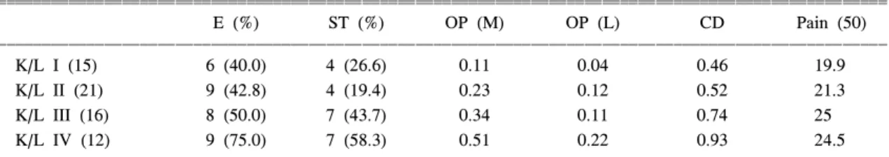
― 161 ― 령은 60.2±10.1세였으며 슬골관절염의 평균이환 기 간은 7.4±8.5년이었다. 단 한 명의 환자에서만 외측 구획 슬골관절염(lateral compartment knee OA)의 소 견을 보이고 있었으며 BMI는 25.1±6.3이었고, K/L grade에 따라 환자를 다시 분류한 결과 grade I은 15 명, II는 21명, III는 16명 그리고 grade IV는 12명이 었다(표 1).
2. 초음파 소견
전체 환자 가운데 삼출액을 보이는 환자는 32명 (50%)이었으며 활막 비후 소견을 보이는 예는 22명 (34.3%)이었다. 활막 비후 소견을 보이는 모든 예에 서 삼출액이 동반되었고 삼출액을 보이는 예 중에서 는 68.7%에서 활막 비후 소견을 보였다. 초음파 소 견 및 통증 정도(pain score)의 K/L grade에 따른 결 과는 표 1과 같다.
K/L grade가 높아짐에 따라 내측 골돌기의 길이와 신전된 관절낭의 길이가 각각 증가함을 확인할 수 있었다. Group 1 (K/L I, II)과 2 (K/L III, IV)의 두 그룹으로 나누어 비교해본 결과 두 그룹 간에 통증 과 내측 골돌기의 길이 그리고 신전된 관절낭 간에 유의한 차이를 보였다(그림 2).
관절연골의 두께를 초음파상 정확히 측정하기 어 려운 경우들이 많았는데 이는 골관절염이 진행되서 연골의 앞선(anterior margin)을 설정하기 어려운 경우, 연골이 균일하지 않고 불규칙한 두께를 보이는 경우, 부종으로 인한 슬관절의 굴곡제한 등 때문이었다.
그 외 소견으로 거위다리 주머니(anserine bursa)에 점액낭염 소견을 보이는 예는 한 예가 있었고 슬와 부 불편감을 호소하는 환자의 2명에서 슬와부 낭종 소견이 관찰되었다.
3. 통증과 상관관계
삼출액 유무와 관련하여 삼출액이 있는 경우가 통증 지수가 의미있게 높았지만 그 삼출액의 정도 와 통증 간에 통계학적인 연관성은 없었다. 활막 비후 여부와 통증 간에는 의미있는 관련성을 보이 지 않았다.
전체 환자를 대상으로 통증과 각 변수 간의 관련 성을 살펴보았는데(삼출액의 경우 삼출액이 없는 경 우 ‘0'으로, 있는 경우 ‘1'로 설정하여 상관관계분석 에 포함시켰음), 이환 기간과 삼출액, 내측골돌기의 길이 그리고 신전된 관절낭 길이가 상관관계를 나타 내었다(Pearson 상관계수: 각각 r=0.279, r=0.415, r=
0.537, r=0.608, p<0.05, 표 2).
K/L grade에 따라 두 그룹으로 나누어 통증에 관 련된 인자를 조사하였더니 Group 1(K/L I, II)에서는 Table 1. Ultrasonographic findings and pain severity according to the K/L grades
ꠧꠧꠧꠧꠧꠧꠧꠧꠧꠧꠧꠧꠧꠧꠧꠧꠧꠧꠧꠧꠧꠧꠧꠧꠧꠧꠧꠧꠧꠧꠧꠧꠧꠧꠧꠧꠧꠧꠧꠧꠧꠧꠧꠧꠧꠧꠧꠧꠧꠧꠧꠧꠧꠧꠧꠧꠧꠧꠧꠧꠧꠧꠧꠧꠧꠧꠧꠧꠧꠧꠧꠧꠧꠧꠧꠧꠧꠧꠧꠧꠧꠧꠧꠧꠧꠧꠧꠧꠧꠧꠧꠧꠧꠧꠧꠧꠧꠧꠧꠧ
E (%) ST (%) OP (M) OP (L) CD Pain (50)
ꠏꠏꠏꠏꠏꠏꠏꠏꠏꠏꠏꠏꠏꠏꠏꠏꠏꠏꠏꠏꠏꠏꠏꠏꠏꠏꠏꠏꠏꠏꠏꠏꠏꠏꠏꠏꠏꠏꠏꠏꠏꠏꠏꠏꠏꠏꠏꠏꠏꠏꠏꠏꠏꠏꠏꠏꠏꠏꠏꠏꠏꠏꠏꠏꠏꠏꠏꠏꠏꠏꠏꠏꠏꠏꠏꠏꠏꠏꠏꠏꠏꠏꠏꠏꠏꠏꠏꠏꠏꠏꠏꠏꠏꠏꠏꠏꠏꠏꠏꠏ
K/L I (15) 6 (40.0) 4 (26.6) 0.11 0.04 0.46 19.9
K/L II (21) 9 (42.8) 4 (19.4) 0.23 0.12 0.52 21.3
K/L III (16) 8 (50.0) 7 (43.7) 0.34 0.11 0.74 25
K/L IV (12) 9 (75.0) 7 (58.3) 0.51 0.22 0.93 24.5
ꠏꠏꠏꠏꠏꠏꠏꠏꠏꠏꠏꠏꠏꠏꠏꠏꠏꠏꠏꠏꠏꠏꠏꠏꠏꠏꠏꠏꠏꠏꠏꠏꠏꠏꠏꠏꠏꠏꠏꠏꠏꠏꠏꠏꠏꠏꠏꠏꠏꠏꠏꠏꠏꠏꠏꠏꠏꠏꠏꠏꠏꠏꠏꠏꠏꠏꠏꠏꠏꠏꠏꠏꠏꠏꠏꠏꠏꠏꠏꠏꠏꠏꠏꠏꠏꠏꠏꠏꠏꠏꠏꠏꠏꠏꠏꠏꠏꠏꠏꠏ E: effusion, ST: synovial thickening, OP (M): length of medial osteophyte (cm), OP (L): length of lateral osteophyte (cm), CD: length of capsular distension (cm)
Fig. 2. Comparison of capsular distension, medial osteo- phyte and pain score between Group 1 (K/L grade I, II) and Group 2 (K/L grade III, IV) (*p<0.01).
― 대한류마티스학회지 제 10 권 제 2 호 2003 ―
― 162 ― 이완기간, 내측골돌기 길이와 신전된 관절낭 길이가 의미있는 상관관계를 보였고(Pearson 상관계수: 각각 r=0.420, r=0.544, r=0.567, p<0.05), 이를 다시 다중회 귀분석(mutiple regression analysis; stepwise method)한 결과 내측골돌기만이 통증과 연관성이 있었다(r2= 0.396, p<0.05, 그림 3). Group 2 (K/L III, IV)에서는 통 증과 각 변수 간의 상관관계 분석상 신전된 관절낭만 이 의미있는 결과를 보였다(r=0.609, p<0.002, 그림 4).
Group 2에서 내측골돌기 길이를 통제(controlling) 한 편상관관계상 통증과 신전된 관절낭 간에 순상관 관계를 보였다(r=0.445, p<0.05).
ESR, CRP는 삼출액, 활막 비후, K/L grade 각각에 대해 어떠한 상관관계도 보이지 않았다.
고 찰
슬골관절염에서의 초음파의 유용성11-13)은 최근 널 리 보편화되고 있다. 초음파가 엑스선 촬영보다 유 용한 점은 삼출액 여부, 점액낭염 여부, 활막 증식의
여부 및 연골의 이상소견 등을 알 수 있다는 점을 들 수 있다. 그리고 견관절 등의 관절의 경우 초음 파 유도하에 주사 치료 시 그 성공률이 높아진다는 점 또한 있다. 초음파를 이용하여 기존 엑스선 촬영 에서 확인할 수 없었던 관절연골 손상여부를 직접 관찰할 수도 있었으나 본 연구에서는 슬골관 절염 환자에 있어서 통증과 초음파 소견과의 연관성에 초 점을 맞추었다.
본 연구에서는 골관절염의 소견을 가지고 있는 환 자 가운데 전체 50% (K/L I; 40%, K/L IV; 75%)에서 삼출액을, 그리고 34.3%에서 활막비후의 소견을 보 였다. 삼출액의 경우 다른 논문5,14)과 비슷한 결과를 나타낸 반면 활막 비후의 소견은 낮게 나타났다.
Fernandez 등14)은 슬골관절염 환자에서 MRI를 검사하 여 활막비후가 73%에서, Hill 등5)은 삼출액이 없는 경 우 45%, 삼출액이 많을 때는(large effusion) 80%에서 활막비후 소견이 관찰되었다고 보고한 바 있다. 본 연구의 경우 초음파의 특성상 MRI보다 민감도가 더 떨어지며, 초음파 탐색자(probe)의 경사(angulation)에 Table 2. Correlation between pain score and several variables in all patients
ꠧꠧꠧꠧꠧꠧꠧꠧꠧꠧꠧꠧꠧꠧꠧꠧꠧꠧꠧꠧꠧꠧꠧꠧꠧꠧꠧꠧꠧꠧꠧꠧꠧꠧꠧꠧꠧꠧꠧꠧꠧꠧꠧꠧꠧꠧꠧꠧꠧꠧꠧꠧꠧꠧꠧꠧꠧꠧꠧꠧꠧꠧꠧꠧꠧꠧꠧꠧꠧꠧꠧꠧꠧꠧꠧꠧꠧꠧꠧꠧꠧꠧꠧꠧꠧꠧꠧꠧꠧꠧꠧꠧꠧꠧꠧꠧꠧꠧꠧꠧ
Length of medial Length of capsular Effusion Disease duration
osteophyte distensio ꠏꠏꠏꠏꠏꠏꠏꠏꠏꠏꠏꠏꠏꠏꠏꠏꠏꠏꠏꠏꠏꠏꠏꠏꠏꠏꠏꠏꠏꠏꠏꠏꠏꠏꠏꠏꠏꠏꠏꠏꠏꠏꠏꠏꠏꠏꠏꠏꠏꠏꠏꠏꠏꠏꠏꠏꠏꠏꠏꠏꠏꠏꠏꠏꠏꠏꠏꠏꠏꠏꠏꠏꠏꠏꠏꠏꠏꠏꠏꠏꠏꠏꠏꠏꠏꠏꠏꠏꠏꠏꠏꠏꠏꠏꠏꠏꠏꠏꠏꠏ
Pain 0.279* 0.415** 0.537** 0.608**
ꠏꠏꠏꠏꠏꠏꠏꠏꠏꠏꠏꠏꠏꠏꠏꠏꠏꠏꠏꠏꠏꠏꠏꠏꠏꠏꠏꠏꠏꠏꠏꠏꠏꠏꠏꠏꠏꠏꠏꠏꠏꠏꠏꠏꠏꠏꠏꠏꠏꠏꠏꠏꠏꠏꠏꠏꠏꠏꠏꠏꠏꠏꠏꠏꠏꠏꠏꠏꠏꠏꠏꠏꠏꠏꠏꠏꠏꠏꠏꠏꠏꠏꠏꠏꠏꠏꠏꠏꠏꠏꠏꠏꠏꠏꠏꠏꠏꠏꠏꠏ
*Correlation is significant at the .05 level (2-tailed), **Correlation is significant at the .01 level (2-tailed)
Fig. 3. Correlation between length of medial osteophyte and pain score in Group 1 (Multiple Regression Analysis; Stepwise Method).
Fig. 4. Correlation between capsular distension and pain score in Group 2 (Spearman Rank Correlation Coefficient).
― 강효종 외:Knee Osteoarthritis, Ultrasonography, Osteophyte, Joint Capsule ―
― 163 ― 따른 오차(error) 때문에 활막 비후의 소견이 확실한 경우만 양성으로 판정하였으므로 활막비후의 정도가 낮게 측정되었다고 생각해 볼 수 있다. 또한 Hill 등 은 활막비후가 있는 경우 그렇지 않는 경우보다 통 증정도가 의미있게 높다고 보고하였는데 본 연구에 서는 그렇지 않은 결과를 나타내었는데 이 역시 대 상 환자 수가 적고 활막비후의 소견이 낮게 측정되 어 다른 결과를 얻었다고 생각된다.
골관절염 시 활막염의 증거는 많은 논문에서 발표 된 바있다. Mayer 등15)은 단핵구 세포의 활막 내침 윤 정도는 활막 표면의 세포층(synovial lining cell layer)의 두께와 관련된다고 하였고 Smith 등16)도 다 양한 시기에 있는 63명의 골관절염 환자의 활막 조 직에서 혈관의 분포가 증가하고 염증성 세포 침윤이 보이며 표면세포 층이 비후되어 있음을 확인하였다.
Smith 등16)은 동시에 IL-1 alpha, IL-1 beta, TNF-alpha 도 증가되어 있는 것을 확인하여 proinflammatory cytokines과 관련된 만성 염증성 변화가 엑스선 촬영 상 변화가 보이지 않는 초기 골관절염을 가진 환자 의 활막에서 보이는 소견이라고 보고한 바 있다.
본 연구에서 엑스선 촬영상 슬관절염이 진행된 환 자에서 초음파상 활막 증식 소견이 많이 관찰되고 K/L grade I인 경증 슬관절염 환자에서도 26.6%에서 활액막 증식의 소견이 관찰되었다. 활막 비후의 정 도도 다양하여 어떤 환자에서는 류마티스 관절염 환 자처럼 융모결절 모양(villonodular type)을 보이기도 하고, 어떤 경우는 미만성 비후(diffuse thickening) 양 상으로 내측이나 외측의 어느 한측 내지 양측 모두 에서 관찰되기도 하였다. Spector 등17)은 초기 슬골 관절염을 가진 여성에서 CRP 정도가 조금 의미있게 (modestly and significantly) 증가하며 더 높은 수치를 보이는 경우 질환이 더 진행할 것을 예측할 수 있다 고 보고하였으나 본 실험에서는 활막 비후의 소견과 ESR, CRP 간에는 어떤 상관관계를 찾기가 어려웠 다. 앞으로 초기 골관절염의 환자에서 활막 증식의 소견이 동반된 경우 그 예후에 관해서는 더 많은 연 구가 필요하다고 생각된다.
전체 대상 환자에서 통증과의 상관관계상 삼출액 의 존재, 이환기간, 내측 골돌기 길이 그리고 신전된 관절낭 길이가 양의 상관관계를 보였는데 이를 다시 다중회귀분석(mutiple regression analysis)으로 분석한
결과 통계학적으로 의미있는 결과를 보이지 않았다.
이는 아마도 전체 환자의 분포가 다양하고, 골돌기 와 관절낭 신전 사이에 연관이 깊기 때문으로 추측 된다. 따라서 환자를 엑스선 촬영상 심하지 않는 군 (group 1; K/L grade I, II)과 심한 군(group 2; K/L grade III, IV)의 두 그룹으로 나누어 다시 통증과 초 음파 소견과의 관련성을 살펴보았다.
현재까지 슬골관절염에서 통증의 원인으로 골돌 기, 관절낭 이상, 인대 손상, 반월상 연골의 파열, 골 내 압력증가, 삼출액, 활막증식 등이 이야기되고 있 는데 본 연구에서는 여러 원인인자 가운데 환자의 상태에 따라 어느 해당 변수가 통증과 연관이 깊은 지를, 특히 초음파를 이용하여 밝혀 보고자 하였다.
그 결과 K/L grade I, II인 경한 골관절염 환자군에서 는 다중회귀분석상 내측 골돌기 길이만이 통증과 의 미있는 관계를 보였고, K/L grade III, IV인 진행된 골관절염군에서는 신전된 관절낭이 통증과 연관성을 나타내었다.
골돌기 자체가 골막을 들어올림으로써 통증을 유 발할 수 있으며 신전된 관절낭의 경우도 통증의 원 인으로 보고된 바 있다18). 관절낭은 진한 섬유성 결 합조직(dense fibrous connective tissue)으로 이루어져 있고 관절의 안정화를 유지하는 데 중요하다19). Acker- mann 등20)은 인대와 건, 그리고 관절낭에서 감각성 신경펩티드(sensory neuropeptides)를 분석하여 substance P, calcitonin gene-related peptide (CGRP), neurokinin A, galanin 그리고 somatostatin에 반응하는 신경섬유 가 아킬레스 건, 무릎의 측방인대와 무릎의 관절낭 에서 관찰됨을 보고한 바 있다. 신전된 관절낭의 경 우 관절낭 자체에도 통증신경섬유가 있어 통증이 유 발되며 또 관절낭이 신전되면 동시에 내측측방인대 도 긴장이 되어 통증이 유발된다고 생각된다. 관절 막이 신전되는 이유는 골돌기나 삼출액 이외에도 관 절간극 협착(joint space narrowing)이 심할 경우 생기 는 내측 반월상 연골의 돌출과 슬관절염에 동반되는 부정정렬(malalignment)의 영향이 있는 것으로 생각 된다21,22).
Group 2 (K/L grade III, IV)에서 신전된 관절막만 이 통증과 상관관계를 보였는데 이는 실험 방법상 골돌기의 경우 이환기간에 따라 수직 및 수평성장을 보이는데 본 연구에서는 골에 수직인 길이만을 측정
― 대한류마티스학회지 제 10 권 제 2 호 2003 ―
― 164 ― 하여 심한 골관절염 환자에서 골돌기가 상대적으로 낮게 평가되지 않았나 생각해 볼 수 있다. 역시 이 연구의 제한점으로 대상 환자수가 많지 않았으며, 3 차 의료기관을 방문한 환자의 특성상 고식적인 치료 에 반응하지 않는 대체로 심한 환자들이 많이 포함 되었으리라는 점을 생각해 볼 수 있다. 통증의 정량 화 방법으로 사용된 WOMAC의 경우 원형식(original format)에서는 100-mm visual analog scale (VAS)을 이용하였으나 본 연구에서는 0부터 10까지의 VAS을 사용하였다. 이는 다른 저널들의 방법을 인용한 것
이었으며7,10) 본 연구에서 자체적으로 타당성 조사
(Validation study)를 시행하지는 않았다23,24). 그리고 WOMAC이 교육정도나 무력감(helplessness) 같은 인 자의 영향을 받을 수 있는데 이를 고려하지 않은 점 도 이 연구의 제한점으로 들 수 있겠다25).
결 론
슬관절염 환자의 초음파 소견상 많은 예에서 삼출 액, 활막 비후 소견 및 골돌기의 소견이 관찰되었다.
삼출액이 있는 경우 통증지수가 의미있게 높았으며 활막 비후 소견과 통증 지수 사이에는 상관관계가 존재하지 않았다. 슬골관절염 초기에는 골돌기의 생 성이 중요한 통증 기전으로 작용하리라 생각되며, 슬골관절염이 진행할수록 즉, 관절간극 협착(joint space narrowing)이 진행될수록 신전된 관절낭이 슬 관절 통증에 있어서 중요한 인자로 생각된다.
감사의 글
설문을 도와준 백승인 간호사에게 감사의 마음을 전합니다.
REFERENCES
1) Hochberg MC, Lawrence RC, Everett DF, Cornoni- Huntley J. Epidemiologic associations of pain in osteoarthritis of the knee. Semin Arthritis Rheum 1989;18(4 Suppl 2):4-9.
2) Wojtys EM, Beaman DN, Glover RA, Janda D. Inner- vation of the human knee joint by substance-P fibers.
Arthroscopy 1990;6:254-63.
3) Felson DT, Chaisson CE, Hill CL, Totterman SM, Gale ME, Skinner KM, et al. The association of bone marrow lesions with pain in knee osteoarthritis. Ann Intern Med 2001;134:541-9.
4) Hill CL, Gale DG, Chaisson CE, Skinner K, Kazis L, Gale ME, et al. Knee effusions, popliteal cysts, and synovial thickening: association with knee pain in osteoarthritis. J Rheumatol 2001;28:1330-7.
5) 홍성환, 공근영, 정혜원, 최영호, 송영욱, 강흥식. 슬관절 대퇴연골의 초음파 검사. 대한방사선의학회지 2000;
42:983-7.
6) Grobbelaar N, Bouffard JA. Sonography of the knee, a pictorial review. Semin Ultrasound CT MR 2000;
21:231-74.
7) Angst F, Aeschlimann A, Steiner W, Stucki G. Res- ponsiveness of the WOMAC osteoarthritis index as compared with the SF-36 in patients with osteoa- rthritis of the legs undergoing a comprehensive reha- bilitation intervention. Ann Rheum Dis 2001;60:
834-40.
8) Vignon E, Conrozier T, Piperno M, Richard S, Car- rillon Y, Fantino O. Radiographic assessment of hip and knee osteoarthritis. Osteoarthritis Cartilage 1999;
7:434-6.
9) Bellamy N, Buchanan WW, Goldsmith CH, Campbell J, Duku E. Signal measurement strategies: Are they feasible and do they offer and advantage in outcome measurement in osteoarthritis. Arthritis Rheum 1990;
33:739-45.
10) Wolfe F. Determinants of WOMAC fuction, pain and stiffness scores: evidence for the role of low back pain, symptom counts, fatigue and depression in osteoarthritis, rheumatoid arthritis and fibromyalgia.
Rheumatol 1999;38:355-61.
11) Chhem RK, Cardinal E. Guidelines and gamuts in musculoskeletal ultrasound. 1st ed. P. 166, New York, Wiley-Liss, 1999.
12) Hashimoto BE, Kramer DJ, Wiitala L. Applications of musculoskeletal sonography. J Clin Ultrasound 1999;
27:293-318.
13) Newman JS, Adler RS. Power doppler sonography:
applications in musculoskeletal imaging. Semin Mus- culoskelet Radiol 1998;2:331-40.
14) Fernandez-Madrid F, Karvonen RL, Teitge RA, Miller PR, Negendank WG. MR features of osteoarthritis of the knee. Magn Res Imaging 1998;12:703-9.
15) Myers SL, Brandt KD, Ehlich JW, Braunstein EM, Shelbourne KD, Heck DA, et al. Synovial inflam- mation in patients with early osteoarthritis of the knee.
― 강효종 외:Knee Osteoarthritis, Ultrasonography, Osteophyte, Joint Capsule ―
― 165 ― J Rheumatol 1990;17:1662-9.
16) Smith MD, Triantafillou S, Parker A, Youssef PP, Coleman M. Synovial membrane inflammation and cytokine production in patients with early osteoar- thritis. J Rheumatol 1997;24:365-71.
17) Spector TD, Hart DJ, Nandra D, Doyle DV, Mackillop N, Gallimore JR, et al. Low-level increases in serum C-reactive protein are present in early osteoarthritis of the knee and predict progressive disease. Arthritis Rheum 1997;40:723-7.
18) Creamer P, Hunt M, Dieppe P. Pain mechanisms in osteoarthritis of the knee; effect of intraarticular anesthetic. J Rheumatol 1996:23:1031-6.
19) Ralphs JR, Benjamin M. The joint capsule: structure, composition, ageing and disease. J Anat 1994;184:
503-9.
20) Ackermann PW, Finn A, Ahmed M. Sensory neuro- peptidergic pattern in tendon, ligament and joint capsule. A study in the rat. Neuroreport 1999;10:
2055-60.
21) Grobbelaar N, Bouffard JA. Sonography of the knee,
a pictorial review. Semin Ultrasound CT MR 2000;21:
231-74.
22) Cerejo R, Dunlop DD, Cahue S, Channin D, Song J.
The influence of alignment on risk of knee osteoa- rthritis progression according to baseline stage of disease. Arthritis Rheum 2002;46:2632-6.
23) Bae SC, Lee HS, Yun HR, Kim TH, Yoo DH, Kim SY. Cross-cultural adaptation and validation of Korean Western Ontario and McMaster Universities (WOMAC) and Lequesne Osteoarthritis Indices for Clinical Research. Osteoarthritris Cartilage. 2001;9:746-50.
24) Bellamy N, Buchanan WW, Goldsmith CH, Campbell J, Stitt LW. Validation study of WOMAC: a health status instrument for measuring clinically important patient relevant outcomes to antirheumatic drug therapy in patients with osteoarthritis of the hip or knee. J Rheumatol. 1988;15:1833-40.
25) Creamer R, Cejku ML, Hochberg M. Determinants of pain severity in knee osteoarthritis:effect of demo- graphic and psychosocial variables using 3 pain mea- sures. J Rheumatol 1996;26:1785-92.