79
ABBREVIATIONS: PA, Phellodendron amurense; BER, berberine;
AD, Alzheimer’s disease; ACh, acetylcholine; SCO, scopolamine;
CNS, central nervous system; IL-1β, interleukin-1β; TNF-α, tumor necrosis factor-α; PAT, passive avoidance test; MWM, Morris water maze; TA, tacrine; ChAT, Cholineacetyltransferase; AchE, Acethyl- cholinesterase; PBS, phosphate-buffered saline; ABC, avidin–biotin–
peroxidase complex; DAB, 3,3’-diaminobenzidine; ELISA, Enzyme- Linked Immunosorbent Assay; BDNF, Brain-derived neurotrophic factor; CREB, cAMP-response element-binding protein; COX-2, cyclooxygenase-2; RT-PCR, reverse transcription-polymerase chain reaction; GAPDH, glyceraldehyde-3-phosphate dehydrogenase;
ANOVA, analysis of variance; NF-κB, nuclear factor-kappaB; PGE2, prostaglandins E2.
Received December 8, 2011, Revised February 17, 2012, Accepted March 3, 2012
Corresponding to: Bombi Lee and Dae-Hyun Hahm, Acupuncture and Meridian Science Research Center, College of Oriental Medicine, Kyung Hee University, 1, Hoegi-dong, Dongdaemun-gu, Seoul 130-701, Korea. (Tel) 82-2-961-0943, (Fax) 82-2-963-2175, (E-mail) bombi@
khu.ac.kr and [email protected]
*These authors are equally contributed to this study.
This is an Open Access article distributed under the terms of the Creative Commons Attribution Non-Commercial License (http://
creativecommons.org/licenses/by-nc/3.0) which permits unrestricted non-commercial use, distribution, and reproduction in any medium, provided the original work is properly cited.
Phellodendron amurense and Its Major Alkaloid Compound, Berberine Ameliorates Scopolamine-Induced Neuronal Impairment and Memory Dysfunction in Rats
Bombi Lee
1,*, Bongjun Sur
2, Insop Shim
1,2, Hyejung Lee
1,2, and Dae-Hyun Hahm
1,2,*
1
Acupuncture and Meridian Science Research Center,
2The Graduate School of Basic Science of Oriental Medicine, College of Oriental Medicine, Kyung Hee University, Seoul 130-701, Korea
W e examine whether Phellodendron amurense (PA) and its major alkaloid compound, berberine (BER), improved memory defects caused by administering scopolamine in rats. Effects of PA and BER on the acetylcholinergic system and pro-inflammatory cytokines in the hippocampus were also investi- gated. Male rats were administered daily doses for 14 days of PA (100 and 200 mg/kg, i.p.) and BER (20 mg/kg, i.p.) 30 min before scopolamine injection (2 mg/kg, i.p.). Daily administration of PA and BER improved memory impairment as measured by the passive avoidance test and reduced the escape latency for finding the platform in the Morris water maze test. Administration of PA and BER significantly alleviated memory-associated decreases in cholinergic immunoreactivity and restored brain-derived neurotrophic factor and cAMP-response element-binding protein mRNA expression in the hippocampus. PA and BER also decreased significantly the expression of proinflammatory cytokines such as interleukin-1β, tumor necrosis factor-α and cyclooxygenase-2 mRNA in the hippocampus. These results demonstrated that PA and BER had significant neuroprotective effects against neuronal impairment and memory dysfunction caused by scopolamine in rats. These results suggest that PA and BER may be useful as therapeutic agents for improving cognitive functioning by stimulating cholinergic enzyme activity and alleviating inflammatory responses.
Key Words: Scopolamine, Memory, Cholinergic neurons, Brain-derived neurotrophic factor, Proinflam- matory cytokines
INTRODUCTION
Alzheimer’s disease (AD), which is characterized by a pro- gressive decline in cognitive functioning due to degeneration of the cholinergic nervous system and neuronal dysfunction, is one of the most common forms of dementia in the aging population [1]. In discussions of the neuropathological fea- tures of memory loss and cognitive dysfunction, as observed primarily, in patients with AD, cholinergic deficits and neu- ronal dysfunction have been considered a primary causes [2]. Accordingly, various cholinergic drugs have been appro- ved to treat AD, and they exert their therapeutic effects
by counteracting acetylcholine (ACh) deficits and consequen- tly enhancing the ACh levels in the brain [3].
In the present study, intraperitoneal (i.p.) administration
of the muscarinic antagonist scopolamine (SCO), a blocker
of muscarinic ACh receptors, was exploited as a pharmaco-
logical model for AD in rats. SCO induces dysregulation
of the cholinergic neuronal pathway and memory circuits
in the central nervous system (CNS), resulting in serious
impairments in learning, acquisition, and short-term reten-
tion of spatial memory tasks [4]. Because such lesions in
cholinergic neuronal circuits result in decreased ACh release
and subsequent dysfunction in learning and memory, the
SCO-induced amnesic model has been widely used to provide
a model system of memory dysfunction to screen for phar- macological agents with memory-enhancing activities [5].
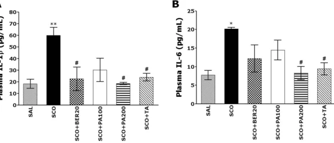
Inflammation, as well as cholinergic neuronal degener- ation, may play an important role in the pathogenesis of the degenerative changes and cognitive impairment asso- ciated with AD. Proinflammatory cytokines, such as inter- leukin-1β (IL-1β) and tumor necrosis factor-α (TNF-α) are upregulated in the AD brain [6]. These inflammatory cyto- kines may play a role as neurotoxic agents in the patho- logical cascade of AD [7].
Many studies have reported that the use of herbal medi- cine or natural products in treating Alzheimer type-de- mentia patients improved their memory related symptoms [8,9]. However, there has been little scientific evidence re- ported regarding their effectiveness or mechanisms of their action.
Phellodendron amurense (PA) has been widely used in Korean traditional medicine to treat bacterial infection of the respiratory system and chronic inflammatory diseases [10]. PA and its major alkaloid compound, berberine (BER), have multiple pharmacological actions and produce a varie- ty of biological effects in the CNS [11]. In particular, BER has been reported to improve diabetes-induced memory im- pairment and to reduce oxidative stress [12]. Several stud- ies have shown that BER was effective in ameliorating spa- tial memory impairment and decreasing the expression of IL-1β in a rat model of AD [13], suggesting that the anti-in- flammatory effects of PA and BER may result from their ability to inhibit the degeneration of cholinergic neurons, thereby alleviating deficits in spatial learning ability in the memory-impairment animal model [14].
The aim of the present study was to evaluate the ability of PA and BER to improve learning and memory in rats exposed to repeated SCO-induced memory deficits as meas- ured by performance on the passive avoidance test (PAT) and the Morris water maze (MWM) test. Moreover, we ex- amined how these effects were related to the cholinergic system and to anti-inflammatory effects to determine the neural mechanisms underlying the memory-enhancing acti- vity of PA and its main alkaloid compound, BER.
METHODS Animals
Adult male Sprague-Dawley (SD) rats weighing 260∼280 g were obtained from Samtako Animal Co. (Seoul, Korea).
The rats were housed in a limited-access rodent facility with up to five rats per polycarbonate cage. The room con- trols were set to maintain the temperature at 22±2
oC and the relative humidity at 55±15%. Cages were lit by artificial light for 12 h each day. Sterilized drinking water and stand- ard chow diet were supplied ad libitum to each cage during the experiments. The animal experiments were conducted in accordance with the Guide for the Care and Use of Labo- ratory Animals (NIH Publications No. 80-23), revised in 1996, and were approved by the Kyung Hee University Ins- titutional Animal Care and Use Committee. All animal ex- periments began at least 7 days after the animals arrived.
Main reagent
Scopolamine hydrobromide, tacrine (TA) and BER were obtained from standard commercial suppliers (Sigma-Aldrich
Chemical Co., St Louis, MO, USA). PA, used in this study, was purchase from an oriental drug store (Health maxi- mum Co., Jecheon, Korea). A voucher specimen of PA has been deposited at the herbarium located at the College of Oriental Medicine, Kyung Hee University (Code number KH-PA01 for PA), according to described previous study [15].
One hundred grams of PA were cut into small pieces and extracted three times with 4 l of 85% methanol by soni- cation in a reflux condenser for 24 h at room temperature (25±2
oC). The extracted solutions were combined, filtered through Whatman No. 1 filter paper, concentrated using a rotary vacuum evaporator (EYELA CCA-1110, Tokyo Rikakikai Co., Tokyo, Japan) to obtain concentrated extracts, and then lyophilized (EYELA FD-800). The final yield of PA in a powder form was 13.7% (w/w).
Experimental groups
To develop learning and memory deficits, male rats were intraperitoneally injected at 2 mg/kg body weight with SCO, dissolved in physiological saline solution, once a day for 14 days. Normal animals received saline instead of SCO as a vehicle. Different rats in an experimental group were sub- jected to either behavioral testing or immunohistochemistry.
The rats were randomly divided into seven groups of seven individuals as follows: normal group (SAL group, n=7), the saline-induced plus 200 mg/kg PA-treated group (PA group, n=7), the SCO-induced and saline-treated group (SCO group as a control, n=7), the SCO-induced plus 20 mg/kg BER- treated group (SCO+BER20 group, n=7), the SCO-induced plus 100 mg/kg PA-treated group (SCO+PA100 group, n=7), the SCO-induced plus 200 mg/kg PA-treated group (SCO+
PA200 group, n=7), and the SCO-induced plus 0.2 mg/kg TA-treated group (SCO+TA group, n=7). TA, a centrally acting cholinesterase inhibitor, was used as a positive control. The rats were intraperitoneally administrated with PA and BER once a day for 14 days, and PA and BER were dissolved in 0.9% physiological saline. Thirty minutes after PA and BER administration, all rats except SAL group were received the SCO injection. When starting 9th day af- ter SCO injection, rats performed to take the MWM task.
The experimental schedule of all drug administration and behavioral tests are shown in Fig. 1.
Passive avoidance test
All animals were subjected to a passive avoidance test.
The test was basically performed according to the step-
through method described previously [16]. The Gemini
Avoidance System (SD Instruments., San Diego, CA, USA)
was used for this experiment. Basically, the step-through
passive avoidance apparatus consists of a two-compartment
acrylic box with a lightened compartment connected to a
darkened one by an automatic guillotine door. Electric shock
was delivered to the grid floor of both compartment, made
of stainless steel rods (3 mm diameter) spaced 1 cm apart,
by an isolated shock generator (Behbood Pardaz Co., Ghaem,
Iran). First, rats were taken trials to acquisition test in the
apparatus. In this trial, rats were placed in a lightened
compartment for 300 s, and then the guillotine door was
opened. Rats have native preference to the dark environ-
ment. Immediately upon entering the dark compartment,
the door was closed. Acquisition test was recorded the la-
tencies times for entering the dark compartment. After 30
min, rats were again placed in the lightened compartment.
Fig. 1. Experimental schedules of scopolamine-induced spatial memory impairments in the rats. Exp 1 was designed to explore the efficacy of PA and BER administration for amelioration of memory impairment in an animal model using a passive avoidance test (PAT) and immuno- histochemistry. Exp 2 was designed to explore the efficacy of PA and BER administration for amelioration of memory impairment in an animal model using the Morris water maze (MWM) test, ELISA and RT-RCT analyses.
Rats had spontaneously entered the dark compartment, the guillotine door was closed and a mild electrical shock (0.5 mA) was applied for 3 s. Exactly 24 h after the acquisition trial for training, the retention test was performed. The rat was again placed in the lightened compartment and the guillotine door was opened. The retention test was meas- ured the latencies times for entering the dark compartment in a same method with acquisition test. The maximum en- try latency allowed in the retention test was 180 s [17].
Morris water maze test 1. Morris water maze apparatus
The MWM test was performed using a polypropylene cir- cular pool (painted white internally, 2.0 m in diameter and 0.35 m high). The pool contained water maintained at a temperature of 22±2
oC. The water was made opaque by adding 1 kg of skim milk powder. During the MWM test, a platform 15 cm in diameter was located 1.5 cm below the water in one of four sections of the pool, approximately 50 cm from the sidewalls. The pool was surrounded by many cues external to the maze. The pool was divided into four quadrants of equal area. A digital camera was mounted to the ceiling above the pool and was connected to a compu- terized recording system equipped with a tracking program (S-MART: PanLab Co., Barcelona, Spain), which permitted on- and off-line automated tracking of the paths taken by the rats.
2. Hidden platform trial for acquisition test
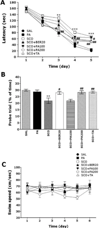
The MWM test was initiated on the 9
thday after the PA, BER and SCO administration commenced. The animals re- ceived three trials per day. The rats were trained to find the hidden platform, which remained in a fixed location throughout the test. The trials lasted for a maximum of 180 s, and the time it took to find the submerged platform was recorded each time. The animals were tested three tri- als per day for 5 days, and they received a 60-s probe trial on the sixth day. Finding the platform was defined as stay- ing on it for at least 4 s before the acquisition time of 180 s ended. If the rat failed to find the platform in the allotted
time in first trial of hidden platform test, those rats should placed onto the platform for 20 s and assigned a latency of 180 s. Between one trail and the next, water was stirred to erase olfactory traces of previous swim patterns. The en- tire procedure took sixth consecutive days, and each animal had three training trials per day, with a 30- to 40-min inter- trial interval.
3. Probe trial for retention test
For the probe trial, each rat was placed into the water diagonally from the target quadrant, and for 60 s, was al- lowed to search the water from which the platform had been removed. The probe trial was expressed by the ratio of the time spent for searching the platform in the target quadrant to total duration spent for swimming in the pool.
Cholineacetyltransferase (ChAT) and Acethylcholine- sterase (AchE) immunohistochemistry
For immunohistochemical studies, the animals were deeply anesthetized with sodium pentobarbital (80 mg/kg, by in- traperitoneal injection) and perfused through the ascending aorta with normal saline (0.9%) followed by 300 ml (per rat) of 4% paraformaldehyde in 0.1 M phosphate-buffered saline (PBS). The brains were removed, post-fixed over-night, and cryoprotected with 20% sucrose in 0.1 M PBS at 4
oC.
Coronal sections 30 μm thick were cut through the septal region and hippocampus using a cryostat (Leica CM1850;
Leica Microsystems Ltd., Nussloch, Germany). The sections were immunostained for ChAT expression using the avi- din-biotin-peroxidase complex (ABC) method. Briefly, the sections were rinsed three times for 5 min each in PBS and then incubated with primary rabbit anti-ChAT antibody (1:2,000 dilution; Cambridge Research Biochemicals Co., Bellingham, UK), or goat anti-AchE antibody (1:2,000 di- lution; Santa Cruz Biotechnology Inc., CA, USA) in PBST (PBS plus 0.3% Triton X-100) for 72 h at 4
oC. Both secon- dary antibodies were obtained from Vector Laboratories Co.
(Burlingame, CA, USA) and diluted 1:200 in PBST contain-
ing 2% normal serum. To visualize immunoreactivity, the
sections were incubated for 90 min in ABC reagent (Vecta-
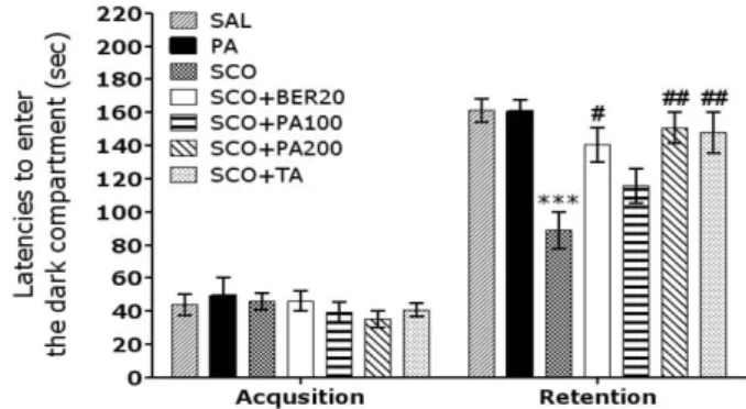
Fig. 2. Effect of PA and BER on the latencies to enter the dark compartment in the acquisition trial and in the retention test during the PAT test. ***p<0.001 vs. the SAL group;
#p<0.05 and
##