Yonsei Med J http://www.eymj.org Volume 51 Number 6 November 2010 978
There are various clinical manifestations of neurocysticercosis (NCC) that depend on not only the topography, number, and size of the lesions, but also the status of the host’s immune response to the parasite infection.1Only a few reports have described third cranial nerve palsy in patients with NCC involving, especially, the midbrain.2-4Meanwhile, Claude’s syndrome is a distinctive brainstem syndrome characterized by ipsilateral third cranial nerve palsy with contralateral hemiataxia and is due to an intrinsic or extrinsic lesion in the midbrain.5We report a case of Claude’s syndrome caused by NCC infection.
A 68 year-old man was admitted to our hospital because of ataxia, left ptosis, and diplopia started two days prior. His previous medical history was unremarkable. He did not travel overseas before this event. The left eye pupil was 1.5 mm larger than the right side with impaired pupillary light reflex. Extraocular movements were normal in the right eye. In contrast, there was limited movement (adduction, eleva-
Case Report
DOI 10.3349/ymj.2010.51.6.978pISSN: 0513-5796, eISSN: 1976-2437 Yonsei Med J 51(6):978-979, 2010
Claude’s Syndrome Associated with Neurocysticercosis
Tae-Jin Song,
1Sang Hyun Suh,
2Hanna Cho,
3and Kyung-Yul Lee
31Department of Neurology, JungAng General Hospital, Jeju;
Departments of 2Radiology and 3Neurology, Gangnam Severance Hospital, Yonsei University College of Medicine, Seoul, Korea.
Claude’s syndrome is a distinctive brainstem syndrome characterized by ipsilateral third cranial nerve palsy with contralateral hemiataxia and is due to an intrinsic or extrinsic lesion in the midbrain. We report a case of Claude’s syndrome caused by neurocysticercosis infection. A 68 year-old Asian man was admitted to our hospital because of ataxia, left ptosis, and diplopia. Brain magnetic resonance imaging (MRI) showed a cystic lesion in the midbrain, which was surrounded by ring enhancement and peripheral edema. Neurocysticercosis infection was diagnosed by the cerebral spinal fluid study. The patient was treated with albendazole and steroids. A follow-up brain MRI three months later demonstrated the disappearance of a surrounding brain edema and rim enhancement. The most common cause of Claude’s syndrome is cerebrovascular disease and malignancy.
However, there is no report caused by neurocysticercosis infection. Therefore, if we encounter Claude’s syndrome, we should consider neurocysticercosis infection as one of the etiologic factors.
Key Words: Neurocysticercosis, Claude’s syndrome, ataxia
Received: December 6, 2008 Revised: February 27, 2009 Accepted: March 15, 2009
Corresponding author: Dr. Kyung-Yul Lee, Department of Neurology,
Gangnam Severance Hospital, Yonsei University College of Medicine, 712 Eonju-ro, Gangnam-gu,
Seoul 135-720, Korea.
Tel: 82-2-2019-3325, Fax: 82-2-3462-5904 E-mail: [email protected]
∙The authors have no financial conflicts of interest.
© Copyright:
Yonsei University College of Medicine 2010 This is an Open Access article distributed under the terms of the Creative Commons Attribution Non- Commercial License (http://creativecommons.org/
licenses/by-nc/3.0) which permits unrestricted non- commercial use, distribution, and reproduction in any medium, provided the original work is properly cited.
INTRODUCTION
CASE REPORT
tion, depression) in the left eye and fourth cranial nerve func- tion was considered normal in the left eye because intorsion of the left eye was preserved. The gait was ataxic and dysdia- dochokinesia was present on examination of the right arm.
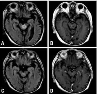
Brain magnetic resonance imaging (MRI) showed a cystic lesion in the midbrain, which was surrounded by ring enhancement and peripheral edema (Fig. 1A and B).
Enzyme-linked immunosorbent assay for an anticysti- cercal antibody in serum was negative, but it was positive in cerebral spinal fluid of 0.38 (normal range < 0.18). A lumbar puncture yielded clear cerebral spinal fluid under normal opening pressure, and cytochemical analysis show- ed 3 mononuclear cells per mm3, 90 mg/dL proteins, and normal glucose contents. Cerebral spinal fluid cytospin test for malignancy was negative.
The patient was treated with oral albendazole 15 mg/kg/
day in two divided doses for two weeks, and intravenous methylprednisolone 1g/day for six days. Paresis of the third cranial nerve and ataxia resolved completely within one week of treatment. A follow-up brain MRI three months later demonstrated the disappearance of surrounding brain edema and ring enhancement (Fig. 1C and D).
NCC is a common parasite affecting the central nervous system. Brain parenchyma involvement is the most com- mon and usually presents with seizures. Other clinical
manifestations may occur, depending upon the localization and viability of the parasite. There have been several re- ports that NCC occurred within the intrinsic midbrain, mani- fested by isolated third cranial nerve palsy with poor pro- gnosis,2isolated bilateral ptosis,4accompanying hydrocep- halus,6and recurrent third cranial nerve palsy.7,8 However, none of these patients presented with ipsilateral third cranial nerve palsy accompanying contralateral hemiataxia, that is, Claude’s syndrome. The Claude described a house painter who developed right third cranial nerve palsy with contra- lateral gait ataxia.9The pathological examination revealed a paramedian mesencephalic infarction on the right involv- ing the superior cerebellar peduncles, the medial half of the red nucleus, and some reports describe the syndrome with Claude’s original red nucleus involvement.5,10In this case, the lesion of NCC involved the left superior cerebellar pe- duncle but not the red nucleus. This finding supports the report that suggests the lesion of the superior cerebellar peduncle just below and medial to the red nucleus can be a cause of Claude’s syndrome.11The most common cause of Claude’s syndrome is cerebrovascular disease and malig- nancy.11There is no report caused by NCC infection. There- fore, we should consider NCC infection as an etiologic factor if we encounter Claude’s syndrome.
1. Del Brutto OH, Rajshekhar V, White AC Jr, Tsang VC, Nash TE, Takayanagui OM, et al. Proposed diagnostic criteria for neuro- cysticercosis. Neurology 2001;57:177-83.
2. Katz B. Central American mesencephalopathy. Surv Ophthalmol 1994;39:253-9.
3. Lath R, Rajshekhar V. Solitary cysticercus granuloma of the brainstem. Report of four cases. J Neurosurg 1998;89:1047-51.
4. Sawhney IM, Singh G, Lekhra OP, Mathuriya SN, Parihar PS, Prabhakar S. Uncommon presentations of neurocysticercosis. J Neurol Sci 1998;154:94-100.
5. Leigh RJ, Zee DS. The neurology of eye movements. 2nd ed.
New York: Oxford University Press;1991.
6. Wadley JP, Shakir RA, Rice Edwards JM. Experience with neu- rocysticercosis in the UK: correct diagnosis and neurosurgical management of the small enhancing brain lesion. Br J Neurosurg 2000;14:211-8.
7. Mokta JK, Mahajan S, Machhan P, Mokta KK, Patial RK, Pra- shar BS. Recurrent oculomotor nerve palsy: a rare presentation of neurocysticercosis. Neurol India 2004;52:402.
8. Kim JS, Jeong SM, Moon SY, Park SH. Third cranial nerve palsy from midbrain neurocysticercosis: repeated exacerbation on taper- ing corticosteroids. J Neuroophthalmol 2004;24:217-20.
9. Claude H, Loyez M. Ramollissement du noyau rouge. Rev Neurol 1924;41:417-23.
10. Kremer C, Baumgartner RW. Aortic embolism in Claude’s syn- drome. Cerebrovasc Dis 2002;13:142-3.
11. Seo SW, Heo JH, Lee KY, Shin WC, Chang DI, Kim SM, et al.
Localization of Claude’s syndrome. Neurology 2001;57:2304-7.
Claude’s Syndrome Accompanying Neurocysticercosis
Yonsei Med J http://www.eymj.org Volume 51 Number 6 November 2010 979
DISCUSSION
Fig. 1. Brain MRI. (A) Initial FLAIR image shows high signal intensity on left midbrain and perilesional edema. (B) Initial contrast enhanced T1-weighted image shows cystic lesion with peripheral ring enhancement. The lesion involves dorsal midbrain tegmentum, left third cranial nerve fascicle, and superior cerebellar peduncle, but not the red nucleus. The lesion is located posterior and inferior to the red nucleus. (C and D) Follow up MRI after three months, reveals disappearance of perilesional edema and ring enhancement.