110 Copyright 2005 by the Korean Society for Clinical Neurophysiology
대한임상신경생리학회지 7(2):110~113, 2005 ISSN 1229-6414There are many kinds of complications about chronic alcoholism such as myopathy, nutritional problems, neuropathy, withdrawal seizure and encephalopathies. Among them, it is well known that alcoholic myopathies have characteristic pathological features according to their types.
1Acute alcoholic myopathy is caused by severe alcoholic binges. It shows rapid onset muscle pain and swelling with myoglobulinuria. On the other hand, chronic alcoholic myopathy complicates years of alcohol abuse. It is characterized by slowly progressive and painless proximal weak- ness correlated with lifetime consumption of e t h a n o l .
1We report a case of acute alcoholic myopathy superimposed on chronic alcoholic myopathy ini- tially presented as acute reversible encephalopa- thy showing transient abnormalities of brain magnetic resonance images (MRI).
CASE REPORT
A 59-year-old woman admitted due to acute mental change without definite history of seizure episodes because she was found by her family member. On first neurological examinations in regional hospital, she showed stuporous mentality for several minutes. Her mental status recovered fully after one or two hours during transfer to our hospital.
She was a chronic alcoholic for about 10 years.
In history, she noticed progressive motor weak- ness especially during walking and standing since 6 months ago. And she experienced several episodes of much more severe disability with pain
급성으로 발병한 가역성 뇌병증을 동반한 알코올 근육병증
고신대학교 의과대학 신경과학교실, 동아대학교 의과대학 신경과학교실 , 경상병원 신경과
김종국・이지현・김민정・유봉구・김광수・천상명 ・조희영 ・이상원
Alcoholic Myopathy Accompanied with Acute Reversible Encephalopathy
Jong Kuk Kim, M.D., Ji-Hyun Lee, M.D., Min-Jeong Kim, M.D., Bong-Goo Yoo, M.D., Kwang-Soo Kim, M.D., Sang-Myung Cheon, M.D.*,
Hee Young Jo, M.D.*, Sang Won Yi, M.D.
†Department of Neurology, Kosin University College of Medicine Department of Neurology, Dong-A University College of Medicine*
Department of Neurology, Gyeong Sang Hospital
†Patients of chronic alcoholism may show many kinds of complications such as myopathy, nutritional problems, peripheral neuropathy, withdrawal seizure and encephalopathies. We report an unusual case of alcoholic myopathy diagnosed with typical laboratory and pathological findings initially manifested as acute reversible encephalopathy showing transient abnormalities on brain MRI.
Key Words: Alcohol, Myopathy, Encephalopathy
Address for correspondence Jong Kuk Kim, M.D.
Department of Neurology, Kosin University College of Medicine 34 Amnam-dong, Seo-gu, Busan, 602-702, Korea
Tel: +82-51-990-6461 Fax: +82-51-990-3077
E-mail : [email protected]
on legs remitted for spontaneously. From the before 3 days on admission, she reduced amount of alcohol consumption due to exacerbated weak- ness and pain on her legs.
Neurological examinations revealed quadripare- sis dominant on proximal limbs muscles (MRC Grade IV+). Laboratory findings showed markedly elevated serum creatine kinase (CK) and lactate dehydrogenase level (over 16000 and 5068 U/L, respectively) and positive urine myoglobin.
Routine complete blood counts, electrolytes and chemistry including renal function tests showed no definite abnormal finding except for elevated hepatic enzymes. Thyroid function tests, rheuma- tological laboratories (RA, ANA, Anti-Ro and Anti-La) and vitamin levels (thiamine, folate, pyridoxine, cobalamin) were within normal limits.
Cerebrospinal fluid examinations revealed no other abnormal finding.
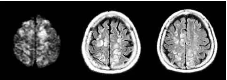
Electroencephalography (EEG) on admission showed mild diffuse slowing without epileptiform discharges. Needle electromyography (EMG) stud- ies revealed chronic active myopathic discharges especially in proximal limb muscle group. Brain MRI of diffusion weighted and FLAIR sequences images showed multiple diffuse high signal intensities in bilateral frontoparietal cortical and subcortical areas (Fig. 1) and these findings were regressing rapidly in follow-up images (Fig. 2).
Pathological findings from right biceps muscle revealed severe atrophic changes and size varia- tion of type Ⅱ muscle fibers compatible with chronic alcoholic myopathy (Fig. 3).
On education and psychiatric support, after abstaining from alcohol, her motor functioning was improved slowly and she was discharged without any other complication.
D I S C U S S I O N
Patients with alcoholic myopathy show a variety of manifestations. They are classified into two types of myopathies, acute and chronic.
1Acute alcoholic myopathy with myonecrosis develops suddenly in the context of binge drink- ing with muscle pain and swelling. Its severity ranges from asymptomatic transient elevation of creatine kinase to frank rhabdomyolysis with myoglobulinuria. Occasionally, it is associated with acute alcohol withdrawal. Frequently, EMG
findings show no abnormality. Pathological find- ings show scattered type Ⅰ predominant muscle fibers (high oxidative, low glycolytic, slow twitch) necrosis suggesting mechanisms of oxidative d a m a g e s .
1 , 2Chronic alcoholic myopathy is a gradually evolving syndrome of proximal weakness and atrophy characterized by slowly progressive proximal weakness and muscle atrophy. In pathology, there are striking muscle atrophy and degeneration of type Ⅱ fibers, especially type ⅡB muscle fibers (high glycolytic, low oxidative, fast twitch). EMG may show myopathic potentials, neuropathic features, or both. In some cases, acute muscle injury, manifested by muscle ten- derness and creatine kinase elevation, may be superimposed on chronic alcoholic myopathy.
3Our patient showed typical history and findings of chronic alcoholic myopathy but, she was mani- fested with acute alcoholic myonecrosis. It is explained by the fact that chronic alcoholic myopathy is accompanied by acute type in some- times if there is abrupt reduce or abstinence of alcohol consumption.
Unusual findings of brain MRI in this patient are interesting. Though there are many kinds of abnormalities associated with alcoholism, we cannot explain the exact nature of these acute transient multiple cortical and subcortical abnor- malities of brain images because we meet this kind of findings infrequently.
4In concerning the time relationships, it may be proposed that multi- ple high signal intensities of brain MRI are closely related with acute mental change and it may be provoked by recent alcohol abstinence or alcoholic rhabdomyolysis not by vitamin deficiency.
There are several kinds of complications mani- fested as encephalopathy in chronic alcoholics.
Among them, there is a rare type of disease such as subacute encephalopathy with seizures in chronic alcoholism (SESA syndrome). It is charac- terized by focal seizures in chronic alcoholics and can be diagnosed by periodic lateralized epilepti- form discharges (PLEDs) in the EEG with or without abnormalities in brain images.
5 , 6Same as reported cases of SESA syndrome, our patient’ s mentality was recovered without remained neu- rological deficits except for muscle weakness.
But, there was no definite evidence of seizure episode in our patient by history. Also we could
급성으로 발병한 가역성 뇌병증을 동반한 알코올 근육병증
J Korean Society for Clinical Neurophysiology / Volume 7 / November, 2005 111
not get adequate information from EEG though this may be due that it was done in recovering state.
There are other possibilities that the findings
are results of ischemia associated with hyper- myoglobinemia, hyperCKemia or alcohol with- drawal seizures because the patterns of images resemble the one of watershed infarction. But
김종국・이지현・김민정・유봉구・김광수・천상명・조희영・이상원
112 J Korean Society for Clinical Neurophysiology / Volume 7 / November, 2005 Figure 2. Follow up images after 3 days showed decreased amounts of the abnormal high signal intensities on the same regions.
Figure 3. Pathological findings from right biceps muscle showed severe atrophic changes and size variations of type Ⅱ muscle fibers (H&E, × 100, ATPase 9.4, × 200).
Figure 1. Diffusion and FLAIR images of brain MRI showed diffuse multiple high signal intensities in bilateral frontoparietal corti-
cal and subcortical areas mimicking cerebral infarctions.
급성으로 발병한 가역성 뇌병증을 동반한 알코올 근육병증