약학희지 제39권 제2호 169-" 174(1995) Yakhak Hoeji Vol. 39, No. 2
유산균에 의한 장내미생물효소의 저해
김동현* • 한명주*
경희대학교 약 학 과 , * 식 품 영 양 학 과 (Received March 8, 1995)
Inhibition of Intestinal Bacterial Enzymes by Lactic Acid Bacteria
D ong-H yun, K im 화 and M y u n g Joo, H an*
College of Pharmacy, * Department of Food Sciences and Nutrition.
Kyung-Hee University, Seoul 130-701, Korea
Abstract —By coculturing
E. coli
HGU-3 with Bifidobacterium KH-2 orStreptococcus faecalis
HGO-7with
Bifidobacterium
KH-2, the productivity of p-glucuronidase and p-glucosidase was inhibited. When lactulose, growth factor of lactic acid bacteria, was added into this medium, the productivity of these enzymes and pH of the medium were dramatically decreased. When intestinal microflora of human and rat were inoculated in the medium containing lactulose, the enyzme productivity and pH of the medium were dramatically decreased. By s.c. injecting DMH into mice, p-glucuronidase of intestinal bacteria was induced, but the production of the enzymes was inhibited by adminstering lactulose.
복미, 서유럽등대장암발생 고빈도 지역과아시아, 아프리카등대장암발생 저빈도지역외 장내세균총을 비교해 보면 저빈도지역은 B acteriodes가적은반면
에 Streptococcus가 많으며 호기성 균주에 대한 혐
기성 균주의 비율이 낮다. 이러한 것을 제외하면 대 장암의 발생번도가 높은 복미, 서유럽 지역의 사람 들과 발생빈도가 낮은 지역 주민과 별차이가 없다. 그럼에도 불구하고대장암발생 고빈도지역에서는 장 내미생물이 글루쿠론산포합체를가수분해 시키거나
steroid를대사시키는능력이 증가되어 있었다.* 이
러한 반응에 3-glucuronidase, p-glucosidase, nitro
reductase, steroid 7ct-dehydroxylase 등이 관여하여
carcinogenic aglycone을 방줄시킴으로써 암형성을
일으키는 것으로 알려져 있다. 결국 발암물질이 간 에서 포합체롤 형성하고 담관을 통헤서 분비된 후 장내 미생물에 의해서 포합체가 가수분헤 됨으로써 발암작용에 기여하는 것으로 사려되고있다."^" 이외
에도장내 pH 외번화는 세균성 효소에 의한 이물(록
t o의대사에 영향을 주며, 대장암의 발생과 밀접한
* 본 논문에 관한 문외는 이 저자에게로
관계를 갖고 있다."« 특히 흰쥐에서, 장내세균에 대
사되어 유기산을 생성하는 인공다당류인 lactulose를 투여하므로써 암발생율을 감소시켰다고보고하고 있 다.™
대장암발생의 역학연구는 앞에서 언급한 바와 같 이 아시아, 아프리카보다 복미, 서유럽이 더 높음을 보여준다. 그것은 높온 고지방육류과 낮은 섬유소로 구성되는 서앙 식이가 주요 원인으로 생각되어지고 었다. 특히 핀란드는 1인당 지방 섭취량이 높은 나 라중에서 비교적 대장암의 발생율이 낮은 예외적인 나라이다, 이핀란드인들은 요구르트를 많이 섭취한 다. 그 결과 핀란드인들외 장내에는 L actobacillus가
많이존재할 것으로생각되고있으며 G oldin 등은 이
역학적 조사를 뒷받침 하기 위해 L actobacillus aci- dophillus 투여에 외한장내세균의 3-glucuronidase, nitroreductase, azoreductase 등의 효소활성 변화를 조사한 결과 P-glucuronidase, n itro red u ctase의 활
성이 저하됨을 보고하였다."
최근 저자등은이러한 장내미생물효소가 산성보다 는 중성 또는 알카리성에서 잘 유도됨을 밝혔으며
170 김동현• 한명주
사람의 장내미생물로부터 이러한효소들을생산하는 균주들을 보 고 하 였 다 . 그 래 서 여기에서는 pH에 의헤서 유도되어지는 P - g lu c o s id a s e를 생산하는 H G 0 -7 균주및 P-glucuronidase를생산하는 균주인
HGU-3균주와 유산균을 동시배양하는 과정에서 유
산균증식제를첨가하는정도에따라이들효소활성에 미처는효과를 조사하였다.
설 험
실험재료
General anaerobic medium(GAM ) broth, Glucose blood liver broth(BL) agar, T ryptic soy(TS) agar-b 일본 니수이제약주식최사로부터 구입하였다. p-Nit- rophenyUp-D-glucopyranoside, p-nitrophenyl-P-D- glucuronide, p-nitorphenylsulfate는 머국외 Sigma 사로부터 구입하였으며, 1 ,2 - d im e th y lh y d r a z in e (DM H )는미국의 Aldrich사로부터 구입하였으며, 혈 액은 한국메디아로부터 구입하여 사용하였다.
실험균주
HGU-3 균주 및 H G 0-7 균주는 이미 보고한 분리
균주를 사용하였으며 Bifidobacterium KH-2 균주는 건강한 잠식성 20대남자의 분편으로부터 Mitsuoka 방 에따라 분리한 균주를 사용하였다.
실험 동물
국립보건 안전 연구원에서 분앙받은 W istar계흰 쥐(male, 약 200 g)을사용하였으며 사료는대한사료 (주)의 곡물사료로 사육하였고 물은 충분히 공급하 면서 사육하였다.
D M H에의한흰쥐 장내미생물효소 유도시험을위
해서는 흰쥐(Wistar 200g m ale)를 6마리썩 4군으로
나누어 (A)군은 대조군으로 일반식(삼양사, 한국)을
먹이는군, (B)군은 lactulose와일반식을흔합한식을 투여하면서 D M H를투여한군, (C)군은 일반식을먹
이면서 D M H를 투여한 군, (D)군은 일반식과 함께
lactulose을투여한군으로 나눠 각군에 대하여 일반
식과물은자유롭게 먹이면서 사육하였다. Lactulose 는 7%를 포함하도록 일반식(대한사료)파 흔합하여 투여하였으며, DM H는 20 mg/kg을 3일마다 한번씩 피하투여 하였다.
설험방법
효소황성측정'*' — P-Glucuronidase 외효소 활성은 10 mM p-nitrophenyl-p-D-glucuronide 20 |il, O.IM phosphate완충액 0.38 m l , 효소액 0.1 m/를 넣온 후 37°C 에서 50분간 반응시키고 0.25N NaOH를 0.5 m/
넣어 반응을 중지시킨 후 흡광도를 40 5nm에서 측
정하였다. P-Glucosidase의효소활성은 2 m M p-nit- rophenyl-P-D-glucopyranoside 0.2 m l , O.IM phos- phate완충액 0.3 m/, 효소액 0.1 m/를 넣은 후 위와 동일한 방범으로 활성을 측정하였다. 효소활성저해 시험은효소활성을측정할때기질과함께일정농도의 저해제를 첨가한후효소를 가하여효소활성을 측정 하는 방법을 이용하였다.
유산균과 동시 배앙시 효소활성측정 ~ H G U -3와 H G0-7 균주 각각 5 X 10 "개를 G A M배지에이식하여 각각의 균주에 대하여 4개군으로 분리하고, 여기에 사람외 장내세균으로부터 분리한 Bifidobacterium KH-2균주를 5X 10", 5X10", 5X 10M ) 를 각군에 이 식하고 Bifidobacterium KH-2균주만을 이식한 것을 대조군으로 하여 37°C 에서 24시간 배양한후효소활
성, 배지외 pH, 생균수을측정하였다. 생균수의 측정
은 적당히 희석한 검액을 B L 한천배지에 이식하고
steel wo이법에따라 2 ~ 3일간배양한후측정하였다. 장내세균층에 유신균증식제첨가시 효소황성측정
~ 사람및흰쥐의신선한장내세균총을 lactulose함유
G A M배지에 이식하고 24시간 혐기적으로 배양하여
유산균수, 배지외 pH, 효소활성을측정하였다.
사람과흰쥐의분현탁액의조제■곡물사료와물을 충분히 공급해 준흰쥐로부터 직접얻은신선한분을
중류수를 사용하여 10% (w/v)로 잘현탁시킨 후상
등액을 취하여 효소활성 측정에 이용하였다. 결과 및고찰
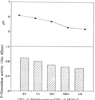
유산균과 동시배앙시 유산균에 의한 효소황성의
변화" 유산균과 HGU-3균주를 동시배앙시 효소활성
를비교한 걸과는 Fig. 1과같다. 그림에서 보는 것과 같이 HGU-3균주만을 배양했을 때보다 Bifidobacte-
rium과함께 배양했을때 HGU-3 균수는감소했으며,
유산균수를 10배, 100배증가시킴에 따라 HGU-3균 수는 더 많이 감소했다. 유산균수가 증가함에 따라 배지의 pH에도영향을미쳐 유산균수의증가와함께
/.
Pharm. Sco. Korea
Concentration of Lactulose (%)
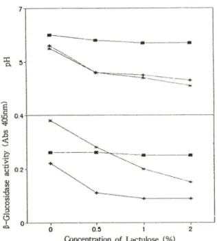
Fig. 3 — Effect of lactulose on p-glucuronidase produ
ctivity and pH of the cultured medium by cu
lturing HGU-3 only or with Bifidobacterium KH-2: ■, cultivation of Escherichia coli HGU-3:
*, cocultivation of Escherichia coli HGU-3 with Bifidobacterium KH-2.
배지의 pH는 감소하였다. 더욱이 이유산균수외 증 가는 HGU-3균주의 P-glucuronidase 효소활성에도 영향을 미쳐유산균을 HGU-3균주의 100배많이 이 식했을경우효소활성은유산균을동시배양하지 않은
대조군에 비해 24%로효소활성이 현저히 감소했다.
H G 0 -7균주는유산균과동시 배양한경우에배지의
pH는 HGU-3균주의 경우와 비슷하게 저하하였으나
효소활성에 었어서는 대조군외 79 %로 2 1 %정도 감
소하였다(Fig, 2), 그러나, 유산균수는 현저하게 증가
하였다. 여기에 결과를나타내지 않았지만 Bifidoba
cterium KH-2 유산균만 배양했을 경우도 P-glucosi
dase 를 생산하고 있으며 pH가 낮아짐에 따라 효소
활성이 낮아졌으나, 이유산균이 직접적으로 P-glu
cosidase 효소를 저해하지는 않았다.
이러한 결과는 Bifidobacterium KH-2 유산균에 생산된 유기산은 배지외 pH를 저하시키고 그 결파 Bifidobacterium KH-2외 P-glucosidase 및 HG0-7
균주의 P-glucosidase의효소활성이 모두 감소됐다고
생각된다.
유산균과 동시배양시 유산균증식제(lactulose)의 영항~유산균증식제로 알려진 lactulose함유배지에 사람외 장내세균 분리균주인 HGU-3과 H G0-7 균주
Vol. 39. No. 2, 1995
유산균에 의한 장내미생물효소외 저해 171
Fig.
CFU of Bifidobacterium/CFU of HGU
-31 — Change of p-glucuronidase productivity and pH of the cultured medium by coculturing Esche
richia coli HGU-3 with Bifidobacterium KH-2.
Fig.
CFU of Bifidobacterium/CFU of HGO
-72 —Change of p-glucosidase productivity and pH of the cultured medium by coculturing Esche
richia coli HGO-7 with Bifidobacterium KH-2.
Concentration of Lactulose (%)
Fig. 4—Effect of lactulose on |3-glucuronidase produ
ctivity and pH of the cultured medium by co- culturing Escherichia coli HGO-7 with Bifido
bacterium KH-2: cultivation of Streptococ
cus faecalis HGO-7: +, cultivation of Bifido
bacterium KH-2; cocultivation of Escheri
chia coli HGU-3 with Bifidobacterium KH-2.
를 유산균과 동시배양시 효소의 생산성에 미치는 효과를 조사한 것이 Fig. 3과 Fig. 4 이다.
Lactulose를 첨가한 배지에서 Bifidobacterium
KH -2을 배양함에따라 배지의 pH는 현저하게 저하
하였으며 P-glucosidase효소활성도 배지의 pH와 함 께 감소하였다.
이 Bifidobacterium을 H G 0 -7과함께 배양했을 경 우에도 lactulose가 Bifidobacterium KH -2의 성장을 촉진시킨 결과 배지의 pH가 감소했으며 이에 따른 효소활성이 억제되었다.
HGU-3균주만 단득으로 lactulose배지에서 키운
결과, 배지의 pH 및효소활성에는거의영향을미치지 않았으나, 유산균과동시배양한 경우는배지의 pH가 현저하게 감소했으며 효소활성도 60% 이상 억제되 었다. 그러나, lactulose는 시험관내에서 p-glucosi- dase 또는 P-glucuronidase의 효소저해효과는 없었 다.
이러한 결과는 배지의 lactulose가유산균을 증식
&
Fig. 5 ― Effect of the medium on p-glucuronidase and P-glucosidase activities of intestinal bacteria of human and rat: 1, p-glucuronidase activity of human intestinal bacteria; 2, p-glucuronidase activity of rat intestinal bacteria: 3, p-glucosi- dase activity of human intestinal bacteria; 4.
p-glucosidase activity of rat intestinal bacteria:
The final concentration of lactulose in the me
dium was 0%(H), !%(■) and 5%(®, respecti
vely.
시켜 배지의 pH을 저하시켰으며 P-glucosidase와 P-
glucuronidase효소외 생산성에 영향을 미쳤다고 사
려된다.
장내세균총에 유산균증식제(lactulose)첨가시효소 황성의 변화■장내세균총을 배양하는 배지에 lactu
lose 를 첨가함으로써 p-giucosidase 및 p-glucuroni- dase의활성에미치는영향을조사한 결과톨 Fig. 5에 나타내었다. Lactulose를배지에 첨가한 결과흰쥐의 장내세균총인 경우 1% 첨가시 P-glucuronidase는 86%, P-glucosidase는 73%외효소저해를나타내었고 사람의 장내세균총인 경우 1 ^ 첨가시 P-glucuroni
dase 는 89%, P-glucosidase는 82%외 서해를받았다.
이것으로 보아 lactulose에 의한 대장암 발생억제효
과는 장내외 pH를 간접적으로 저하시킴으로써 나타 났으며, 특히 pH 6.0에서보다 pH 7.0 이상에서현저히 유도되는두효소를 포함한효소군이 대장암 발생에 주요한 인자로 작용한다고 사료된다.
D M H투여에 의한흔!쥐장내세균효소의변화와 la
ctulose 에 의한 영향一/w 에서 D M H를 흰쥐에 투여했을 때장내효소의 활성변화를측정했다(Fig. 6, Fig. 7). 장내미생물외 P-glucuronidase 효소활성은
D M H롤 투여했을 경우 대조군에 비해 30% 정도 증
가하였으나 D M H를 투여하면서 lactulose를 투여한
경우에는 대조군에 비교해서 처음에는 약 70% 감소
하여 산존활성이 30%였으나 차차증가하여 나중에는
]. Pharm. Sco. Korea
172 김동현 • 한명주
=& 5-
d6{mu90t*wqv
} ■로
>p oe
aseplsoonjo
l 삭
유산균에 의한 장내머생물효소외 저해 173
T im e (D a y )
Fig. 6 —Change of P-glucuronidase activity of intestinal bacteria by the administration of DMH(s.c.) and/or lactulose(p.o.): (1), control; (2), DMH only; (3), lactulose only: (4), DMH and lactu
lose.
T im e ( D a y )
Fig. 7 — Change of p-glucosidase activity of intestinal bacteria by the administration of DMH(s.c.) and/or lactulose(p.o.): (1), control: (2), DMH only: (3), lactulose only; (4), DMH and lactu
lose.
대조군외 6 0 ~ 70%까지 회복되었다. 그러나, lactu- lo se만을투여한 경우는대조군의 30 ~ 50 % 수준에서 유지되었다. 이러한 결과는 lactulose에 외헤 장내유
산균이 증식되어 p H가 저하되고 그 걸과효소생산
성이 감소되었다고 생각된다. 이러한 경향은 황산전 이효소의 경우는 나타나지 않았다.
그러나, P-glucosidase효소활성은 lactulose투여후 2 ~ 3일에 효소활성이 약 10 ~ 3 0 % 정도밖에 감소시
키지 못했다. 이러한 결과는 lactulose를 투여한 경
우에 증가한 유산균이 P-glucosidase를 생산하기 때
문이라고 생각된다.
결 론
1. 사람의 장내세균으로부터 분리한 B ifid o bacte
rium KH -2를 (3-glucuronidase 생산하는 H G U -3균 주나 (3-glucosidase를생산하는 H G 0 -7와동시배앙시
Bifidobacterium의 비율을 증가시킴에 따라 배지외
pH 저하와 함께 이균들이 생산하는 효소도 억제되
었다.
2. 유산균증식제인 lactulose는 직접적으로 P-glu
cosidase 나 P-glucuronidase를 거의 저해하지 못했 으나유산균파동시배앙시배지에첨가할 경우배지외 pH와효소의 생산성은 저해되었다.
3. 사람 및 흰쥐의 장내세균총을 배양시 배지에
lactulose룔 첨가한 경우에 농도의존적으로 |3-gluco
sidase 및 P-glucoronidase효소활성이 80% 이상 억 제되었다.
4. 생쥐에 D M H룔 투여함으로써 장내미생물효소
중 P-glucuronidase효소활성이 유도되었으나, lactu-
Isoe를투여한 결과 대조군의 50% 이하까지 억제되
었다.
감사의 말씀
본 연구는 한국과학재단연구비지원(921-1600-001- 2)에 의하여 이루어졌기에 이에 감사드립니다.
문 헌
1) Drasar B. S. and Hill M. J. Human intestinal flora ppl23 Academic Press (1974).
2) Burkitt D. P. Epidemiology of cancer of the colon and rectum,
Cancer
28(1), 3-13, (1971).3) Hill M. J., Drasar B. S., Aries V., Crowther J., Ha- wkesworth G., Williams R.E.O., Bacteria and etio
logy of cancer of the large bowel.,
Lancet
1, 95-100, (1971).4) Faivre, J., Boutron, M. -C., Quipourt, V. Diet and large bowel cancer pp 107-118 (in Advances on Nutrion and Cancer edited by Zappia, V. et al Academic press 1993).
5) Reddy, B. S., Engle, A., Simi, B., Goldman, M. Effect of dietary fiber on colonic bacterial enzymes and bile acids in relation to colon cancer.
Gastroenterol
Vol. 39, No. 2, 1995
174 김동현• 한명주
102, 1475-82, (1992).
6) Reddy, B. S., John, H. W. and Earnest, L. W. Faecal bacterial p-glucuronidase
Science
183, 416-417 (1974).7) Kinoshita, N., Gelvoin, H. V., p-Glucuronidase ca
talyzed hydrolysis of Benzo [a] pyrene-3-glucuro- nide and binding to DNA.
Science
199, 307-309 (1978).8) Walker, A.R.P., Walker, B. F. Faecal pH value and its modification by dietary means in south african black and white schoolchildren
S. AFR. Med. J.
55,495-498 (1979).
9) Thornton, J. R. High colonic pH promotes colorectal cancer.
Lancet
1081-1082 (1982).10) Samelson, S. L, Nelson, R. N. and Nynus, L. M.
Protective role of faecal pH in experimental colon carcinogensis.
J. Royal Society Med.
78, 230-233 (1985).11) Goldin B. R.&Gorbach, S. Alteration of the inte
stinal microflora by diet, oral antibiotics, oral an
tibiotics, and Lactobacillus: Decreased production
of free amines from aromatic nitro compounds, azo dyes, and glucuronides./
Natl Cancer inst.
73,689-695 (1984).
12) Goldin, B., & Gorbach, S. L. Alteration in faecal microflora enzymes relatdd to diet, age, Lactoba
cillus supplements, and DMH.
Cancer
40. 2421-2426 (1977).13) Kim. D. -H., Kang, H. -J., Kim. S. -H. and Kobashi, K. pH-inducible p-glucosidase and p-glucuronidase of intestinal bacteria
Chem. Pharm. Bull.
40, 1667-1669 (1992).
14) Kim, D. -H., Kang, H. -J. Park, S. -H. and Kobashi, K. Characterization of p-glucosidase and p-glu
curonidase of alkalotolerant intestinal bacteria.
Biol.
Pharm. Bull.
17, 423-426 (1992).15) Mitsuoka, T. The method of screening for intestinal flora.
J. Japan. Infect Dis.
45, 406-419 (1971).16) 김동현, 한명주 베타글루코시다제나 메타글루쿠로니 다제를 생산하는 호알카러성장내세균외 검색. 약학 회지 37. 187-192 (1993).
/