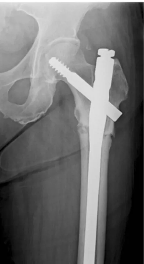
Atypical femoral fractures after anti-osteoporotic medication: a Korean multicenter study
7
0
0
전체 글
(2)
(3)
(4)
(5)
(6)
(7)
수치


관련 문서
‒ 사료생산형은 축산농가가 늘어나면서 조사료나 사일리지 생산을 위해 이루어지는 2모작 재배유형으로, 논에 벼를 재배한 다음 부산물인 볏짚을 조사료로
Percutaneous polymethylmethacrylate vertebroplasty in the treatment of osteoporotic vertebral body compression fractures: technical aspects. Pulmonary embolism caused
Although a large compressive residual stress was generated in the surface layer due to the peening process, shot peening showed –23 MPa, unlike laser... shock peening
이번 수업에서는 바이 오환경전문가가 무슨 일을 하는지 알아보고, 바이오환경전문가가 되어 친환 경기술 개발을
Purpose: Calcaneal fracture is a rare fracture, which accounts for about 2% of all fractures, but is one of the most common fractures in the ankle bone.. There is
Purpose: This study was aimed to compare the functional outcomes of the continuous and discontinuous fractures, retrospectively, in multiple thoracolumbar
High complication rate in locking plate fixation of lower periprosthetic distal femur fractures in patients with total knee arthroplasties.. Management and
The change in active oxygen was decreased after exercise in the exercise group than in the control group, and there was a statistically significant
