---
투고일 : 2/10 수정일 : 3/1 채택일 : 3/6
교신저자 : 김보경, 부산시 진구 양정동 동의대학교 부속한방병원 한방신경정신과.
Tel : 051-850-8809, Fax : 051-867-5162, E-mail : [email protected] 동의신경정신과 학회지
J. of Oriental Neuropsychiatry Vol. 20. No. 1, 2009
疏風順氣元이 mouse의 NMu2Li 간세포와 C2C12 골격근세포에서 PPARs 조절의 분자기전에 미치는 영향
오영진, 신순식*, 윤미정†, 김보경
동의대학교 한의과대학 한방신경정신과학 교실, 방제학교실*, 목원대학교 바이오건강학부†
A Molecular Study of Sopungsungi-won(Shufengshunqiyuan) about Regulation of PPARs in Mouse NMu2Li Liver Cells and C2C12 Skeletal Muscle Myogenic
Progenital Cells
Young-Jin Oh, Soon-Shik Shin*, Mi-Chung Yoon†, Bo-Kyung Kim Dept. of Neuropsychiatry, Dept. of Formula Sciences College of Oriental Medicine, Dongeui University*,
Department of Life Sciences, Mokwon University†
Abstract
Objectives :
We investigated the effects of Sopungsungi-won(Shufengshunqiyuan) (SSEx1, SSEx2) to treat the metabolic syndrome by the molecular mechanism of regulation of PPAR and modulation of mitochondrial MCAD, VLCAD mRNA expression.
Methods :
Mouse NMu2Li liver cells and C2C12 skeletal muscle myogenic progenital cells were transiently transfected with expression plasmids for PPAR(PPARα, PPARδ), a luciferase reporter gene construct containing 3 copies of the PPRE from the rat acyl-CoA oxidase gene and β-galactosidase gene. Cells were treated with several concentrated kinds of SSEx1, SSEx2 at the initial time of culture and analyzed PPARα, PPARδ reporter gene activity using spectrophotometer (405 nm). Total RNA was extracted from SSEx1, SSEx2 and measured mRNA levels of mitochondrial MCAD, VLCAD. Representative RT-PCR bands are shown.
Results :
1. SSEx1 increased the expression of PPARα reporter gene activities at 0.1 μg/ml (p<0.01) and 10 μg/ml (p<0.05), SSEx2 at 0.1 μg/ml (p<0.01) and 1 μg/ml (p<0.05) significantly in NMu2Li liver cell lines.
2. SSEx1 increased the expression of PPARα reporter gene activities at 1 μg/ml (p<0.01), SSEx2 at 0.1 μg/ml (p<0.01) significantly in C2C12 skeletal muscle cells.
3. SSEx1, SSEx2 both showed no significant changes of the expression of PPARδ reporter gene activities in C2C12 skeletal muscle cells.
4. SSEx1 increased the modulation of mitochondrial MCAD mRNA expression (p<0.05) significantly in NMu2Li liver cell lines.
5. SSEx1, SSEx2 both increased the modulation of mitochondrial MCAD mRNA expression (p<0.05) significantly in C2C12 skeletal muscle cells.
Conclusions :
These results show the SSEx1, SSEx2 can be used as therapeutic agent for metabolic syndrome and it's molecular mechanisms of PPAR more contribute to the activation of PPARα then PPARδ reporter gene activities and it's total RNA more contribute to the modulation of mitochondrial MCAD then VLCAD mRNA expression.
Key Words :
Sopungsungi-won(Shufengshunqiyuan) (SSEx1, SSEx2), PPARα, PPARδ, MCAD, VLCAD, metabolic syndrome
Ⅰ. 서 론
대사증후군 (metabolic syndrome)이란 과거 인 슐린 저항성 증후군이라 불린 비만, 내당능장애, 고지혈증, 고혈압 등을 특징적으로 보이는 상태 로1), 현재 NCEP-ATPⅢ (National Cholesterol Education Program Adult Treatment Panel Ⅲ) 에서는 복부비만, 고혈압, 내당능장애나 당뇨병, 고중성지방혈증, 저HDL-cholesterol혈증의 다섯 가지 위험요소 중 3개 이상을 가지고 있는 경우 로 정의하고 있다2). 국내 대사증후군의 유병률 은 2001년 국민건강영양조사에서 남녀 각각 22.5%, 24.1%이지만, 연령증가에 따라 현저히 증가하는 것으로 나타나 60세 이상 노인에서
는 약 30~70%로 매우 높다3).
대사증후군의 존재는 죽상경화증으로 인한 심혈관질환, 관상동맥질환, 뇌혈관 및 말초혈 관질환의 발병위험을 2~3배 증가시키고, 복부 비만과 같은 내장지방의 누적을 야기하며, 적 절한 치료 없이 장기간 방치할 경우 제2형 당 뇨병, 고혈압, 고지혈증 등 각종 퇴행성질환을 초래하여 조기사망을 일으키는 직․간접적인 원인으로 작용하는 것으로 알려지고 있다4).
과도한 에너지의 섭취로 energy homeostasis가 깨지게 되면 비만, 동맥경화증, 제 2형 당뇨병과 같은 대사이상을 일으키는데, 지난 수년간 orphan receptor인 PPAR (peroxisome proliferator-activated receptor)가 energy balance를 조절하는 중요한 인자로 밝
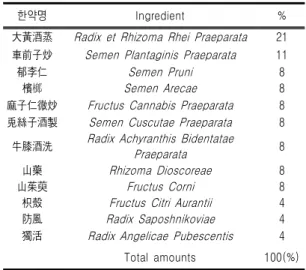
한약명 Ingredient % 大黃酒蒸 Radix et Rhizoma Rhei Praeparata 21 車前子炒 Semen Plantaginis Praeparata 11
郁李仁 Semen Pruni 8
檳榔 Semen Arecae 8
麻子仁微炒 Fructus Cannabis Praeparata 8 兎絲子酒製 Semen Cuscutae Praeparata 8
牛膝酒洗 Radix Achyranthis Bidentatae
Praeparata 8
山藥 Rhizoma Dioscoreae 8
山茱萸 Fructus Corni 8
枳殼 Fructus Citri Aurantii 4
防風 Radix Saposhnikoviae 4
獨活 Radix Angelicae Pubescentis 4
Total amounts 100(%)
Table Ⅰ. The Composition of SSEx1 혀짐으로써 이 분야에 대한 연구가 활발하게
이루어지고 있다5,6).
한의학적으로 대사증후군은 질병의 전단계 인 未病의 개념과 유사한데, 확실한 치료법 없 이 체중감량 등으로 증상의 호전을 목표로 하 기에7), ‘治未病’의 개념에서 접근할 수 있겠다.
『黃帝內經․素問』8)과 『難經』9)에서 “上工 治未病, 中工治已病.”이라 하여 未病의 치료를 중요시하였고, 최근 현대의학에서도 未病은 질 병의 전단계, 반건강 상태, 질환의 전변악화 전 상태 등을 포괄하는 개념으로 이해되며10), 未病에 대한 양생법과 예방적 치료를 중요하 게 생각하고 있다11).
실험에 사용된 疏風順氣元은 『世醫得效方』
12)에 ‘順氣圓’으로 나와 있으며, “治三十六種 風, 七十二般氣, 上熱下冷, 腰脚疼痛, 四肢無力, 惡瘡下疰, 大腸秘澁, 眞良方也...久服, 自然精神 强健, 百病不生.”이라 하였고, 처방구성은 『東 醫寶鑑․大便門』13)의 疏風順氣元과 동일하나 용량에 약간의 차이가 있다. 구성약물 중 가 감한 麻子仁은 老人이나 體虛 및 出産 後에 津 枯血少로 인한 腸燥便秘에 要藥으로14), rat의 혈중 TG, HDL-cholesterol, LDL-cholesterol, LPO (lipid peroxide) 등을 개선시켜 고지혈증 치료 효능이 있다는 보고가 있으나15), 환각성 분인 tetrahydrocannabinol (THC) 등을 함유하 여 한의학 전문가에 의해 주의 깊게 사용되어 져야 한다16-18). 疏風順氣元에 관한 연구로는, 정19)은 중풍환자의 便秘에 사용할 수 있다고 보고하였고, 이20) 등은 공복 및 비공복시 혈당,
당화혈색소, 중성지방의 농도, 소장의
glucosidase 활성 억제, 근육조직의 GLUT4 mRNA양의 감소를 보고하였다. 배21)는 혈당강 하, 체중증가 감소, 총 cholesterol수치 감소, TG 감소 등의 효과가 있다고 발표하였다.
이에 저자는 임상에서 빈용되고 있는 疏風順 氣元 (SSEx1)과 疏風順氣元去麻子仁 (SSEx2)이
대사증후군의 예방과 치료에 미치는 효과와 작 용기전을 실험적으로 연구하였다. 이를 위해 에 너지대사가 활발한 NMu2Li 간세포와 C2C12 골격근세포에서 지방대사와 관련된 bio marker 인 PPARs의 조절을 측정하고, MCAD (medium chain acyl-CoA dehydrogenase)와 VLCAD
(very long chain acyl-CoA dehydrogenase)
의 mRNA 수준을 측정하여, 다소의 지견을 얻 어 이에 보고하는 바이다.
Ⅱ. 실험재료 및 연구방법
1. 실험약물
실험약물은 『東醫寶鑑』13)에 수재된 疏風順 氣元 (SSEx1, SSEx2)을 사용하였으며, 그 구성 약물은 화림제약 (Busan, South Korea)에서 구 입하고, 동의대학교 한의과대학 방제학교실에서 정선한 뒤 분말하고 이 분말을 DW 3000 (South Korea, 대우사)을 이용하여 에탄올 30%
의 용매로 95℃에서 22시간 추출한 뒤에 동결 건조하여 실험에 사용하였다 (Table Ⅰ, Ⅱ).
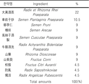
한약명 Ingredient % 大黃酒蒸 Radix et Rhizoma Rhei
Praeparata 22
車前子炒 Semen Plantaginis Praeparata 10.5
郁李仁 Semen Pruni 9
檳榔 Semen Arecae 9
兎絲子酒
製 Semen Cuscutae Praeparata 9 牛膝酒洗 Radix Achyranthis Bidentatae
Praeparata 9
山藥 Rhizoma Dioscoreae 9
山茱萸 Fructus Corni 9
枳殼 Fructus Citri Aurantii 4.5
防風 Radix Saposhnikoviae 4.5
獨活 Radix Angelicae Pubescentis 4.5
Total amounts 100(%)
Table Ⅱ. The Composition of SSEx2
Name Species Source/
Application Morphology Growth Mode NMu2Li mouse liver epithelial adherent
C2C12 mouse muscle fibroblast adherent
2. Cell culture
간세포주인 NMu2Li 세포와 골격근세포주인 C2C12 세포를 사용하였다 (Table Ⅲ). 모든 세포 주는 10% fetal bovine serum (Gibco-Brl, Grand Island, NY, USA), penicillin G (100 Uml-1), streptomycin sulfate (100 ㎍ml-1), amphotericin B (0.25 ㎍ml-1), 그리고 2-mercaptoethanol (50
㎛)이 포함되어 있는 DMEM 배지에서 배양하며, 37℃ 온도와 5% CO2가 공급되는 습윤한 조건을 유지시킨 후, transfection 24 시간 전에 6-well tissue culture plate에 well 당 2.7 X 10⁵개의 세포 수로 seeding 하였다.
Table Ⅲ. Cell Lines Used for Transient Transfection Assays
3. Transient transfection assay
모든 transfection의 경우 well 당 각각 200 ng
의 plasmid가 사용되고, lipofectamine (Life technologies, Rockville, MD)을 사용하여 제조사 의 지시에 따라 transfection하였다. 반응 6시간 후 신선한 배지 (1 ml)를 첨가하고, 해당 화학물인 SSEx1, SSEx2를 처리하며, 처리한지 24시간 후 배 지를 제거하고 배양한 세포를 수확하였다.
Luciferase와 β-galactosidase 활성은 kit (Promega, Madison, WI, USA)를 사용하여 제조 사의 지시에 따라서 측정하며, 간략히 설명하면 다음과 같다. β-galactosidase와 luciferase 분석에 사용되는 시약들은 냉동실에서 꺼내어 녹인 후 내 용물이 잘 섞이도록 흔들어 사용하였다. 6-well의 세포들은 배지를 제거한 후 PBS를 이용해서 2회 세척하며, 각각의 plate에 1 X reporter lysis buffer (300 ㎕)를 넣고 상온에서 15분간 방치해서 세포 를 용해시켰다. 세포가 완전히 용해되면 plate로 부터 세포들을 모으며, 완전히 용해된 세포를 원 심분리 (12,000 rpm, 4℃, 5분)하여 용해성 단백질 을 포함한 상층액을 분리하였다. Luciferase 활성 은 luciferase assay system을 이용하여 분석하며, luciferase를 포함한 상등액 (20 ㎕)과 substrate (30 ㎕)를 혼합하여 luminometer로 측정하였다.
Luciferase 활성을 보정하기 위해 β-galactosidase 활성을 조사하였다 (Promega's β-galactosidase enzyme assay system). 96 well plate에 분리된 상 층액과 2 X assay buffer를 20 ㎕씩 동량으로 분주 하여 37℃에서 1시간 반응시킨 후, 반응색이 노랗 게 변하면 1 M sodium carbonate (50 ㎕)를 넣고 반응을 정지시켜 spectrophotometer (405 nm)로 흡광도를 측정하였다.
4. C2C12 differentiation
Mouse myogenic C2C12 cell을 10% fetal bovine serum (Gibco-BRL, Grand Island, NY), penicillin G (100 U/ml), streptomycin sulfate (100 μg/ml), amphotericin B (0.25 μ g/ml)과 2-mercaptoethanol (50 μM)이 포함된
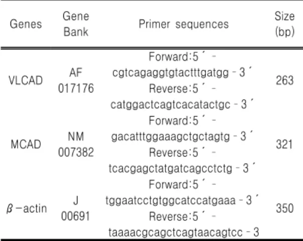
Genes Gene
Bank Primer sequences Size
(bp)
VLCAD AF
017176
Forward:5´
cgtcagaggtgtactttgatgg3´
Reverse:5´ 263 catggactcagtcacatactgc3´
MCAD NM
007382
Forward:5´
gacatttggaaagctgctagtg3´
Reverse:5´ 321 tcacgagctatgatcagcctctg3´
β-actin J
00691
Forward:5´
tggaatcctgtggcatccatgaaa3´
Reverse:5´ 350 taaaacgcagctcagtaacagtcc3
DMEM medium에서 배양하였다. C2C12 cell 의 분화를 유도하기 위하여 cell이 70%
confluence에 도달하였을 때 10% fetal bovine serum을 2% horse serum으로 교체하였다. 4 일간 배양 후 cell들은 다핵성 myotube로 분화 되었으며, SSEx1, SSEx2 등의 chemical 처리 후 RNA isolation을 위해 사용되었다.
5. Total RNA 분리
Total RNA는 Trizol (Invitrogen, Avenue Carlsbad, USA)을 사용하여 간, 지방, 근육조직으 로부터 추출하며, 간단히 설명하면 다음과 같다.
Total RNA을 추출하기 위해서 Trizol 1 ml에 조 직 100 mg을 넣고 homogenizer를 이용하여 20초 동안 조직을 갈며, 균질화된 용액을 상온에서 5분 간 방치하였다. 여기에 chloroform (Sigma, USA) 200 ㎕를 첨가한 후에 chloroform이 전체적으로 잘 섞이도록 15초 동안 혼합하며, 상온에서 3분간 방치한 후 원심분리 (13,000 rpm, 4℃, 15분) 하였 다. 원심분리 된 용액에서 맑은 상층액 만을 새로 운 튜브에 분리하고 여기에 동량의 isopropanol (Sigma, USA)을 첨가하여 상온에서 10분간 방치 한 후 원심분리 (13,000 rpm, 4℃, 10분) 하였다.
원심분리로 튜브 바닥에 형성된 RNA pellet에 70% ethanol 1 ml을 첨가하여 2회 세척하였으며 (4,500 rpm, 4℃, 5분), RNA pellet이 완전히 건조 되면 0.01% DEPC로 처리된 증류수를 150 ㎕ 첨 가하여 RNA pellet을 용해시켰다.
6. Reverse transcription-polymerase chain reaction
Reverse transcription-polymerase chain reaction (RT-PCR)을 이용하여 mRNA의 양을 측 정하였다. Complementary DNA는 total RNA 2
㎍과 reverse primer 0.5 ㎍을 혼합하여 최종 양 14 ㎕를 준비하여 75℃에서 15분 동안 열처리 (heating)한 후, 5분 동안 얼음 속에 보관하였다.
여기에 5X M-MLV reaction buffer, 10 mM dNTP mixture, 200 units M-MLV RT (Promega, Madison, WI, USA)를 첨가하여 최종 양이 25 ㎕ 가 되게 한 후, 42℃에서 1시간 동안 반응시켰다.
RT reaction 5 ㎕에 10X reaction buffer (Mg2+포 함), 10 mM dNTP, 5 unites Taq polymerase (Solgent, Taejon, South Korea), 그리고 10 μM primer를 첨가하여 최종 50 ㎕가 되게 한 후, PTC-100TM Programmable Thermal Controller (MJ Research, Inc., Waltham, MA, USA)를 이용 하여 PCR을 실시하였다. Table Ⅳ는 RT-PCR에 사용된 primer와 product size이다.
Table Ⅳ. Sequences of Primers Used for the RT-PCR Assays
7. Expression plasmids
Expression vector로 사용한 pSG5-mPPARα 와 PPRE3-tk-luc reporter 유전자는 Dr. Frank J. Gonzalez (National Cancer Institute, NIH, Bethesda, MD, USA)로 부터 얻어 사용하였다.
8. 통계분석
모든 값은 mean ± standard deviation (SD)으 로 표시하며, SigmaPlot 2001 (SPSS Inc, Chicago, IL)의 unpaired, student's t-test를 이용하여 통계 적 유의성을 검증하였다.
Ⅲ. 실험 결과
1. NMu2Li 간세포에서 PPARα reporter gene 발현 Mouse 간세포주인 NMu2Li 세포에 rat acyl-CoA oxidase 유전자의 PPRE를 포함하고 있는 luciferase reporter 발현벡터 (PPRE3-tk-luc)에 PPARα와 PPARα parter인 RXRα 발현벡터를 co-transfection 시켰다.
SSEx1 처리군은 0.1 μg/ml과 10 μg/ml 농 도에서 PPARα reporter 유전자 발현을 유의하 게 증가시켰는데, 각각 43% (p<0.01), 42%
(p<0.05)의 증가율을 나타냈다 (Fig. 1).
SSEx2 처리의 경우, 0.1 μg/ml과 1 μg/ml 농도에서 유의하게 증가시켰으며, 증가정도는 각각 46% (p<0.01)와 34% (p<0.05)였다 (Fig.
2).
Fig. 1. Regulation of PPARα reporter gene expression by SSEx1 in NMu2Li liver cells.
Cells were transiently transfected with expression plasmids for PPARα, a luciferase reporter gene construct containing 3 copies of the PPRE from the rat acyl-CoA oxidase gene and β-galactosidase gene. Cells were treated with several kinds of SSEx1 at the initial time of culture. Following incubation for 24 h, cells were harvested, lysed and were subsequently assayed for luciferase and β-galactosidase activities. All values are expressed as the mean ± SD of relative luciferase units/β-galactosidase activity. Experiments were performed at least three times.
* p<0.05, ** p<0.01 Significantly different from vehicle.
Fig. 2. Regulation of PPARα reporter gene expression by SSEx2 in NMu2Li liver cells.
Cells were transiently transfected with expression plasmids for PPARα, a luciferase reporter gene construct containing 3 copies of the PPRE from the rat acyl-CoA oxidase gene and β-galactosidase gene. Cells were treated with several kinds of SSEx2 at the initial time of culture. Following incubation for 24 h, cells were harvested, lysed and were subsequently assayed for luciferase and β-galactosidase activities. All values are expressed as the mean ± SD of relative luciferase units/β-galactosidase activity. Experiments were performed at least three times.
* p<0.05, ** p<0.01 Significantly different from vehicle.
2. C2C12 골격근세포에서 PPARα reporter gene 발현
Mouse 골격근 전구세포주인 C2C12 세포에 PPARα와 RXRα expression construct 그리고 rat ACOX 유전자의 PPRE luciferase reporter construct (PPRE3-tk-luc)를 transfection 시킨 후 PPARα reporter 유전자 발현을 측정하였다.
SSEx1 처리군은 1 μg/ml에서 luciferase activity를 유의하게 증가시켰으며, 증가정도는 69% (p<0.01)였다 (Fig. 3).
SSEx2 처리의 경우 0.1 μg/ml 농도에서 유 의하게 증가시켰으며, reporter gene activity의 증가정도는 65% (p<0.01)였다 (Fig. 4).
Fig. 3. Regulation of PPARα reporter gene expression by SSEx1 in C2C12 cells, a skeletal muscle myogenic progenital cell line.
Cells were transiently transfected with expression plasmids for PPARα, a luciferase reporter gene construct containing 3 copies of the PPRE from the rat acyl-CoA oxidase gene and β-galactosidase gene. Cells were treated with several kinds of SSEx1 at the initial time of culture. Following incubation for 24 h, cells were harvested, lysed and were subsequently assayed for luciferase and β-galactosidase activities. All values are expressed as the mean ± SD of relative luciferase units/β-galactosidase activity. Experiments were performed at least three times.
* p<0.01 Significantly different from vehicle.
Fig. 4. Regulation of PPARα reporter gene expression by SSEx2 in C2C12 cells, a skeletal muscle myogenic progenital cell line.
Cells were transiently transfected with expression plasmids for PPARα, a luciferase reporter gene construct containing 3 copies of the PPRE from the rat acyl-CoA oxidase gene and β-galactosidase gene. Cells were treated with several kinds of SSEx2 at the initial time of culture. Following incubation for 24 h, cells were harvested, lysed and were subsequently assayed for luciferase and β-galactosidase activities. All values are expressed as the mean ± SD of relative luciferase units/β-galactosidase activity. Experiments were performed
at least three times.
* p<0.01 Significantly different from vehicle.
3. NMu2Li 간세포에서 PPARδ reporter gene 발현 간세포주인 NMu2Li 세포에 PPARδ와 RXRα expression construct 그리고 rat ACOX 유전자의 PPRE luciferase reporter construct (PPRE3-tk-luc)를 transfection 시킨 후 PPARδ reporter 유전자 발현을 측정하였다.
SSEx1과 SSEx2 모두 PPARδ reporter 유전 자 발현에 유의성 있는 영향을 주지 못하였으 나, 10 μg/ml SSEx1 과 0.1 μg/ml SSEx2는 각각 19%와 33%씩 luciferase activity를 증가 시켰다 (Fig. 5, 6).
Fig. 5. Regulation of PPARδ reporter gene expression by SSEx1 in NMu2Li liver cells.
Cells were transiently transfected with expression plasmids for PPARδ, a luciferase reporter gene construct containing 3 copies of the PPRE from the rat acyl-CoA oxidase gene and β-galactosidase gene. Cells were treated with several kinds of SSEx1 at the initial time of culture. Following incubation for 24 h, cells were harvested, lysed and were subsequently assayed for luciferase and β-galactosidase activities. All values are expressed as the mean ± SD of relative luciferase units/β-galactosidase activity. Experiments were performed at least three times.
Fig. 6. Regulation of PPARδ reporter gene expression by SSEx2 in NMu2Li liver cells.
Cells were transiently transfected with expression plasmids for PPARδ, a luciferase reporter gene construct containing 3 copies of the PPRE from the rat acyl-CoA oxidase gene and β-galactosidase gene. Cells were treated with several kinds of SSEx2 at the initial time of culture. Following incubation for 24 h, cells were harvested, lysed and were subsequently assayed for luciferase and β-galactosidase activities. All values are expressed as the mean ± SD of relative luciferase units/β-galactosidase activity. Experiments were performed at least three times.
4. C2C12 골격근세포에서 PPARδ reporter gene 발현
Mouse 골격근 전구세포주인 C2C12 세포에 PPARδ와 RXRα expression construct 그리고 rat ACOX 유전자의 PPRE luciferase reporter construct (PPRE3-tk-luc)를 transfection 시킨 후 PPARδ reporter 유전자 발현을 측정하였다.
SSEx1, SSEx2 처리군의 PPARδ reporter gene 발현이 유의성 있는 변화를 보이지 않았 다 (Fig. 7, 8).
Fig. 7. Regulation of PPARδ reporter gene expression by SSEx1 in C2C12 cells, a skeletal muscle myogenic progenital cell line.
Cells were transiently transfected with expression plasmids for PPARδ, a luciferase reporter gene construct containing 3 copies of the PPRE from the rat acyl-CoA oxidase gene and β-galactosidase gene. Cells were treated with several kinds of SSEx1 at the initial time of culture. Following incubation for 24 h, cells were harvested, lysed and were subsequently assayed for luciferase and β-galactosidase activities. All values are expressed as the mean ± SD of relative luciferase units/β-galactosidase activity. Experiments were performed at least three times.
Fig. 8. Regulation of PPARδ reporter gene expression by SSEx2 in C2C12 cells, a skeletal muscle myogenic progenital cell line.
Cells were transiently transfected with expression plasmids for PPARδ, a luciferase reporter gene construct containing 3 copies of the PPRE from the rat acyl-CoA oxidase gene and β-galactosidase gene. Cells were treated with several kinds of SSEx2 at the initial time of culture. Following incubation for 24 h, cells were harvested, lysed and were subsequently assayed for luciferase and β-galactosidase activities. All values are expressed as the mean ± SD of relative luciferase units/β-galactosidase activity. Experiments were performed at least three times.
5. NMu2Li 간세포에서 미토콘드리아 MCAD mRNA 발현
간세포주인 NMu2Li 세포에 PPARα와 RXR α expression construct 그리고 rat ACOX 유 전자의 PPRE luciferase reporter construct (PPRE3-tk-luc)를 transfection 시킨 후 SSEx1, SSEx2를 각각 처리한 후, total RNA를 분리하 고 RT-PCR을 이용하여 mRNA 수준을 정량하 였다.
PPARα의 강력한 activator인 Wy14,643은 MCAD mRNA를 69% (p<0.05)증가시켰으며, SSEx1은 MCAD mRNA를 28% (p<0.05)정도 유의하게 상승시켰으나, SSEx2는 MCAD mRNA 발현에 특별한 변화를 나타내지 않았 다 (Fig. 9).
Fig. 9. Modulation of mitochondrial MCAD mRNA expression by SSEx in NMu2Li cells.
(A) Cells were transiently transfected with expression plasmids for PPARα and a luciferase reporter gene construct containing 3 copies of the PPRE from the rat acyl-CoA oxidase gene. Cells were treated with SSEx at the initial time of culture.
Following incubation for 24 h, cells were harvested and subsequently processed for RNA extraction. mRNA levels of mitochondrial MCAD and β-actin were measured as described in Materials and Methods. All values are expressed as mean ± SD of R.D.U. (relative density units) using β-actin as a reference.
(B) Representative RT-PCR bands from one of three independent experiments are shown.
* p<0.05 compared with vehicle. MCAD, medium chain acyl-CoA dehydrogenase.
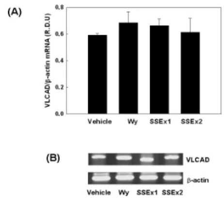
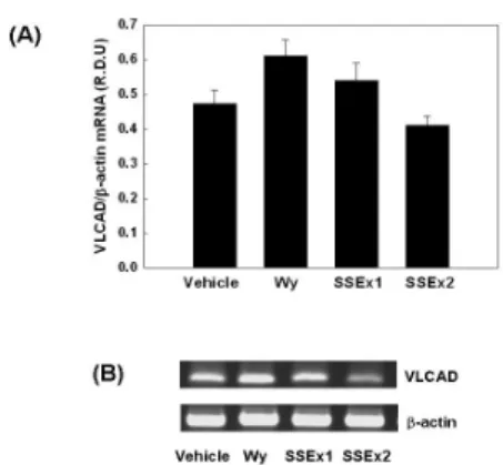
6. NMu2Li 간세포에서 미토콘드리아 VLCAD mRNA 발현
간세포주인 NMu2Li 세포에 PPARα와 RXR α expression construct 그리고 rat ACOX 유 전자의 PPRE luciferase reporter construct (PPRE3-tk-luc)를 transfection 시킨 후 SSEx1, SSEx2를 각각 처리한 후, total RNA를 분리하 고 RT-PCR을 이용하여 mRNA 수준을 정량하 였다.
Wy14,643은 15%. SSEx1은 10% VLCAD mRNA를 증가시켰으나 유의성은 없었고, SSEx2는 VLCAD mRNA 발현에 특별한 변화 를 나타내지 않았다 (Fig. 10).
Fig. 10. Modulation of mitochondrial VLCAD mRNA expression by SSEx in NMu2Li cells.
(A) Cells were transiently transfected with expression plasmids for PPARα and a luciferase reporter gene construct containing 3 copies of the PPRE from the rat acyl-CoA oxidase gene. Cells were treated with SSEx at the initial time of culture.
Following incubation for 24 h, cells were harvested and subsequently processed for RNA extraction. mRNA levels of mitochondrial VLCAD and β-actin were measured as described in Materials and Methods. All values are expressed as mean ± SD of R.D.U. (relative density units) using β-actin as a reference.
(B) Representative RT-PCR bands from one of three independent experiments are shown. VLCAD, very long chain acyl-CoA dehydrogenase.
7. C2C12 골격근세포에서 미토콘드리아 MCAD mRNA 발현
골격근 전구세포주인 C2C12 myoblast를 myotube로 분화시켜 SSEx1, SSEx2를 처리하 여 MCAD mRNA 발현을 측정한 결과, 각각 Wy14,643은 86% (p<0.05), SSEx1은 126%
(p<0.05), SSEx2는 106% (p<0.05)로 유의하게 증가시켰다 (Fig. 11).
Fig. 11. Modulation of mitochondrial MCAD mRNA expression by SSEx in differentiated C2C12 cells.
(A) C2C12 myoblasts were differentiated into myotubes and then myotubes were treated with SSEx for 24 h. RNA was extracted from differentiated cells and mRNA levels of mitochondrial MCAD and β-actin were measured as described in Materials and Methods. All values are expressed as mean ± SD of R.D.U. (relative density units) using β-actin as a reference.
(B) Representative RT-PCR bands from one of three independent experiments are shown.
* p<0.05 compared with vehicle. MCAD, medium chain acyl-CoA dehydrogenase.
8. C2C12 골격근세포에서 미토콘드리아 VLCAD mRNA 발현
골격근 전구세포주인 C2C12 myoblast를 myotube로 분화시켜, SSEx1, SSEx2를 처리하여 VLCAD mRNA 발현을 측정한 결과, 각각 Wy14,643는 28%, SSEx1은 13% 증가하는 경향을 나타냈으며, SSEx2는 감소하는 경향이 나타났지 만, SSEx1, SSEx2 모두 유의성은 없었다 (Fig. 12).
Fig. 12. Modulation of mitochondrial VLCAD mRNA expression by SSEx in differentiated C2C12 cells.
(A) C2C12 myoblasts were differentiated into myotubes and then myotubes were treated with SSEx for 24 h. RNA was extracted from differentiated cells and mRNA levels of mitochondrial VLCAD and β-actin were measured as described in Materials and Methods. All values are expressed as mean ± SD of R.D.U. (relative density units) using β-actin as a reference.
(B) Representative RT-PCR bands from one of three independent experiments are shown. VLCAD, very long chain acyl-CoA dehydrogenase.
Ⅳ. 고 찰
의학의 발달과 함께, 뇌졸중과 같은 순환기 질환으로 인한 사망률은 감소하였으나, 환자 수는 오히려 늘고 있는 추세이며, 이로 인하여 자신의 건강을 스스로 지키려는 예방의학적 태도가 사회전체에서 인지되고 있다. 동양의 예방의학적 개념은 기공, 요가 등을 포괄하는 養生論으로 잘 확립되어 있는데10), 『黃帝內 經․素問』8)에서는 “聖人不治已病, 治未病”이 라는 기록이 있고, 『難經』9)에는 “上工治未 病, 中工治已病”이란 개념으로, 질병의 이전 단계, 未病 상태에서의 예방적 치료를 중요시 해온 것을 알 수 있다.
未病의 현대의학적 개념으로 有地滋는 장기 와 관련된 未病, 면역유전학적, 유전대사학적 질환의 未病을 들었으며, 翁維良은 ‘반건강의 예방과 치료’의 개념에서 ‘건강상태에서 생리 기능 감퇴, 단일증상 출현, 여러 증상의 출현 또는 특정 해부학적 부위 또는 장기의 증상 출현, 자각 및 타각 증상의 동시출현, 특정 질 병상태로 진행’의 순으로 未病의 역동적 단계 를 설명하였다. 永田勝太郞은 ‘건강, 기능적 병 태(제 1상), 기질적 병태(제 2상), 치사적 병태 (제 3상), 죽음’ 등의 순서에서 현대의학은 제 2상에서 역할을 하고 있으나, 1상과 3상에서는 무기력함을 주장하였고, Balint는 “의료 현장 에서 의사의 가장 중요한 역할은 병이 아직 병리학적으로 미완성 상태에서 진단 치료할 필요가 있다”고 하였으며, 本多勝一은 未病을
‘죽음으로 이르는 길, 또는 건강의 길로 가는 병’으로 양면성이 있음을 말하였다11).
한의학에서의 질병 전단계, 반건강 상태, 질 환의 전변악화 전 상태 등의 未病 개념은 현 대의학에서의 비만, 당대사장애, 지질대사장애 및 고혈압, 심혈관계 질환에 영향을 미쳐 총 체적 장기부전의 발생과 관련되는 대사증후군 과 유사한 점이 있을 것으로 생각되었다.
대사증후군 (metabolic syndrome)은 1988년 Reaven1) 등이 인슐린 저항성, 고지혈증, 고혈 압이 한꺼번에 나타나는 경우를 ‘Syndrome X’
로, 1989년 DeFronzo에 의해 인슐린 저항성 증후군, 1989년 Kaplan 등은 비만, 내당능장애, 고지혈증, 고혈압이 있는 경우를 Deadly Quarter로 지칭된 후 1998년 이후부터 명명되 었다. NCEP-ATPⅢ (National Cholesterol Education Program Adult Treatment Panel
Ⅲ)에서는 복부비만, 고혈압, 내당능장애나 당 뇨병, 고중성지방혈증, 저HDL-cholesterol혈증 의 다섯 가지 위험요소 중 3개 이상을 가지고 있는 경우 대사증후군이라 정의하였다2).
국내 대사증후군의 유병률은 2001년 국민건 강영양조사에서 남녀 각각 22.5%, 24.1%이지 만, 연령증가에 따라 현저히 증가하며, 60세 이상 노인에서는 약 30~70%로 훨씬 높게 나 타났다3).
대사증후군을 확실히 치료할 수 있는 방법 은 아직 개발되지 않았으나, 비만한 사람의 체 중을 감소시키면 인슐린 저항성 및 이와 동반 된 당뇨병, 고혈압, 고지혈증 등이 호전되며, 영양요법, 행동수정, 인지교정, 운동훈련을 병 행하여 심혈관기능을 호전시키는 한편 체지방 분포를 유리한 방향으로 전환시킬 것을 권장 하고 있다7).
Energy homeostasis는 흡수한 에너지와 소 비된 에너지 사이의 정밀한 균형을 나타낸다.
그러나 과도한 에너지의 섭취로 이러한 균형 이 깨지게 되면 비만이나 동맥경화증, 그리고 제 2형 당뇨병과 같은 대사이상을 일으키는데, 지난 수년간 orphan receptor인 PPAR (peroxisome proliferator-activated receptor)가 energy balance를 조절하는 중요한 인자로 밝 혀짐으로써 이 분야에 대한 연구가 활발하게 이루어지고 있다5,6).
PPAR는 조직내 지질 전달, 지질 섭취, 지방 산 대사(acyl-CoA oxidase, acyl-CoA synthetase, long-chain acyl-CoA Dehydrogenase) 등의 광범위한 지질 대사에 관여하는 유전자의 발현을 조절하는 것으로 알려져 있는데22-25), 고지혈증 치료제인 fibrate 류, 당뇨병 치료제인 thiazolidinedione, 지방 산, 가소제, 화학 산업에서 사용되는 유기용매, 살충제, 그리고 제초제 등과 같은 peroxisome proliferator (PP)에 의해 활성화되며26), PP에 의해 활성화된 PPAR는 9-cis-retinoic acid receptor (RXR)와 heterodimer를 이룬 후, target gene들의 upstream에 있는 PPAR response elements (PPREs)에 결합하여 target
gene들의 발현을 조절한다27). PPARα는 간과 같이 지방산의 물질대사가 활발히 일어나는 조직에서 주로 많이 발현되며, 이 외에도 신장, 심장, 근육 등 다양한 조직에서 발견된다.
PPARδ는 다양한 조직에서 분포되지만 주로 장, 신장, 심장에서 발현된다. PPARα에 대한 연구에서 고지혈증 치료제인 fibrate류의 화합 물들이 PPARα activator 임이 밝혀졌다.
fibrate는 triglyceride (TG)의 혈중 농도를 낮 추고, 사람의 경우 high density lipoprotein (HDL)의 농도를 증가시키는 효과적인 약물로 써 심혈관질환 (cardiovascular disease)을 치 료하기 위해 점차 그 사용이 증가되고 있는데
28), 분자수준에서는 lipoprotein과 지방산 대사 에 관여하는 유전자를 조절하는 PPARα activator로써 작용한다29). 그러나 triglyceride 와 cholesterol 대사에 이상이 있는 PPARα-KO mouse는 fibrate에 반응하지 못하고, target enzyme의 유전자들을 발현시키지 못하며, 궁 극적으로 시간이 지남에 따라 비만이 된다30). 또한 당뇨병, 비만, 인슐린 저항성을 가진 실 험동물과 고지방식을 섭취한 C57BL/6 mouse 의 몸무게에 대한 fenofibrate의 효과가 보고됨 으로써, PPARα는 세포내 뿐만 아니라 세포외 지방대사의 중요한 조절인자로 보고되고 있다
31-33).
다양한 조직에서 발현되는 PPARδ는 지방조 직, 태반, 피부, 소장과 같은 여러 조직의 발달 과 생리에 중요한 역할을 하는 것으로 알려져
있다34-38). 최근들어 PPARδ synthetic ligand를
이용한 연구를 기초로, 지질대사에서의 PPARδ 의 기능에 관한 연구가 활발하게 이루어지고 있다. PPARδ agonist를 비만 mouse에 처리했 을 때 지방무게가 감소되고 인슐린민감성이 향상되었으며, 비만형 당뇨동물의 혈당과 인슐 린 농도를 정상화시키고 고밀도지단백질의 농 도를 증가시켰다39,40). 특히 PPARδ는 골격근육
과 갈색지방조직에서 지방산 산화를 촉진함으 로써 비만을 효과적으로 조절하는 것으로 보 인다. PPARδ는 골격근육에서 가장 많이 발현 되는 PPAR isotype이며 L6 myotube나 C2C12 세포에 PPARδ agonist를 처리하면 지질대사에 관련된 유전자들의 발현이 증가하고 지방산 산화가 촉진된다41,42). 또한 PPARδ는 갈색지방 조직에서 지방산 분해와 에너지 uncoupling을 촉진시키고 지방무게를 감소시켰으며, 고지방 식에 의한 비만과 고지혈증을 억제하였다40). 이러한 결과로 보아 PPARδ는 비만을 비롯한 metabolic syndrome의 조절에 유용한 것으로 보고되었다43,44).
疏風順氣元은 『世醫得效方』12)에 ‘順氣圓’으 로 나와 있으며, “治三十六種風, 七十二般氣, 上熱下冷, 腰脚疼痛, 四肢無力, 惡瘡下疰, 大腸 秘澁, 眞良方也...久服, 自然精神强健, 百病不 生.”이라 하였고, 처방구성은 『東醫寶鑑․大 便門』13)의 疏風順氣元과 동일하나 용량에 약 간의 차이가 있다. 또한 疏風順氣元은 『東醫 寶鑑․大便門』의 老人秘結과 『東醫寶鑑․風 門』의 風秘에는 疏風順氣元으로 『東醫寶 鑑․風門』의 手足癱瘓에는 秘方疏風順氣湯으 로 『東醫寶鑑․身形門』의 老人治法에는 疏 風順氣丸으로 『東醫寶鑑․風門』의 중풍전구 증상에서는 搜風順氣丸으로 이름을 달리하여 기재되어 있으나 처방구성은 동일하다13). 구성 약물은 攻積, 瀉火, 逐瘀하는 大黃, 利水滲濕하 는 車前子, 潤燥滑腸, 下氣, 利水하는 郁李仁, 殺蟲, 行氣, 消積, 利水하는 檳榔, 緩下通便, 滋 養補虛하는 麻子仁, 固精, 明目, 止瀉하는 兎絲 子, 活血去瘀, 引血下行하는 牛膝, 健脾, 補肺, 固腎, 益精하는 山藥, 補益肝腎, 澁精固脫하는 山茱萸, 行氣寬中하는 枳殼, 解表去風, 勝濕, 止痛하는 防風, 去風除濕, 解表止痛하는 獨活 이다14). 疏風順氣元의 구성약물 중 麻子仁은 老人이나 體虛 및 出産後에 津枯血少로 인한
腸燥便秘에 要藥으로14), 구성 지방산 중에서 strearic acid, docohexaenoic acid, linoleic acid, arachidonic acid는 체내에서 cholesterol 치를 저하시키므로 성인병 예방에 도움이 될 것으로 생각된다45). 또한 rat의 혈중 TG, HDL-cholesterol, LDL-cholesterol, LPO (lipid peroxide) 등을 개선시켜 고지혈증 치료 효능 이 있다는 보고가 있으나15), 대마에는 마취성
이나 환각작용을 일으키는
tetrahydrocannabinol (THC), cannabinol, cannabidinol 등이 포함되어 의약전문가에 의 해 주의깊게 사용되어져야 한다16-18).
疏風順氣元에 대한 연구를 살펴보면, 정19)은 3일 이상의 대변불통을 호소하는 뇌졸중 환자 들의 변비에 복통이나 설사 등의 부작용이 적 고, 하루 최대 21g의 복용은 간이나 신장에 무 리를 주지 않아서 중풍환자의 便秘에 무난히 사용할 수 있다고 보고하였다. 이20) 등은 疏風 順氣元이 db/db mouse의 포도당수송체를 증 가시켜 혈중 포도당 유입이 증가하고 그로 인 해 혈당치가 감소되는 기전으로 공복 및 비공 복시 혈당, 당화혈색소, 중성지방의 농도를 유 의하게 감소시키고, 소장의 glucosidase 활성억 제와 근육조직의 GLUT4 mRNA양의 감소를 보고하였다. 배21)는 고지방식이로 고혈당을 유 발한 mouse에 疏風順氣元을 투여하여 혈당강 하, 체중증가 감소, 총 cholesterol수치 감소, TG 감소 등의 효과가 있다고 발표하였다.
이에 저자는 상기의 실험결과를 바탕으로 疏 風順氣元을 고혈당, 비만, 혈관병증, 지방대사 장애 등을 보이는 대사증후군 치료제로 활용할 수 있을 것으로 착안하여, 분자수준에서의 효율 과 작용기전에 대한 연구를 우선 시행하기로 하였다. 또한 장복 시에 환각작용에 대한 우려 의 소지가 있는 麻子仁을 제거한 疏風順氣元의 효능을 실험적으로 확인하고자 하였다.
본 실험에서는 대사가 활발하게 일어나는
mouse 간세포주인 NMu2Li 세포에 rat acyl-CoA oxidase 유전자의 PPRE를 포함하고 있는 luciferase reporter 발현벡터 (PPRE3-tk-luc)와 PPARα와 PPARα parter인 RXRα 발현벡터를 co-transfection 시킨 후, PPARα reporter 유전자 발현 정도를 측정하였 다.
SSEx1 실험군은 0.1 μg/ml과 10 μg/ml 농 도에서 PPARα reporter 유전자 발현을 각각 43% (p<0.01), 42% (p<0.05) 유의하게 증가시 켰으며 (Fig. 1), SSEx2 실험군은 0.1 μg/ml와 1 μg/ml 농도에서 유의하게 증가시켰으며 증 가 정도는 각각 46% (p<0.01)와 34% (p<0.05) 였다 (Fig. 2).
또한 mouse 골격근 전구세포주인 C2C12 세 포에 PPARα와 RXRα expression construct 그리 고 rat ACOX 유전자의 PPRE luciferase reporter construct (PPRE3-tk-luc)를 transfection 시킨 후 PPARα reporter 유전자 발현을 측정하였다.
SSEx1 실험군은, 1 μg/ml농도에서 69%
(p<0.01) 유의하게 증가시켰으며 (Fig. 3), SSEx2 실험군은, 0.1 μg/ml농도에서 65%
(p<0.01) 유의하게 증가시켰다 (Fig. 4).
Mouse NMu2Li 간세포와 C2C12 골격근세 포에 각각 PPARδ와 RXRα expression construct 그리고 rat ACOX 유전자의 PPRE luciferase reporter construct (PPRE3-tk-luc)를 transfection 시키고, PPARδ reporter 유전자 발현을 측정하 였다.
NMu2Li 간세포에서 10 μg/ml SSEx1과 0.1 μg/ml SSEx2 실험군에서 각각 19%와 33%씩 luciferase activity를 증가시켰으나 (Fig. 5, 6), 유의성은 없었고, C2C12 골격근세포에서는 SSEx1, SSEx2 실험군 모두 PPARδ reporter gene 발현에 영향을 주지 않았다 (Fig. 7, 8).
Mouse NMu2Li 간세포와 C2C12 골격근세 포에서 SSEx1, SSEx2가 PPARα 표적 유전자
발현에 어떠한 영향을 나타내는지 알아보고자 미토콘드리아에서 fatty acid-β oxidation을 촉 진하는 PPARα 표적유전자인 medium chain acyl-CoA dehydrogenase (MCAD)와 very long chain acyl-CoA dehydrogenase (VLCAD)의 mRNA 수준을 측정하였다.
간세포주인 NMu2Li 세포에 PPARα와 RXRα expression construct 그리고 rat ACOX 유전자의 PPRE luciferase reporter construct (PPRE3-tk-luc)를 transfection 시킨 후 SSEx를 처 리하였으며, total RNA를 분리한 후 RT-PCR을 이용하여 mRNA 수준을 정량하였다.
MCAD mRNA의 경우 SSEx1 처리군은 28%
(p<0.05) 유의하게 증가하였으나, SSEx2는 별다 른 변화를 나타내지 않았다 (Fig. 9). VLCAD mRNA의 경우 SSEx1 처리군은 10% 증가하였 으나, 유의성은 없었고, SSEx2는 별다른 변화 를 나타내지 않았다 (Fig. 10).
골격근세포에서 SSEx에 의한 PPARα target mRNA 발현을 조사하고자 mouse 골격근 전 구세포주인 C2C12 myoblast를 myotube로 분 화시켜 사용하였다. 분화된 C2C12 세포는 3종 류의 PPAR 모두를 발현하는 것으로 알려져 있으므로 PPARα expression plasmid를 transfection 시키지 않은 상태에서 분화된 myotube에 직접 SSEx1, SSEx2를 처리하여 MCAD와 VLCAD의 mRNA 발현을 조사하였 다.
MCAD mRNA의 경우 SSEx1, SSEx2 처리 군 모두 유의하게 증가하였으며 증가 정도는 각각 126% (p<0.05)와 106% (p<0.05)였다 (Fig.
11). VLCAD mRNA의 경우 SSEx1 처리군은 13% 증가하였으나, 유의성은 없었다 (Fig. 12).
이상의 결과를 총괄하면 SSEx1과 SSEx2는 mouse 간세포와 골격근세포에서 PPARα 활성 화를 촉진하고, PPARδ 활성화에는 영향을 주 지 않는 것을 확인하였다. SSEx1은 간세포와
골격근세포 모두에서 MCAD, VLCAD의 mRNA 발현을 증가시켰으며, SSEx2는 골격근 세포에서 MCAD의 mRNA 발현을 증가시키는 것으로 보였다. 따라서 SSEx1은 SSEx2와 비교 하여 지방산 산화를 좀 더 효율적으로 촉진할 것으로 생각된다. 그러므로, 疏風順氣元은 궁극 적으로 지방대사와 비만 등을 포함하는 대사증후 군에 치료적 효용이 있을 것으로 사료되며, 향후 더 자세한 기전에 대한 분석과 임상연구가 필요 하리라 생각 된다.
Ⅴ. 결 론
대사증후군에서 疏風順氣元(SSEx1)과 疏風順 氣元去麻子仁(SSEx2)의 활용가능성과 작용 기 전을 규명하기 위하여, mouse NMu2Li 간세 포와 C2C12 골격근세포에서 PPARα reporter gene, PPARδ reporter gene의 발현과, MCAD mRNA, VLCAD mRNA 발현을 측정한 결과 다음의 결론을 얻었다.
1. NMu2Li 간세포에서 SSEx1 처리군은 0.1 μ g/ml에서 43%, 10 μg/ml에서 42% PPARα reporter 유전자 발현을 유의하게 증가시켰 다. SSEx2 처리군은 0.1 μg/ml와 1 μg/ml 농도에서 각각 46%와 34% 유의하게 증가 시켰다.
2. C2C12 골격근세포에서 PPARα reporter gene 발현을 측정한 결과, SSEx1 처리의 경 우, 1 μg/ml 농도에서 luciferase activity를 69% 유의하게 증가시켰으며, SSEx2의 경우, 0.1 μg/ml 농도에서 65% 유의하게 증가시 켰다.
3. NMu2Li 간세포와 C2C12 골격근세포에서 PPARδ reporter gene 발현을 측정한 결과, SSEx1과 SSEx2는 모두 유의성 있는 영향을 주지 못했다.
4. NMu2Li 간세포에서 미토콘드리아 MCAD mRNA 발현을 측정한 결과, SSEx1은 28%
정도 유의하게 증가시켰다.
5. C2C12 골격근세포에서 미토콘드리아 MCAD mRNA 발현을 측정한 결과, SSEx1 은 126%, SSEx2는 106%로 유의하게 증가시 켰다.
이상의 결과로 미루어 보아 疏風順氣元이 지방대사와 비만 등을 포함한 대사증후군 치 료에 활용할 수 있는 실험적 근거를 제시한다 고 사료된다.
참 고 문 헌
1. Reaven GM. Role of insulin resistance in human disease. Diabetes.
1999;37:1595-607.
2. Journal of the American Medical Association(JAMA). National Cholesterol Education Program:Executive summary of the third report of the national cholesterol education program(NCEP) expert panel on detection, evaluation and treatment of high blood cholesterol in adults(adult treatment panel Ⅲ). Circulation.
2001;285:2488-97.
3. 임수, 이은정, 구보경, 조성일, 박경수, 장학 철, 김성연, 이홍규. 국민건강영양조사에 근
거한 한국인의 대사증후군의 증가양상. 당 뇨병학회지. 2005;19(5):432-9.
4. 권혁상. 대사증후군의 역학 및 병인. 대한당 뇨병학회지. 2006;7(1):12-9.
5. Kliewer SA, Lehmann JM, Willson TM.
Orphan nuclear receptors:shifting endocrinology into reverse. Science.
1999;284:757-60.
6. Gustafsson JA. Seeking ligands for lonely orphan receptors. Science. 1999;284:1285-6.
7. 이기업. 대사증후군의 치료. 한국지질동맥경 화학회지. 2002;12(1):31-5.
8. 양유걸. 황제내경소문역해. 서울:성보사.
1980:19.
9. 진월인. 난경입문. 서울:법인문화사.
1998:338-40.
10. 전국한의과대학 신경정신과 교과서편찬위 원회. 한의신경정신과학. 서울:집문당.
2007:630-1.
11. 武市昌士. 미병의 동서의학. 서울:군자출판 사. 2008:23-5.
12. 危亦林. 世醫得效方. 北京:中國中醫藥出版 社. 1996:98.
13. 허준. 동의보감. 서울:법인문화사.
1999:140,470,945,948,962,964.
14. 전국한의과대학 본초학교수 공편저. 본초 학. 서울:영림사. 1999:131,242,246,247, 260,313,351,378,427,537,568,626.
15. 任汉陽, 等. 火麻仁油的降脂及對過氧化脂質 作用的實驗硏究. 中國中醫藥科技.
1997;4(4):200.
16. Luthra YK, Rosenkrantz H, Braude MC.
Cerebral and cerebellar neurochemical changes and behavioral manifestations in rats chronically exposed to marijuana smoke. Toxicology and Applied Pharmacology. 1976;35(3):455-65.
17. Barnett G, Chiang CW, Perez-Reyes M, Owens SM. Kinetic study of smoking marijuana. J. Pharmacokinetics and Biopharmaceutics. 1982;10(5):495-506.
18. Mason AP, McBay AJ.
Cannabis:pharmacology and interpretation of effects. J. Forensics Sciences.
1985;30(3):615-31.
19. 정재욱. 중풍환자의 변비에 대한 소풍순기 원의 임상적 연구. 동의대학교 석사학위논 문. 2005.
20. 이성현, 안세영, 두호경, 정성현. db/db 마 우스에서 수풍순기환의 혈당강하 및 기전연 구. 응용약물학회지. 1999;7:335-41.
21. 배미라, 정성현. 고지방식이 섭취 마우스에 서 수풍순기환의 혈당강하활성 및 기전. 경 희약대논문집. 2000;28:101-8.
22. McAinch AJ, Lee JS, Burce CR, Tunstall RJ, Hawley JA, Cameron-Smith D. Dietary regulation of fat oxidative gene expression in different skeletal muscle fiber types.
Obesity Res. 2003;11(12):1471-9.
23. Van Der Lee KA, Willemsen PH, Van Der Vusse GJ, Van Bilsen M. Effect of fatty acids on uncoupling protein 2 expression in the rat heart. FASEB J.
2000;14(3):495-502.
24. Wu P, Peters JM, Harris RA. Adaptive increase in pyruvate dehydrogenase kinase 4 during starvation is medicated by peroxisome proliferators-activated receptor alpha. Biochem Biophys Res Commun. 2001;287(2):391-6.
25. Young ME, Goodwin GW, Ying J, Guthrie P, Wilson CR, Laws FA, Taegtmeyer H. Regulation of cardiac and skeletal muscle malonyl CaA
decarboxylase by fatty acids. Am J Physiol Endocrinol Metab.
2001;280(3):471-9.
26. Lake BG. Mechanisms of hepatocarcinogenicity of peroxisome-proliferating drugs and chemicals. Annu Rev Pharmacol Toxicol.
1995;35:483-507.
27. Glass BG. Differential recognition of target genes by nuclear receptor monomers, dimers, and heterodimers.
Endocr Rev. 1994;15(3):391-407.
28. Claudel T, Leibowitz MD, Fiévet C, Tailleux A, Wagner B, Repa JJ, Torpier G, Lo. Reduction of atherosclerosis in apolipoprotein E knockout mice by activation of the retinoid X receptor. Proc Natl Acad Sci U S A. 2001;98(5):2610-5.
29. Schoonjan K, Staels B, Auwerx J. Role of the peroxisome proliferator-activated receptor (PPAR) in mediating the effects of fibrates and fatty acids on gene expression. J Lipid Res. 1996;37(5):907-25.
30. Neve BP, Fruchart J-C, Staels B. Role of the peroxisome proliferator-activated receptors (PPARs) in atherosclerosis.
Biochem Pharmacol. 2000;60(8):1245-50.
31. Chaput E, Saladin R, Silvestre M, Edgar AD. Fenofibrate and rosiglitazone lower serum triglycerides with opposing effects on body weight. Biochem Biophys Res Commun. 2000;271(2):445-50.
32. Guerre-Millo M, Gervois D, Raspe E, Madsen L, Poulain P, Derudas B, Herbert JM. Peroxisome proliferator-activated receptor alpha activators improve insulin sensitivity and reduce adiposity. J Biol
Chem. 2000;275(22):16638-42.
33. Mancini FP, Lanni A, Sabatino L, Moreno M, Giannino A, Contaldo F, Colantuoni C. Fenofibrate prevents and reduces body weight gain and adiposity in diet-induced obese rats. FEBS Lett.
2001;491(1/2):154-8.
34. Grimaldi PA. The roles of PPARs in adipocyte differentiation. Prog Lipid Res.
2001;40(4):269-81.
35. Lim H, Gupta RA, Ma WG, Paria BC, Moller DE, Morrow JD, DuBois RN, Trzaskos JM. Cyclo-oxygenase-2-derived prostacyclin mediates embryo implantation in the mouse via PPARdelta. Genes Dev.
1999;13(12):1561-74.
36. Soon TN, Liliane M, Beatrice D, Walter W. Peroxisome proliferator-activated receptor (PPAR)-beta as a target for wound healing drugs: what is possible?
Am J Clin Dermatol. 2003;4(8):523-30.
37. Poirier H, Niot I, Monnot MC, Braissant O, Meunier-Durmort C, Costet P, Pineau T, Wah. Differential involvement of peroxisome-proliferator-activated receptors alpha and delta in fibrate and fatty-acid-mediated inductions of the gene encoding liver fatty-acid-binding protein in the liver and the small intestine.
Biochem J. 2001;355(Pt 2):481-8.
38. Peters JM, Lee SS, Ward JM, Gavrilova O, Everett C, Reitman ML, Hudson LD.
Growth, adipose, brain, and skin alterations resulting from targeted disruption of the mouse peroxisome proliferator-activated receptor beta(delta).
Mol Cell Biol. 2000;20(14):5119-28.
39. Tanaka T, Yamamoto J, Iwasaki S, Asaba H, Hamura H, Ikeda Y. Activation of peroxisome proliferator-activated receptor delta induces fatty acid beta-oxidation in skeletal muscle and attenuates metabolic syndrome. Proc Natl Acad Sci U S A.
2003;100(26):15924-9.
40. Wang YX, Lee CH, Tiep SB, Yu RT, Ham JY, Kang HJ, Evans.
Peroxisome-proliferator-activated receptor delta activates fat metabolism to prevent obesity. Cell. 2003;113(2):159-70.
41. Muoio DM, MacLean PS, Lang DB, Lis, Houmard JA, Way JM, Winegar DA.
Fatty acid homeostasis and induction of lipid regulatory genes in skeletal muscles of peroxisome proliferator-activated receptor (PPAR) alpha knock-out mice.
Evidence for compensatory regulation by PPAR delta. J Biol Chem.
2002;277(29):26089-97.
42. Dorte H, Serge L, Véronique N, Karsten K, Xavier L, G. Nutritional regulation and role of peroxisome proliferator-activated receptor delta in fatty acid catabolism in skeletal muscle. Biochim Biophys Acta.
2003;1633(1):43-50.
43. Braissant O, Foufelle F, Scotto C, Dauça M, Wahli W. Differential expression of peroxisome proliferator-activated receptors (PPARs):tissue distribution of PPAR-alpha, -beta, -gamma in the adult rat.
Endocrinology. 1996;137(1):354-66.
44. Rangwala, Shamina M, Lazar, Mitchell A.
Peroxisome proliferator-activated receptor gamma in diabetes and metabolism.
Trends Pharmacol Sci. 2004;25(6):331-6.
45. 고대희. 대마(Cannabis sativa L.)씨의 지방 산 조성에 관한 연구. 한국식품영양학회지.
1990;3(2):201-6.