Effects of 17-DMAG Administration on Autophagy Flux in Mouse Skeletal Muscle
Jeong-sun Ju1* and Yoo-Hyun Lee2
1Department of Sports Science, The University of Suwon, Hwaseong, Gyeonggi-do 18323, Korea
2Department of Food and Nutrition, The University of Suwon, Hwaseong, Gyeonggi-do 18323, Korea Received October 14, 2015 /Revised November 21, 2015 /Accepted February 14, 2016
The purpose of this study was to determine if heat shock proteins are involved in autophagy in skel- etal muscle. We used the autophagy flux strategy, which is an LC3 II/p62 turnover assay conducted with and without an autophagy inhibitor, to determine whether 17-DMAG (an Hsp90 inhibitor/Hsp72 activator) stimulates autophagy in skeletal muscle. We treated C2C12 cells with 17-DMAG (500 nM) for 24 hr with and without the autophagy inhibitor (Bafilomycin A1, 200 ng/ml), and we injected C57BL/6 mice i.p. with 17-DMAG (10 mg/kg) daily for 7 days with and without colchicine as an au- tophagy inhibitor (0.4 mg/kg/day, administered on the last 2 days). C2C12 myotubes and tibialis an- terior muscles were harvested for analysis of mTOR-dependent autophagy signaling pathway proteins and autophagic marker proteins (p62 and LC3 II) by Western blot analysis. The blots showed that 17-DMAG upregulated hsp72 and decreased Akt protein levels and S6 phosphorylation in C2C12 cells.
However, an in vitro autophagic flux assay demonstrated that 17-DMAG did not increase LC3 II and p62 protein concentrations to a greater extent than Bafilomycin A1 treatment alone. Similarly, 17- DMAG increased Hsp72 protein levels and decreased the expression of Akt and the phosphorylation of S6 in mouse skeletal muscle. However, unlike the response seen in C2C12 myotubes, the p62 pro- tein levels were significantly decreased in 17-DMAG-treated mouse skeletal muscle (~50%; p<0.05).
The LC3 II protein levels in 17-DMAG-treated mice were increased ~2-fold more when degradation was inhibited by colchicine (p<0.01). This suggests that 17-DMAG stimulates basal autophagy in skel- etal muscle but is not found in C2C12 myotubes.
Key words : 17-DMAG, autophagy, heat shock protein, Hsp72, skeletal muscle
*Corresponding author
*Tel : +82-31-230-2368, Fax : +82-31-230-2563
*E-mail : [email protected]
This is an Open-Access article distributed under the terms of the Creative Commons Attribution Non-Commercial License (http://creativecommons.org/licenses/by-nc/3.0) which permits unrestricted non-commercial use, distribution, and reproduction in any medium, provided the original work is properly cited.
Journal of Life Science 2016 Vol. 26. No. 4. 387~397 DOI : http://dx.doi.org/10.5352/JLS.2016.26.4.387
서 론
세포는 단백질 항상성(protein homeostasis), 세포소기관의 기능과 전환(turnover), 전반적인 세포 생존 등을 관여하는 여 러 항상성 시스템(homeostatic systems)에 의존하고 있다. 세 포는 heat shock protein (HSP) chaperone system을 통해 단백 질을 접고(fold), 조립하고(assemble), 수리(repair) 그리고 분 해(degrade)시켜 단백질 분해 항상성(proteostasis) 기능을 수 행한다[11]. 분자 샤프랑(molecular chaperones)들은 단백질 의 폴딩(folding)을 촉진시키기도 하지만 비정상적인(misfold- ed/abnormal) 단백질을 유비퀴틴-프로테아좀 시스템(ubiqui- tin-proteasomal system, UPS)을 통해 분해시킨다[9]. Hsp70 와 90 kDa HSP (Hsp90) family가 UPS와 협력하여 단백질
분해에 관여하는 것으로 잘 알려져 있다. 따라서 단백질의 운 명은 분자 샤프랑, Hsp70와 Hsp90을 통한 protein-folding과 protein degradation systems의 밀접한 상호작용에 의해 결정 될 수 있을 것 같다. 그 외에 Hsp70와 Hsp90는 세포성장과 염증과 관련된 다양한 세포 신호체계와도 관련되어 있으며 특히 골격근에서는 근육 손상(muscle damage)으로부터 보호 하고, 근육 재생과 회복을 촉진하며, 골격근의 질량과 상태를 유지시킨다고 한다[30]. HSPs은 골격근에서 많은 양이 합성되 며 다양한 스트레스에 대해 단백질의 양이 급격히 증가한다 [32]. Hsp90과 Hsp70의 양은 근육 미사용(muscle disuse)에 의해 생긴 근위축(muscle atrophy)시에 단백질 수준이 급격히 감소하는 것으로 알려져 있다[31]. 이유는 근위축 동안 근육을 보호할 수 있는 충분한 양의 HSPs를 생산하지 못하기 때문인 듯하다. 한 연구에서 근육 미사용에 의한 근위축이 일어나는 근섬유에 Hsp70를 plasmid로 과발현(overexpression)시켰을 때 랫 가자미근(soleus muscle)에서 근위축을 충분히 억제시 킬 수 있었다[4]. 하지만, 이런 근위축이 일어나는 골격근에서 HSPs의 합성을 조절하는 기전은 현재 알려져 있지 않다.
인간은 약 13개 정도의 각기 다른 Hsp70 단백질을 발현시 키며 이 들 중 가장 많이 연구된 것은 항상 발현되고 주로 세포질에 존재하는 heat shock cognate protein 70 (HSC70과
HSC73)와 스트레스에 의해 세포질에서 합성되는 유도성 Hsp70 (Hsp72)가 있다[31]. Hsp72는 HSPA1A와 HSPA1B의 유전인자로부터 HSP70-1과 HSP70-2로 각각 발현되며 이 두 단백질을 합쳐 HSP70 또는 Hsp72라고 한다(여기서부터 Hsp72로 명한다). Hsp72는 UPS를 통해 단백질 분해를 촉진시 키고 관여하는 것으로 보고되고 있다. 예를 들어, Hsp72가 간 에서 저밀도 지단백질을 조립하고 분비시키는 역할을 하는 apolipoprotein B100 (apoB) [10]의 분해를 돕고 낭성 섬유종 (Cystic fibrosis)을 일으키는 돌연변이 유전자(CFTR△F508)에 서 만들어진 단백질의 분해를 촉진시킨다[16, 36]. 세포 내 비 정상적인 단백질들은 Hsp72에 의해 인식되고 UPS에 전달되 어 분해되는 것으로 알려져 있다.
세포 내의 또 다른 분해시스템의 하나인, autophagy는 세포 막 표면 단백질과 세포 밖의 엔도사이토시스 된(endocytosed) 단백질, 세포내부에 불필요한 물질이나 단백질 그리고 미토콘 드리아와 같은 세포소기관을 이중막인 autophagosome으로 둘러싸서 lysosome에서 분해시키는 이화작용 과정이다.
Autophagy는 세포의 항상성을 유지시키고 대사적 스트레스 를 감소시키는 역할을 한다[27]. Autophagy는 30개 이상의 autophagy 조절 유전자들(regulator genes)이 관여하는 다단 계조절 메커니즘으로 정상적인 세포에서 일정한 수준을 유지 하고 있으며, 특히 골격근에서 autophagy는 영양부족, 약물 (eg. rapamycin)과 운동에 의해 활성화 된다[4]. 최근 연구들에 서 HSP가 autophagy와도 깊은 관련이 있음을 보여주고 있다.
예를 들어, 세포 내 지속적으로 Hsp73는 chaperone-mediated autophagy의 구성요소로 KFERQ motif를 갖는 특정한 단백질 들을 리소솜에 전달하는 역할을 한다[6]. Hsp72를 과발현시켰 을 때, starvation과 rapamycin으로 유도된 autophagy를 억제 시키며 이 억제 기전은 Akt-mTOR 신호체계를 활성에 의한 것으로 보고되었다[7]. 또한 methylene blue로 Hsp72-의존적 프로테아좀 시그널을 억제시킬 때, autophagy가 활성화되어 androgen receptor의 분해를 증가시켰다[34]. 따라서 위의 증 거들로 봤을 때 Hsp72가 autophagy를 조절하는 것은 사실인 듯하다. 하지만 Hsp72와 autophagy와 관련된 연구들은 대부 분이 배양세포에서 조사되었고 상반된 결과가 보고되었으며 현재 Hsp72가 골격근에서 autophagy에 미치는 영향을 조사 한 연구는 미비하다. 따라서 본 연구에서 HSp90를 억제시켜 Hsp72를 활성화시키는 약물, 17-DMAG (17-dimethylaminoe- thylamino-17- demethoxygeldanamycin hydrochloride)을 사 용하여 골격근에서 autophagy의 변화를 조사하였다.
17-DMAG은 Geldanamycin 유사물(a benzoquinone ansa- mycin 항생제)로 Hsp90의 ATP-binding을 방해하여 Hsp90- Hsp70 complex의 결합을 억제한다. 따라서 Hsp72를 Hsp90- Hsp72 complex로부터 분리시켜 Hsp72을 활성화시키는 약물 이다[21]. 17-DMAG은 수용성이고 종양세포주에서 강력한 세 포증식 억제제로 사용되며 종양세포주에서 autophagy를 활
성시킨다고 보고되었다[13]. 하지만 현재 17-DMAG이 골격근 에서 autophagy에 미치는 영향을 조사한 연구는 없다. 따라서 이 연구는 선행연구에서 기술된 것처럼[13, 27], “autophag- ometer”라고 불리는 autophagy flux를 측정하여 autophagy 의 유도/활성을 조사하였다. 즉, autophagy를 억제하는 약물 (Bafilomycin A1 또는 colchicine)을 사용하는 경우와 사용하 지 않는 경우를 동시에 두고 autophagosome marker 단백질 들인 LC3 II (microtubule-associated protein light chain 3)와 p62 (Sequestosome 1, SQSTM1)의 turnover assay를 사용하여 17-DMAG 처치가 in vitro와 in vivo에서 autophagy flux가 변 화하는가를 조사하였다. 이 변화된 autophagy가 어떤 signal- ing/기전을 통해 이루어지는가도 조사하였다.
재료 및 방법
배양세포와 실험동물
마우스 myoblast cell line, C2C12 세포(American Type Culture Collection, Rockwille, MD)를 37°C의 10% fetal bo- vine serum과 50 g/ml penicillin, 50 g/ml streptomycin이 들어있는 low-glucose DMEM에서 배양하였다. 6-well plates 에 plate된 myoblasts를 myotubes로 분화시키기 위해 2%의 horse serum이 들어있는 DMEM으로 교체하였다. 본 연구에 서 사용된 실험동물은 생후 8 주된 C57BL/6 마우스(샘타코) 24마리를 구입하여 1주간의 적응을 마친 후, 각각 6마리씩 무 작위로 임의 배정하여 실험에 사용하였다. 사육실의 온도는 22~23°C, 습도는 약 60%, 명암은 12시간 주기로 조절하였다.
사료와 물은 충분히 공급하고, 실험동물 취급법에 따라 실험 하였으며, S대학교 동물실험윤리위원회의 승인을 받아 실시 하였다(USW-IACUC-2013-001).
실험방법
세포와 동물모델에서 17-DMAG이 autophagy를 변화시키 는 가를 알아보기 위해 아래에 설명된 것처럼 in vitro / in vivo autophagy flux assay를 사용하여 autophagy 활성을 측정하 였다.
In vitro autophagy flux assay
In vitro에서 autophagy flux를 측정하기 위해, 분화된 C2C12 myotubes (horse serum이 들어있는 DMEM을 교체된 후 5~6일 뒤)는 17-DMAG (500 nM, LC Laboratories, USA)을 16시간 처치한 후 Bafilomycin A1 (200 ng/ml)이 더해진 후 8시간 더 처치되었다(총 24시간). 세포는 PBS로 wash된 후 ly- sis buffer와 함께 harvest되었다. C2C12 myotube에서 au- tophagy의 활성화를 측정하기 위해 autophagy의 분해, 즉 LC3 II (autophagosome marker)의 분해를 화학약품으로 막는 억제제를 사용할 경우와 사용하지 않을 경우를 동시에 두고
A
B
Fig. 1. A. In vitro and in vivo ex- perimental design. B. Exper- imental design of in vivo auto- phagic flux assay.
LC3 II 단백질 수준을 측정하는 방법인 autophagy flux assay 를 사용하였으며 이 연구에서 Bafilomycin A1 (Baf, 200 ng/
ml, Sigma-Aldrich, #B1793)을 처치하는 그룹과 처치하지 않 는 그룹을 포함시켜 LC3 IIB 단백질을 Western blot으로 측정 하였다. Bafilomycin A1은 autophagosome-lysosome 융합 (fusion)과 lysosomal vascular-type H+-ATPase 억제시켜 세 포 내에서 Autophagy를 억제한다[12]. Fig. 1A에 보여진 것처 럼 Bafilomycin A1 처치그룹과 DMSO 처치그룹을 포함시킨 4개의 그룹을 적용시켰다(H2O+DMSO, H2O+Bafilomycin A1, 17-DMAG+DMSO, 17-DMAG+Bafilomycin A1).
In vivo autophagy flux assay
17-DMAG이 마우스 골격근의 autophagy를 활성화시키는 가를 조사하기 위해, in vitro autophagy flux assay를 동물모델 에 적용시켜 개발된 “in vivo autophagometer”[13] 방법을 사 용하였다. 미세소관 해중합제(microtubule depolymerizing agent)인 colchicine (Col, 0.4 mg/kg/day, Sigma-Aldrich,
#C9754)을 처치하는 그룹과 처치하지 않는 그룹을 포함시켜 LC3 IIB를 Western blot으로 측정하였다. 마우스에 17-DMAG (10 mg/kg/day)을 7일 동안 매일 i.p. 주사였고 쥐를 희생시키 기 전 이틀 동안 colchicine i.p. 주사 그룹과 saline i.p. 주사 그룹을 포함시킨 4개의 그룹을 적용시켰다(vehicle+saline, vehicle+colchicine, 17-DMAG+saline, 17-DMAG+colchicine) (Fig. 1B).
Western blotting 분석
C2C12 myotube와 마우스 골격근에서 autophagy flux의 측정은 전기영동법(Western blot)에 의해서 분석되었다. 이 실 험에서는 Bio-Rad사의 Western blot 시스템을 사용하여 전형 적인 형태의 전기영동법을 사용하여 특정한 단백질의 양을 분석하였다. 간단히 설명하면, 채취된 C2C12 세포와 전경골건
(Tibialis anterior)은 글라스 그라인더에 protease inhibitors cocktail (Sigma-aldrich, #P2714)이 섞인 RIPA buffer 안에서 분쇄되어 lysates로 만들어졌다. BCA assay를 통해 전체 단백 질 양이 조사된 뒤 SDS와 염색약과 함께 섞어 샘플을 준비하 였다. 단백질은 전기영동에 의해 젤에서 분리되고 nitro- cellulose membrane (0.2 μm, Bio-rad)에 전이시킨 후 5%의 우유에 blocking을 하였다. 그런 뒤 primary항체와 함께 over- night 4°C에서 incubation을 시킨 뒤 washing을 하고 secon- dary 항체로 incubation을 시켰다. 다시 washing과정을 거친 뒤 ECL 용액(Pierce Biotechnology)에 incubation 되고 필름에 현상되어 특정 단백질 수준을 분석하였다. Band의 강도는 densitometric scanning을 통해 “ImageJ” (NIH) 프로그램을 통해 단백질 양이 수량화(quantification)되었다. 이 실험에서 사용된 Primary 항체: anti-LC3B (L7543), anti-actin (A2066), Sigma-Aldrich; anti-p62 (1842-1-AP), Proteintech; anti-Hsp72 (ADI-SPA-812), anti-Hsp90 (ADI-SPA-836), Enzo Life Sci- ence; anti-Akt (#9272), anti-S6 (#2317), anti-phospho-S6 (ser/
235/236) (#2211), Cell Signaling Technology.
자료처리
SPSS 18.0을 이용하여 각 측정변인에 대해 평균값과 표준편 차(M±SD)를 산출하여 그룹 간 차이에 대한 유의성을 일원변 량분석(one-way ANOVA)으로 검증하였으며, 사후검증은 Fishers LSD post-hoc으로 실시하였다. 유의 수준은 α=0.05로 설정하였다.
결 과
17-DMAG이 C2C12 myotube에서 Akt-mTOR sig- naling에 미치는 영향
17-DMAG이 C2C12세포에서 Hsp72/90의 유전자 발현을
A
B C
D E
Fig. 2. 17-DMAG increases Hsp72 protein levels and decreases Akt-mTOR signaling pathway in C2C12 myotubes. A: Representative images of Western blot and densitometry of Hsp72 (B), Hsp90 (C), Akt (D), and P-S6 (ser235/236) (E). Values are means±SE;
n=6 in each group. * p<0.05 vs. H2O+DMSO. # p<0.05 vs. H2O+Baf.
변화시키는 가를 알아보기 위해 Western blot을 통해 Hsp72 와 Hsp90의 단백질 수준을 측정하였다. 17-DMAG 처치된 세 포에서 Hsp72가 유의한 증가를 보였다(~600%, p<0.05, Fig.
2A and B). 예상했듯이 rapamycin 처치에 의해 Hsp72 단백질 발현의 증가는 나타나지 않았으며 17-DMAG 처치에 의해 C2C12 세포에서 Hsp90 단백질 발현은 변화되지 않았다(Fig.
2A and C). Hsp90의 억제가 Akt-kinase의 단백질 수준을 유의 하게 감소시켰다(~80%, p<0.05, Fig. 2A and D). 또한 p70S6 kinase의 downstream target인 S6 ribosomal protein의 ser- ine235/236 domain의 인산화가 17-DMAG (Akt 억제에 의해) 또는 rapamycin (mTOR 억제에 의해) 처치에 의해 유의하게
감소하였다(p<0.05, Fig. 2A and E). 17-DMAG이 rapamycin 처럼 mTOR에 의존하여 autophagy를 활성화시키는 가를 알 아보기 위해, rapamycin (10 μg/ml)은 이 실험의 control로 사용되었다.
17-DMAG이 C2C12 myotube에서 autophagy flux에 미치는 영향
17-DMAG이 C2C12세포에서 autophagy flux를 증가시키 는가를 조사하기 위해 autophagy marker인 LC3 II와 p62 단 백질 수준을 autophagy flux assay를 통해 측정하였다. Fig 3에서 보이는 것처럼, Bafilomycin A1 (autophagy 억제제)의
A
B C
Fig. 3. 17-DMAG did not increase autophagy flux in C2C12 myotube. Cells were treated with H2O, 17-DMAG, EtOH, rapamycin for 16 hr, and then DMSO or Bafilomycin A1 was added to the cells and incubated for 8 more hr to measure autophagic flux. A: Representative immunoblot images LC3 and p62. LC3/actin (B) and p62/actin (C) ratios were quantitated via densitometry. Values are means±SE: (n=6) ns: not significant.
처치가 LC3 II 단백질 수준을 유의하게 증가시켰지만, H2O+
Baf 그룹과 비교했을 때, 17-DMAG+Baf 처치 그룹이 LC3 II 단백질 수준을 증가시키지 못하였다(ns). 예상 밖으로 autoph- agy 활성제인 rapamycin 또한 C2C12 세포에서 LC3 II 단백질 수준을 증가시키지 못하였다(EtOH+Baf vs. RAP+Baf, ns).
LC3 II와 비슷하게, C2C12 myotube에서 17-DMAG과 rapa- mycin 처치 그룹 둘 다 p62 단백질 수준의 차이가 나타나지 않았다(H2O+Baf vs. 17-DMAG/RAP+Baf, ns). 이 결과는 C2C12 세포에서 17-DMAG과 rapamycin 처치가 autophagy flux를 증가시키지 않는다는 것을 의미한다.
17-DMAG이 마우스 골격근에서 Akt-mTOR signaling 에 미치는 영향
C2C12 myotube에서 발견된 것처럼, 17-DMAG이 마우스 골격근에서 Hsp72 단백질 수준을 증가시켰으나(~600%, p<
0.05, Fig. 4A and B), Hsp90 단백질 발현은 변화시키지 않았다 (Fig. 4A and C). 또한 S6 ribosomal protein의 phosphor- ylation (ser235/236)이 17-DMAG 또는 rapamycin 처치에 의 해 유의하게 감소하였다(p<0.05, Fig. 4A and E). Autophagy 활성물질인 rapamycin은 17-DMAG 처치처럼, 10 mg/kg/
day로 7일 동안 i.p. 주사되었다. 17-DMAG에 의한 Hsp90의 억제가 Akt-kinase의 단백질 수준을 유의하게 감소시켰으며
(p<0.05, Fig. 4A and D), 놀랍게도 rapamycin처치 또한 total Akt 단백질을 유의하게 감소시켰다(Fig. 4A and E).
17-DMAG이 마우스 골격근에서 autophagy flux를 증가 시킨다.
C2C12세포에서 사용했던 비슷한 방법으로 autophagy marker인 LC3 II와 p62 단백질 수준을 autophagy flux assay 를 통해 측정하였다. Fig 5에서 보이는 것처럼, colchicine (autophagy 억제제)의 처치가 veh+sal과 17-DMAG+sal 그룹 과 비교했을 때, 두 그룹(veh+col과 17-DMAG+col)의 LC3 II 단백질 수준을 유의하게 증가시켰다(Fig. 5A and B). LC3 II 단백질 수준이 veh+col 처치 그룹보다 17-DMAG+col 처치 그룹에서 유의하게 증가되었다(~400%, p<0.05, Fig. 5A and B). 이 LC3 II의 증가는 rapamycin 처치 그룹에서도 발견되었 다. LC3 II와 비슷하게, Fig. 5A and C는 마우스 골격근에서 17- DMAG+col 처치 그룹과 rapamycin+col 처치 그룹 둘 다 veh+col 처치 그룹보다 더 증가된 p62 단백질 수준을 보여준 다. p62 단백질은 autophagosome 막에 결합하여 autophagy 활성화에 의해 분해되기 때문에 17-DMAG 처치가 p62 단백질 수준을 유의하게 감소시켰다(Fig. 5A and C). 따라서 이 결과 들은 17-DMAG이 마우스 골격근에서 autophagy flux를 증가 시킨다는 것을 제시해 준다.
A
B C
D E
Fig. 4. 17-DMAG increases Hsp72 protein levels and decreases Akt-mTOR signaling pathway in mouse skeletal muscle. A:
Representative images of Western blot and densitometry of Hsp72 (B), Hsp90 (C), Akt (D), and P-S6 (ser235/236) (E). Values are means±SE; n=6 in each group. * p<0.05 vs. Veh+Sal. # p<0.05 vs. Veh+Col.
논 의
본 연구는 heat shock protein들이 골격근의 autophagy를 조절하는가를 조사하기 위해, Hsp90을 억제시키고 Hsp72를 활성화시키는 약물인 17-DMAG을 사용하여 근육세포에서 autophagy의 활성화를 측정하였다. In vivo autophagy flux assay를 사용하여, 17-DMAG이 마우스 골격근에서 autoph- agy flux를 증가시킨다는 것을 발견하였다. Fig. 5에서 보인 것처럼, autophagy markers인 LC3 II와 p62 단백질 수준이
colchicine (autophagy 억제제) 처치와 함께 유의하게 증가하 였다. 이 증가된 autophagy는 Hsp72 유전자 발현의 증가와 Akt-mTOR signaling pathway의 감소를 통해 이루어졌다 (Fig. 4). 동물모델 실험과 비슷하게, 17-DMAG이 C2C12 my- otube에서 Hsp72 유전자 발현을 크게 증가시켰고 Akt 단백질 의 분해를 증가시켜 mTOR의 활성을 감소시켰지만(Fig. 2), 17-DMAG 처치에 의한 동물모델에서 발견된 autophagy flux 의 증가는 C2C12 myotube에서는 나타나지 않았다. Bafilomy- cin A1 (autophagy 억제제)과 17-DMAG을 동시에 처치한 그
A
B C
Fig. 5. 17-DMAG increases autophagy flux in mouse skeletal muscle. 8-wk old male mice were treated with vehicle, 17-DMAG and rapamycin for 7 days. Mice were also treated with saline or 0.4 mg/kg/day colchicine in the last 2 days prior to sacrifice.
TA muscles were obtained and aliquots of muscle homogenates were subjected SDS-PAGE A: Representative immunoblot images LC3 and p62. LC3/actin (B) and p62/actin (C) ratios were quantitated via densitometry from 6 mice per treatment conditions. Values are means±SE: (n=6) * p<0.05 vs. vehicle+saline, # p<0.05 vs. vehicle+colchicine.
룹이 Bafilomycin A1+H2O를 처치한 그룹에 비해 유의한 차이 가 없었다(LC3 II와 p62 단백질, Fig. 3). C2C12세포에서 au- tophagy의 유도가 일어나지 않은 것에 대해 꼭 놀랄만한 것도 아닌 듯하다. C2C12세포를 사용하여 autophagy를 측정한 선 행연구에서도 또한 비슷한 결과가 발견되었다[13, 37]. 이들 연구에서 rapamycin (mTOR 억제제/autophagy 활성제)을 처 치하였을 때, C2C12 myotube에서 autophagy의 미미한 활성 화(autophagy marker인 LC3 II의 ~15% 증가)가 발견되었다.
본 연구에서도 rapamycin 처치가 C2C12세포에서 autophagy flux의 유의한 증가는 나타나지 않았다(Fig. 3). 본 연구에서 in vitro와 in vivo의 autophagy flux 결과의 차이는 다른 종류 의 autophagy 억제제의 사용 때문은 아니라고 생각된다. 통제 그룹(vehicle 처치)에 비해, Bafilomycin A1 (in vitro)과 colchi- cine (in vivo), 두 가지 실험 모두에서 LC3 II와 p62 단백질 증가(autophagy의 억제)가 발생하였으며 두 억제제 모두 비슷 하게 autophagy를 억제시킨다. 즉, autophagy 초기 단계인 autophagosome의 형성에 관여하기 보다는 말기 단계인 auto- phagosome과 lysosome의 결합을 방해하여 autophagy를 억 제시킨다. 본 연구(17-DMAG과 rapamycin 처치)와 두 개의 선행 연구(rapamycin 처치)의 결과를 봤을 때, C2C12 세포에 서 autophagy의 활성이 제한적인 듯하다. In vitro 모델에서 미미한 autophagy의 유도는 in vitro와 in vivo간의 차이에 의 한 것이라기 보다는 C2C12 세포가 갖고 있는 고유의 속성일
수 있다. C2C12 세포에서 autophagy의 유도가 충분히 일어나 지 않은 이유는 현재 밝혀져 있지 않다. C2C12 myotube에서 autophagy와 관련된 신호체계 그리고 autophagy를 유도시키 는 중요한 인자가 부족하게 발현되거나 결여되어 있는지도 모르겠다. 이러한 이유 중의 하나가 어떤 약물들은 in vivo에서 효능을 발휘하지만 in vitro model에서 효능이 나타나지 않는 경우의 예가 될 수 있겠다.
세포는 여러 외재적 스트레스들의 영향으로 단백질의 변성 (denaturation), 핵산(nucleic acid)의 손상, 심지어 죽음까지 노출될 수 있다. 또한 운동, 허혈(ischemia), 염증(inflamma- tion) 등과 같은 내재적 요인들에 의해서도 세포 항상성에 영 향을 받게 된다. 이러한 스트레스를 극복하고 생존하기 위해 세포는 특정한 단백질인 HSPs, 항산화(antioxidant), 항염증 (antiinflammatory) 인자들을 발현시켜 스트레스에 대처한다 [28]. Hsp72는 여러 위험 인자들에 대항하여 거대분자, 세포, 전체 조직을 보호해주는 샤프랑 단백질이다[2]. 이미 알려져 있는 Hsp72의 세포 보호 기능 외에도 최근의 연구에 의하면 Hsp72 과발현이 동물의 cytokine의 수준을 낮춰주는 것으로 도 보고되었고[8] autophagy에 관여하는 것으로 알려져 있다 [35].
Autophagy가 스트레스에 대해 반응하여 세포의 또 하나의 보호 시스템으로써 중요한 역할을 한다는 사실은 최근에 와서 명확해진 것 같다. Autophagy는 골격근에서 여러 신호체계에
의해 잘 통제되고 조절된다[18, 26]. Autophagy와 관련된 단백 질(ATG proteins)과 kinases, serine/threonine kinase mam- malian target of rapamycin (mTOR)이 autophagy를 유도하 는 가장 핵심적인 요소들이라고 할 수 있다. mTOR의 활성은 여러 개의 upstream kinase들에 의해 조절이 된다. Akt는 mTOR의 upstream 조절 인자인 tuberous sclerosis complex 1/2 (TSC 1/2)를 활성화시켜 mTOR를 활성화시키고 autoph- agy를 억제시킨다[24]. 비슷하게 IkB kinase complex [5], ERK 1/2 pathway [19, 22], 그리고 AMPK signaling [17]들이 mTOR를 억제시켜 autophagy를 활성화시킨다.
Autophagy는 기본 상태(basal condition)에서 단백질과 세 포기관의 품질관리(quality control)의 기능을 하고 스트레스 에 반응하여 유도되는 것으로 보아 HSPs와 겹치는 기능을 갖 고 있으며 두 시스템간의 연결된 signaling 또는 서로 공유하 는 분자물질을 통한 상호 조절 기전이 있을 수 있을 것 같다.
최근의 보고에 의하면 세포 샤프랑이 autophagy를 조절한다 고 한다. 예를 들어, 화학적 샤프랑인, 4-phenylbutyric acid의 처치가 mTOR pathway를 통해 ER stress를 감소시키고 au- tophagy를 억제시키며[14], Dokladny K. et al. [7]은 Hsp72를 adenovirus를 통해 과발현시켰을 때, starvation과 rapamycin 으로 유도된 autophagy를 Akt-mTOR 신호체계의 활성을 통 해 억제시킨다고 보고하였다. 반대로, ER 스트레스를 유도하 는 화학물질의 처치가 autophagy를 활성화시켰으며 이 효과 는 Akt/TSC/mTOR pathway의 억제에 의한 것이다[23]. 또한 17-AAG (Geldanamycin 유사물, Hsp90 억제제/Hsp72 활성 제)를 파킨슨 질환을 일으키는 배양세포(OLN-93 cells)에 처치 하였을 때 mTOR를 억제시켜 autophagy를 활성화시켰으며 [23], 이 연구에서 17-AAG의 처치가 autophagy를 유도하여 파킨슨 질환과 관련된 α-synuclein aggregate clearing 효과를 보여주었다[25]. 또 다른 연구에서 17-DMAG을 두 종류의 종 양세포(human multiple myeloma cells, MM.1S와 U266 cells) 에 처치한 결과, 17-DMAG이 mTOR를 억제시키고 autoph- agy flux를 증가시켰다[21]. 이러한 연구들로 보아 샤프랑 단 백질과 heat shock protein들이 autophagy의 활성을 조절하는 중요한 역할을 하는 것 같다. 하지만 현재 heat shock protein 들이 autophagy를 증가시키는가 아니면 감소시키는가에 대 한 대답은 명확하지 않다.
Heat shock protein들은 세포가 받는 여러 스트레스의 종 류, 정도, 조건 그리고 상황에 따라 autophagy에 대해 상반되 게 반응하고 작용할 수 있다. 예를 들어, 앞에서 설명한 Doklad- ny K. et al. [7]의 연구 결과와 상반되게, 본 연구에서는 17- DMAG의 처치가 Hsp72 단백질의 발현을 증가시켰고 Akt- mTOR의 활성을 감소시켜 골격근의 basal autophagy를 증가 시켰다. 현 시점에서는 두 연구간의 상반된 차이의 원인은 불 분명하지만, 첫 번째 이유로는 autophagy와 heat shock pro- tein들은 세포 항상성과 보호 시스템을 함께 조절하는 것으로
보아 adenovirus를 통한 강압적인 Hsp72의 지나친 과발현이 autophagy의 활성화를 필요치 않아 오히려 감소시켰을 수 도 있겠다. 이 연구에서 adenovirus로 Hsp72를 과발현시키지 않 고 세포를 42℃로 heat shock시켜 Hsp72을 단백질 발현을 증 가시켰을 때, 반대의 결과인 autophagy flux의 증가(37℃ 온도 + Bafilomycin A1 처치에 비해 42°C 온도 + Bafilomycin A1 처치에서 증가된 LC3 II/actin)가 발견하였다[7]. 이는 Hsp72 의 증가가 autophagy를 활성화시키는, 본 연구의 결과와 일치 한다. 두 번째 이유는 본 연구의 17-DMAG 처치에 의한 골격 근에서 autophagy의 증가는 단순히 Hsp72의 발현에 의한 것 만이 아닐 수 있다.
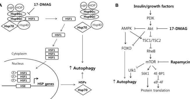
17-DMAG이 어떤 신호체계에 의해 골격근에서 autophagy 를 활성화시키는가는 불분명하지만 2가지 기전에 의해 au- tophagy의 증가시켰을 것이다. 첫째로, Fig. 6A에 묘사되어 있는 것처럼, 17-DMAG은 특정적으로 Hsp90의 ATP-binding site에 결합하여 Hsp90의 ATP-dependent 기능을 방해하여 transcription factor인 heat-shock factor1 (HSF1)을 활성화시 킨다[1, 15]. 스트레스가 낮은 상태에서 HSF1은 Hsp90 dock- ing complex 내에서 inactive 형태로 유지되지만 스트레스 반 응에 의해 HSF1은 Hsp90 complex에서 분리된다. 그리고 나 서 HSF1 단백질은 핵으로 이동하여 인산화된 후 target 유전 자들(주로 heat shock proteins)의 promoter에 결합하여 이들 유전자들(e.g. Hsp72)의 발현을 촉진시킨다[35]. Fig. 2와 4에 서 보여진 증가된 Hsp72 단백질 수준은 이러한 기전에 의한 것일 것이다. HSF1에 의해 증가된 HSPs는 주로 protein fold- ing이나 proteasomal degradation을 촉진시키지만 최근의 연 구에 의하면 HSPs가 autophagy와도 연결되어 있다고 한다.
예를 들어, BAG3 (Bcl2-associated athanogene, Hsp70 co- chaperone)와 결합하는 Hsp72와 HspB8 (small HSP)가 au- tophagy flux를 증가시키고 신경퇴행성 질환에 의해 생기는 단백질 응집체(aggregates)의 제거를 증대시켰다[2, 3]. 또 다 른 small HspB7 또한 autophagy을 이용하여 단백질 응집체의 제거를 촉진시켰다[33]. 따라서 HSF1은 HSPs의 발현을 촉진 시켜 간접적으로 autophagy의 유도를 증가시킬 수 있을 것이 다(Fig. 6A).
둘째로, Hsp90는 Akt-dependent signaling을 조절하는 것 으로 알려져 있다. Akt 단백질은 Hsp90의 client protein으로 Hsp90 단백질과 결합(Akt-Hsp90 complex)되었을 때 Akt- kinase 단백질은 안정화되고 Hsp90와 분리될 경우 UPS에 의 해 분해된다[29]. 따라서 본 연구에서 17-DMAG 처치에 의한 Hsp90의 억제가 Akt 단백질의 UPS에 의한 분해를 촉진시켰 을 것이다. 그 결과 17-DMAG 처치 그룹에서 Akt 단백질 수준 이 유의하게 감소한 것을 알 수 있다(Fig. 2 and 4). 감소된 Akt signaling에 의한 mTOR의 억제(↓ Akt → ↑ TSC2 →
↓ Rheb → ↓ mTOR)가 Ulk1 (unc-51 like autophagy activat- ing kinase 1, 초기 autophagosome 형성에 필요한 단백질)의
A B
Fig. 6. A. Potential model of 17-DMAG action on autophagy via the heat shock response. B. Simplified scheme of Akt-mTOR signaling pathway and the molecular targets of rapamycin and 17-DMAG.
증가를 통해 autophagy의 유도가 일어났을 것이다(Fig. 6B).
본 연구에서 17-DMAG 처치에 의해 autophagy의 활성화는 Hsp72 단백질 발현의 증가 그리고/또는 Akt-mTOR signaling 의 감소를 통해 일어난 것으로 생각된다.
본 연구는 autophagy flux assay (LC3 II와 p62 단백질 turn- over)를 활용하여 17-DMAG (Hsp90 억제제/Hsp72 활성제) 처치가 근육세포에서 autophagy에 미치는 영향을 살펴보았 다. 17-DMAG 처치가 C2C12 myotube와 마우스 골격근에서 Hsp90 억제를 통해 Hsp72 발현을 증가시켰고 Akt 단백질 분 해를 촉진하여 Akt-mTOR signaling pathway를 감소시켜 au- tophagy를 유도시킬 수 있는 잠재성을 보여주었지만 autoph- agy flux의 유의한 증가가 마우스 골격근에서만 관찰되었다.
17-DMAG이 골격근의 Hsp90를 억제시켜 Hsp72를 유전자 발현을 촉진시키고 mTOR를 감소시키는 강력한(potent) au- tophagy inducer라는 것을 보여주었다. 골격근에서 17-DMAG 의 autophagy 조절기전을 더 자세히 연구해보기 위해, 유전자 변형 쥐 모델(예, Hsp90-ATP binding 손상 쥐나 Hsp72-과발 현 쥐 또는 Hsp72-결핍된 쥐)을 이용하여 골격근에서 이 heat shock protein들에 의한 autophagy에 대한 기능과 역할을 더 정확히 연구해 볼 수 있을 것으로 기대해 본다. 본 연구를 기반 으로 골격근에서 17-DMAG의 몇 가지 유용성을 제시해볼 수 있겠다. 17-DMAG 처치를 통해 근육 손상이나 미사용 때 생길 수 있는 근감소증(muscle atrophy)을 Hsp72의 발현을 촉진시 켜 근감소증의 완화를 시도해 볼 수도 있겠으며 골격근에서 autophagy의 감소 현상이 일어날 수 있는 증상(예, 당뇨병, 노화된 근육, 근질환)을 17-DMAG을 처치하여 autophagy 조 절을 통해 골격근의 노화/대사성 증상 등을 완화시킬 수 있는
가능성을 제시해 볼 수 있겠다.
References
1. Bagatell, R., Paine-Murrieta, G.. D., Taylor, C. W., Pulcini, E. J., Akinaga, S., Benjamin, I. J. and Whitesell, L. 2000.
Induction of heat shock factor1-dependnent stress response alters the cytotoxic activity of HSP90-binding agents. Clin.
Cancer Res. 6, 3312-3320.
2. Carra, S., Cripppa, V., Rusmini, P., Boncoraglio, A., Minoia, M., Giorgetti, E., Kampinga, H. H. and Poletti, A. 2012.
Alteration of protein folding and degradation in motor neu- ron diseases: Implications and protective functions of small heat shock proteins. Prog. Neurobiol. 97, 83-100.
3. Carra, S., Brunsting, J. F., Lambert, H., Laudry, J. and Kampinga, H. H. 2009. HspB8 participates in protein quality control by a non-chaperone-like mechanism that requires eIF2alpha phosphorylation. J. Biol. Chem. 284, 5523-5532.
4. Ching, J. K., Ju, J. S., Pittman, S. K., Margeta, M. and Weihl, C. C. 2013. Increased colchicine-induced muscle toxicity.
Autophagy 12, 2115-2125.
5. Criollo, A., Senovilla, L., Authier, H., Maiuri, M. C., Morselli, E., Vitale, I., Kepp, O., Tasdemir, E., Galluzzi, L., Shen, S., Tailer, M., Delahaye, N., Tesniere, A., De Stefano, D., Younes, A. B., Harper, F., Pierron, G., Lavandero, S., Zitvogel, L., Israel, A., Baud, V. and Kroemer, G. 2010. The IKK complex contributes to the induction of autophagy.
EMBO J. 29, 619-631.
6. Cuervo, A. M. and Wong, E. 2014. Chaperone-mediated au- tophagy: roles in disease and aging. Cell Res. 24, 92-104.
7. Dokladny, K., Zuhl, M. N., Mandell, M., Bharttacharya, D., Schneider, S., Derectic, V. and Moseley, P. L. 2013.
Regulatory coordination between two major intracellular homeostatic systems: heat shock response and autophagy.
J. Biol. Chem. 288, 14959-14972.
8. Ferat-Osorio, E., Sanchez-Anaya, A., Gutierrez-Mendoza, M., Bosco-Garate, I., Wong-Baeza, I., Pastelin-Palacios, R., Pedraza-Alva, G., Bonifaz, L. C., Cortes-Reynosa, P., Perez- Salazar, E., Arriaga-Pizano, L., Lopez-Macias, C., Rosenstein, Y. and Isibasi, A. 2014. Heat shock protein 70 down-regu- lates the production of toll-like receptor-induced pro-in- flammatory cytokines by a heat shock factor-1/constitutive heat shock element-binding factor-dependent mechanism. J.
Inflamm. 11, 1476-1492.
9. Goldberg, A. L. 2003. Protein degradation and protection against misfolded or damaged proteins. Nature 426, 895-899.
10. Gusarova, V., Caplan, A. J., Brodsky, J. L. and Fisher, E.
A. 2001. Apoprotein B degradation is promoted by the mo- lecular chaperones hsp90 and hsp70. J. Biol. Chem. 276, 24891-24900.
11. Hartl, F. U. and Hayer-Hartl, M. 2002. Molecular chaperones in the cytosol: from nascent chain to folded protein. Science 295, 1852-1858.
12. Ju, J. S., Fuentealba, R. A., Miller, S. E., Jackson, E., Piwnica- Worms, D., Baloh, R. H. and Weihl, C. C. 2009. Valosin-con- taining protein(VCP) is required for autophagy and is dis- rupted in VCP disease. J. Cell Biol. 187, 875-888.
13. Ju, J. S., Varadhachary, A. S., Miller, S. E. and Weihl, C.
C. 2010. Quantitation of “autophagic flux” in mature skel- etal muscle. Autophagy 6, 929-935.
14. Kim, D. S, Li, B., Rhew, K. Y, Oh, H. W, Lim, H. D, Lee, W., Chae, H. J. and Kim, H. R. 2012. The regulatory mecha- nism of 4-phenylbutyric acid against ER stress-induced au- tophagy in human gingival fibroblasts. Arch. Pharm. Res. 35, 1269-1278.
15. Lu, A., Ran, R., Parmentier-Batteur, S., Nee, A. and Sharp, F. R. 2002. Geldanamycin induces heat shock proteins in brain and protects against focal cerebral ischemia. J.
Neurochem. 81, 355-364.
16. Meacham, G. C., Lu, Z., King, S., Sorscher, E., Tousson, A.
and Cyr, D. M. 1999. The Hdj-2/Hsc70 chaperone pair facili- tates early steps in CFTR biogenesis. EMBO J. 18, 1492-1505.
17. Meley, D., Bauvy, C., Houben-Weerts, J. H. Dubbelhuis, P.
F., Helmond, M. T., Codogno, P. and Meijer, A. J. 2006.
AMP-activated protein kinase and the regulation of auto- phagic proteolysis. J. Biol. Chem. 281, 34870-34879.
18. Mizushima, N., Yoshimori, T. and Ohsumi, Y. 2011. The role of Atg proteins in autophagosome formation. Annu. Rev.
Cell Dev. Biol. 27, 107-132.
19. Ogier-Denis, E., Pattingre, S., El Benna, J. and Codogno, P.
2000. Erk1/2-dependent phosphorylation of Gα-interacting protein stimulates its GTPase accelerating activity and au- tophagy in human colon cancer cells. J. Biol. Chem. 275, 39090-39095.
20. Palacios, C., Lopez-Peres, A. I. and Lopez-Rivas, A. 2010.
Down-regulation of RIP expression by 17-dimethylamino- ethylamino-17-demethoxygeldanamycin promotes TRAIL- induced in breast tumor cells. Cancer Lett. 287, 207-215.
21. Palacios, C., Martin-Perez, R., Lopez-Perez, A. I., Pandiella, A. and Lopez-Rivas, A. 2010. Autophagy inhibition sensi- tizes multiple myeolma cells to 17-dimethylaminoethylamino- 17-demethoxygeldanamycin-induced apoptosis. Leuk. Res.
34, 1533-1538.
22. Pattingre, S., Bauvy, C. and Codogno, P. 2003. Amono acids interfere with the ERK1/2-dependent control of macro- autophagy by controlling the activation of Raf-1 in human colon cancer HT-29 cells. J. Biol. Chem. 278, 16667-16674.
23. Qin, L., Wang, Z., Tao, L. and Wang, Y. 2010. ER stress negatively regulates Akt/TSC/mTOR pathway to enhance autophagy. Autophagy 6, 239-247.
24. Ren, Y., Huang, F., Liu, Y., Yang, Y., Jiang, Q. and Xu, C.
2009. Autophagy inhibition through PI3K/Akt increases apoptosis by sodium selenite in NB4 cells. BMB Rep. 42, 599-604.
25. Riedel, M., Goldbau, O., Schwartz, L., Schmitt, S. and Richter-Landsberg, C. 2010. 17-AAG induces cytoplasmic α- synuclein aggregate clearnce by induction of autophagy.
Plos One 5, e8753.
26. Rubinsztein, D. C., Codogno, P. and Levine, B. 2012.
Autophagy modulation as a potential therapeutic target for diverse diseases. Nat. Rev. Drug Discov. 11, 709-730.
27. Rubinsztein, D. C., Cuervo, A. M., Rauikuma, B., Sarkar, S., Korolchuk, V., Kaushik, S. and Klionsky, D. J. 2009. In search of an “autophagomometer”. Autophagy 5, 585-589.
28. Ryter, S. W. and Choi, A. M. 2013. Autophagy: An integral component of the mammalian stress response. J. Biochem.
Pharmacol. Res. 1, 176-188.
29. Sato, S., Fujita, N. and Tsurun, T. 2000. Modulation of Akt kinase activity by binding to Hsp90. Proc. Natl. Acad. Sci.
USA 97, 10832-10837.
30. Senf, S. M. 2013. Skeletal muscle heat shock protein 70: di- verse functions and therapeutic potential for wasting disorders. Front Physiol. 4, 330.
31. Senf, S. M., Dodd, S. L., McClung, J. M. and Judge, A. R.
2008. Hsp70 overexpression inhibits NFkB and Foxo3a tran- scriptional activities and prevents skeletal muscle atrophy.
FASEB J. 22, 3836-3845.
32. Soti, C., Nagy, E., Giricz, A., Vigh, L., Csermely, P. and Ferdinandy, P. 2005. Heat shock protein s as emerging ther- apeutic targets. Br. J. Pharmacol. 146, 769-780.
33. Vos, M. J., Zijlstra, M. P., Kanon, B., Van Waarde-Verhagen, M. A., Brunt, E. R., Oosterveld-Hut, H. M., Carra, S., Sibon, O. C. and Kampinga, H. H. 2010. HSPB7 is the most potent polyQ aggregation suppressor within the HSPB family of molecular chaperones. Hum. Mol. Genet. 19, 4677-4693.
34. Wang, A. M., Morishima, Y., Clapp, K. M., Peng, H. M., Pratt, W. B., Gestwicki, J. E., Osawa, Y. and Lieberman, A.
P. 2010. Inhibition of hsp70 by methylene blue affects signal- ing protein function and ubiquitination and modulates pol- yglutamine protein degradation. J. Biol Chem. 285, 15714- 15723.
35. Yang, J., Carra, S., Zhu, W. G. and Kampinga, H. H. 2013.
The regulation of the autophagic network and its im- plications for human disease, Int. J. Biol. Sci. 9, 1121-1133.
초록:17-DMAG이 마우스 골격근에서 autophagy flux에 미치는 영향
주정선1*․이유현2
(1수원대학교 스포츠과학과, 2수원대학교 식품영양학과)
본 연구는 17-DMAG이 골격근에서 autophagy에 관여하는 가를 조사하기 위해, C2C12세포와 마우스 골격근에 서 17-DMAG (Hsp90 억제제/Hsp72 활성제)을 처치하는 그룹과 autophagy 억제제(Bafilomycin 또는 colchicine) 를 처치하는 그룹과 처치하지 않는 그룹을 동시에 두고 autophagy flux를 측정하였다. C2C12 배양세포에서 17-DMAG이 Hsp90 억제/hsp72 활성화시켰으며 Akt-mTOR 신호체계를 유의하게 감소시켰지만(p<0.05) autoph- agy marker 단백질인 LC3 II와 p62를 증가시키지 않았다. In vivo 모델의 경우 17-DMAG 처치가 배양세포에서 발견된 것처럼 Hsp90억제/hsp72를 활성화시켰고 Akt-mTOR 신호체계를 유의하게 감소시켰다(p<0.05). 반면 LC3 II와 p62 단백질 수준은 autophagy 억제제(colchicine) 처치 수준보다 더 높게 증가되었다. 이는 17-DMAG이 골격 근에서 autophagy를 증가시키지만 C2C12 배양세포에서는 autophagy의 활성화가 제한적임을 암시한다. 현재 이 러한 in vitro와 in vivo 모델에서의 차이는 불분명하다.
36. Yang, Y., Janich, S., Cohn, J. A. and Wilson, J. M. 1993. The common variant of cyctic fibrosis transmembrane con- ductance regulator is recognized by hsp70 and degraded in a pre-Golgi nonlysosomal compartment. Proc. Natl. Acad.
Sci. USA 90, 9480-9484.
37. Zhao, J , Brault J. J., Schild A., Cao, P., Sandri, M., Schiaffino, S, Lecker, S. H. and Goldberg, A. L. 2007. FoxO3 coordin- ately activates protein degradation by the autophagic/lyso- somal and proteasomal pathways in atrophying muscle cells. Cell Metab. 6, 472-483.