Korean J. Environ. Biol. 34(3) : 141~150 (2016) http://dx.doi.org/10.11626/KJEB.2016.34.3.141
IntroductIon
Marine dinoflagellates are a major component of phyto- plankton communities and play an important role as prima- ry producer in marine ecosystems. Some of these species can cause red tides and some produce neuro-toxins that can accumulate in the food chain (Hallegraeff 1993). According to Kim et al. (2013), to date, a total of 153 planktonic dino- flagellates have been described in Korean waters. However, the morphological characteristics of the dinoflagellates were not described in most studies, and few taxonomic studies on
dinoflagellates, apart from harmful species, have been con- ducted.
To observe dinoflagellates, sea water samples collect- ed along the Korean coasts have typically been fixed in formaldehyde and Lugol’s iodine solution. According to Stoecker et al. (1996), Lugol’s solution minimizes cell loss, however causes cell shrinkage and distortion. In addition, for phytoplankton in particular, the fixative stains the cells dark brown, whereas the use of formaldehyde causes only minor shrinkage and distortion, however results in the loss of cells, at a rate ranging from less than 20% (Stoecker et al. 1989) to more than 70% (Leakey et al. 1994). This indi- cates that the use of samples fixed with the commonly used Lugol’s solution and formaldehyde may cause difficulties in
* Corresponding author: Hyeon Ho Shin, Tel. 055-639-8429, Fax. 055-639-8440, E-mail. [email protected]
ⓒ 2016. Korean Society of Environmental Biology.
Morphological Features of Marine Dinoflagellates from Jangmok Harbour in Jinhae Bay, Korea:
A Case of 30 Species in the Orders Prorocentrales, Dinophysiales, Gonyaulacales and Gymnodiniales
Hyeon Ho Shin*, Eun Song Kim, Zhun Li, Joo Yeon Youn, Seul Gi Jeon and Seok Jin Oh
1Library of Marine Samples, Korea Institute of Ocean Science and Technology,
Geoje 53201, Republic of Korea
1
Pukyung National University, Busan 48513, Republic of Korea
Abstract - Most previous studies on dinoflagellates in Korean coastal areas were conducted without morphological descriptions and illustrations of the observed dinoflagellates. This indicates that the species and diversity of dinoflagellates may have been respectively misidentified and underestimated in the past, probably due to cell shrinkage, distortion and loss caused by sample fixation. This study provides information on the morphological observations of four dinoflagellate orders (Prorocentrales, dinophysiales, Gonyaulacales and Gymnodiniales) from Jangmok Harbour in Jinhae Bay, Korea. The unfixed samples were collected weekly from December 2013 to February 2015. A total of 13 genera and 30 species were identified using light and scanning electron microscopy, although some samples were not clarified at the species level. Harmful dinoflagellates, Prorocentrum donghaiense, Tripos furca, Alexandrium affine, A. fundyense, Aka
shiwo sanguinea and Cochlodinium polykrikoides, were identified based on the morphological observations. The results also reflect the occurrence and identification of dinoflagellates that had not been previously recorded in Jangmok Harbour.
Key words : dinoflagellate, morphological observation, Jangmok Harbour, Jinhae Bay
<Review paper>
optical microscopy, for quantitative and qualitative analyses of phytoplankton as well as species identification.
The taxonomic identification of dinoflagellates is gener- ally based on a unique combination of characteristics such as the size and shape of the cells, their colour, surface orna- mentation, cingulum displacement, position of the nucleus, the shape of the chloroplasts and thecal plate tabulation and variation for armored or thecate species (e.g. Balech 1995).
These features of dinoflagellates may not be observed clear- ly in fixed samples, suggesting that dinoflagellates observed using fixed samples can be misidentified. Many ecological studies on dinoflagellates in Korean coastal areas have been undertaken, which have concentrated on understanding the relationship between the dinoflagellate community and environmental factors, with an emphasis on harmful algal species (e.g. Yoo 1982; Yoo and Lee 1987; Lee et al. 1992;
Lee et al. 1998; Park et al. 2009; Baek et al. 2011; Park et al. 2012). However, the samples used in these studies were mostly fixed in formaldehyde and Lugol’s solution, and some of the identified dinoflagellates have only ever been described and illustrated in Korean coastal areas.
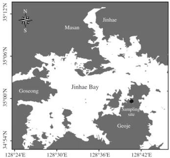
Jinhae Bay is a semi-enclosed embayment with minimal advection and restricted water circulation (Fig. 1). Since the early 1980s, red tides have occurred annually in this Bay, causing the mortality of fishery organisms with major eco- nomic consequences (Han et al. 1992). Jangmok Harbour is located on the northern side of Geoje Island in Jinhae Bay, where red tides caused by Akashiwo sanguinea have occasionally occurred, although these have not been report- ed in the scientific literature. Morphological descriptions and illustrations of the dinoflagellates in this bay have also rarely been reported. To determine the morphological char- acteristics of naturally occurring members of dinoflagellate community, we analysed unfixed water samples collected in Jangmok Harbour and documented dinoflagellates of the orders Prorocentrales, Dinophysiales, Gymnodiniales and Gonyaulacales.
MAterIAls And Methods
Dinoflagellates were collected weekly from December 2013 to February 2015 in Jangmok Harbour, Jinhae Bay (34°
59′39.1″N, 128°40′28.8″E), by using a plankton net with a 20-μm mesh (Fig. 1). One millilitre of the sample was placed in a Sedgwick-Rafter chamber, and the cells were isolat- ed under an inverted microscope (Primo vert, Carl Zeiss, Göttingen, Germany), and then identified and photographed using a Zeiss AxioCam MRc digital camera on an Axio Imager 2 upright microscope (Carl Zeiss, Göttingen, Ger- many). The identified cells were transferred into the indi- vidual wells of 96 well tissue plates filled with F/2 culture medium and a 30 mL culture flask by micropipetting using a capillary pipette and maintained at 20℃ and ca 100 μmol photons m
-2s
-1cool-white illumination under a 14L : 10D photo-cycle. The dinoflagellates reported in this study, ex- cluding heterotrophic species, were deposited at the Library of Marine Samples, Korea Institute of Ocean Science and Technology.
For scanning electron microscopy (SEM), the cells were collected with a micropipette and fixed with Lugol’s io- dine solution. Fixed cells were rinsed twice with deionised water, transferred onto filters and then left to dry at room temperature. These filters were mounted on stubs, coated with platinum-palladium and examined in a field emission scanning electron microscope JEOL JSM 7600F (JOEL Ltd., Tokyo, Japan).
35°12′N35°06′N35°00′N34°54′N
128°24′E
Jinhae Masan
Goseong
Geoje Sampling
site
Jinhae Bay
NS
128°30′E 128°36′E 128°42′E